2. Gram (+) use sugar as (E) and saliva
as(C)
Bacteria that predominate (mature
plaque)
Anaerobic & Asaccharolytic
(Fe) for P. gingivalis
3. Active periodontitis
Susceptible host
The presence of pathogenic species
The absence or a small proportion of
beneficial bacteria
4. Nonspecific Plaque
Hypothesis
Only certain plaque is pathogenic
Because of producing substances that
mediate the destruction of host
tissues
5. Complicating factors
Identification of bacterial pathogen has
been difficult
Periodontal disease (periodic episodes)
Presence of pathogens
6. Criteria for identification of
periodontal pathogens
Robert Koch
MUST BE:
1. Routinely isolated
2. Grown in pure culture
3. Produce similar disease
4. Recovered from lesion
7. Periodontal health & disease
Coccal cell , motile Rod , Spirochetes
In culture:
In healthy area ( gram + of Rod &
coccal )
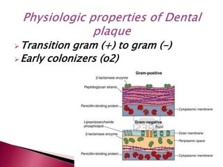
8. Gram + facultative & Streptococcus &
Actinomyces (S. sanguis , S. mitis , A.
viscosus , A. naeslundii)
Gram – (p. intermedia , F. nucleatum ,
Capnocytophaga , Neisseria ,
Veillonella )
9. Studied in model referred to
experimental gingivitis
Gram +….. (56%)
Gram – ….. (44%)
Gingivitis due to pregnancy (steroid
&p.intermedia in GCF)
10. Found in adult population
High rate of Spirochetes
Anaerobic….. (90%)
Gram (-)……. (75%)
In addition :
C.rectus , F.nuclutum , p.gingivalis ,
p.intermdia , A.a
11. MICROBIAL SHIFT DURING DISEASES
In health , gingivitis , periodontitis
1. From G (+) to G (–)
2. From Cocc to Rod
3. From non motile to motile
4. From facultative to obligate anaerobic
5. From fermenting to proteolytic
13. P.intermedia & Spirochete in NUG
Penetrate necrotic tissue in unaffected
connective tissue
14. Increase of periodontal pocket
P.intermedia , P.gingivalis , F. Nuclueatum
T.forsthiya , p.micros
15. Infection around implants
Microbial shifts same as periodontitis
High rate of Gram – Rod , Motile
organism , Spirochete
Also other microorganism are exist
19. causes of gingival inflammation :
1. Bacterial plaque & Calculus
2. Faulty restoration & orthodontic problems
3. Self inflicted injuries
4. Use of tobacco
5. Poor oral hygiene
20. Calculus consist of mineralized
bacterial plaque that forms on the
surfaces of natural teeth and dental
prosthesis
Its classified as :
Supragingival & subgingival
21. Located coronally
Visible in oral cavity
White or whitish yellow
Hard with claylike
Espicially :
1. Lingual area of mandible (anterior)
2. Buccal area of first maxillary molar
22. Located below the crest of the marginal
gingiva
Not visible & hard ,dense
Dark brown or greenish black
Firmly attach to the tooth
23.
24. Buccal area of maxillary molars
Anterior lingual area of mandibular
Both of them seen in Radiography
Interproximal calculus (Radio Opacity)
25. Inorganic content:
About 70%-90%
Calcium phosphate , calcium
carbonate , magnesium phosphate
and other minerals
26. Four main crystals
1. Hydroxyapatite (58%)
2. Magnesium whitlokite (21%)
3. Octacalcium phosphate (12%)
4. Brushite (9%)
27. Complex of protein –polysaccharide
EP cell
Leukocytes
Carbohydrates
Lipid & Amino acid
28. Four mode of attachment :
1. Organic pellicle
2. Mechanical locking (pits)
3. Close adaptation
4. Penetration of calculus bacteria into
cementum
calculocementum
29. Its dental plaque undergone mineralization
1st – 14th days
Calcifying plaques 50% in 2 days & 60%-90%
in 12 days
Source of mineralization :
1. supragingival (saliva)
2. subgingival (GCF)
30. Concentration of :
Microorganism , leukocytes , Epithelial
cell mixture of salivary protein and
lipid
its removable
Pigmented deposit on tooth surface
called Dental stain