
More Related Content
Viewers also liked
Viewers also liked (15)
Similar to Pet
Similar to Pet (20)
Pet
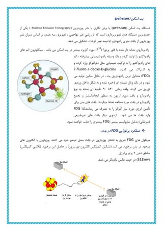
- 1. ( اسکن پتpet-scan) ( اسکن پت دستگاهpet-scanبرش یا )( پوزیترون نشر با نگاریPositron Emission Tomographyاز یکی ) نشر میزان اساس بر بعدی سه تصویری ، تهاجمی غیر روشی با که است تصویربرداری های دستگاه جدیدترین .دهد می تشکیل ، )کوتاه عمر نیمه (با رادیودارو حاوی بافت از پوزیترون ( پرتوزا فلور با شده دار نشانه رادیودارویF18 کاربرد مورد )بیشترباشد می اسکن پت در.های اتم سیکلوترون اتم ، پیشرفته رادیوشیمیایی وسیله یک و کرده تولید را رادیواکتیو و کرده وارد دئوگلوکز مثل شیمیایی ترکیب به را رادیواکتیو های .گذارد می اشتراک به2-fluoro-2-deoxy-D-glucose (FDGمی تولید سالین حالل در ، پت رادیوداروی ترین متداول ) د و شودوریدی داخل شکل به و شده ذخیره ای شیشه ویال یک ر .گردد می تزریقزمانی وقفه30تا90نوع به بسته ای دقیقه تجمع و ایجاد،انتشار منظور به آزمون مورد بافت و رادیودارو .میگردد لحاظ مطالعه مورد بافت در رادیوداروبرای بدن های بافت به را گلوکز نیاز مورد انرژی تأمینرسانند،لذا می مصرفFDG غیرطبیعی های بافت دیگر ازسوی . شود می ها بافت وارد ،بیشتر متابولیسم بدلیل )(سرطانیFDG.نمود خواهند جذب را بیشتری پرتوزایی عملکردFDG: بدن در های مولکولFDGپوزیترون .کنند می خود تجمع محل بافت در پوزیترون انتشار به شروعبالکترون اهای الکترون کمپلکس (تشکیل کند می برخورد بدن در موجود-)پوزیترون)کمپلکس (تالشی برخورد این حاصل و شدن ساطع2پرانرژی پرتو (511kevمی یکدیگر عکس جهت در ).باشد
- 2. همه در بدن از پرتوها جفت از بیشماری تعداد تونل دیواره به و شوند می منتشر جهات دستگاهبه ها پرتو این ثبت . کنند می برخورد تع هدفو تجزیه و یک هر انتشار مرکز یین سیستم بکمک تصاویر انطباق و کردن تحلیل ایجاد را بعدی سه تصویری ، کامپیوتری های کند می. :سیکلوترون دستگاه سیکلوترونشکل به خالی تو و بزرگ مغناطیسی اهنربای دو از که است خطی شتابدهنده یکDتولید برای : از عبارتند آن دهنده تشکیل اجزای .کند می استفاده یکنواخت مغناطیسی میدان 1.الکتریکی نوسانگر(Oscillator):حدود در پتانسیل اختالف ایجاد نوسانگر این وظیفه105می ولت جهت و باشد.شود می عوض بار میلیونها ثانیه هر در پتانسیل اختالف این 2.چشمهیونی(Ion source):هیدروژن بمباران با آن در که دارد قرار سیکلوترون مرکز در یونی چشمه ها سیکلوترون وارد یونها این و شود می تولید )(دوترون مثبت های یون انرژی پر الکترونهای توسط سنگین .گیرد می شتاب باال پتانسیل اثر در و شده 3.(قوی بسیار الکتریکی آهنربای6/1:)تسالم آهنرباها این.شود می ها دوترون دورانی و انحراف وجب 4.خال مخزن(vacuum tank):میگیرد بر در را اجزا همه کهجیوه میلیمتر میلیونیم یک تا را هوا فشار و .باشند داشته را هوا های مولکول با برخورد حداقل یونها تا .دهد می کاهش :پت آشکارسازهای
- 3. کریستال زیادی تعداد از پت سیستم در( آشکارساز8000تا12000مهم دالیل از یکی خود که شود می استفاده ) هر و .شود می محسوب بیمارستانی تجهیز این بودن گران باالتری وضوح و کیفیت باشد بیشتر آشکارسازها تعداد چه .داد خواهد ارائه را اینهای کریستالآشکارسازبلوک مجموعه صورت به که دیواره در آشکارساز های،دارند قرار دستگاه)(فوتون پرتو را ورودیکرده تبدیل الکتریکی سیگنال یک بهومجموعه سیگنالهایالکتریکیبرایتصویرسازیبهکارمیروند.
- 4. ر:آشکارسازی وش روش دراین .است )فرودی انطباقی(هم پت،آشکارسازی سیستم آشکارسازی روش2یکدیگر مقابل در آشکارساز قرار( شوند می داده180گسیل پرتو که گردد می تصویر زمانی )الکترون/پوزیترون (تالشی واقعه یک و )درجه .گردد دریافت ، هم مقابل ساز آشکار دو هر توسط همزمان طور به شدهفوتون ناچیز خطای درصد با روش این . کند می فیلتر را هدف غیر های :نوین های اسکن پت آشکارساز های حلقههستند حلقه طول در هایی شکاف دارای که اند شده ساخته مکعبی بلوک چندین از متداول شناسایی بدون ها فوتون از تعدادی میدهند اجازه وگروه ،کنند عبورپزشکیIFAبلوک شکل تغییر با خالی فضای ،ای ذوزنقه ساختاری به آشکارسازهااز استفاده طرفی از و کرده پر راسنسورctdeباجامد ساختار موجب متداول کریستالی سنسورهای بجایکه اشتباه های سیگنال و یابد بهبود انرژی پذیری تفکیک تا گشته از/الکترون پیونداند نگرفته سرچشمه پوزیتروناثر بی.شوند :اسکن پت تصویربرداری کاربردهای کاربردPET: سرطان در از قبل بیماریها و نارسائیها بیشتر دررخ متابولیسم در تغییراتی شود ایجاد بافت در آناتومیک تغییرات اینکه این از میدهدیعنی مرسوم تصویربرداری های سیستم خالف بر روCTوMRIیا و آناتومیکی تغییرات که SPECTسیستم از استفاده با کند می گیری اندازه را فیزیولوژیکی تغییرات کهPETکه بود خواهیم قادر ما می چنانچه و بپردازیم آن درمان به و جلوگیری بیماری پیشرفت از و دهیم تشخیص اولیه مراحل در را بیماری می شنیده بسیار است برخوردار حیاتی اهمیت از درمان برای هنگام زود تشخیص سرطان مورد در دانیمکه شود مجد و کرده رشد دوباره تومور ، مغز از سرطانی تومور جراحی از پسوضعیت این .کند می مشکل دچار را بیمار ًاد داد تشخیص تومور توده میتوان فقط موجود تصویربرداری های سیستم از استفاده با که آید می پیش دلیل این به از استفاده با صورتیکه در .سازد می خارج را تومور توده تنها مزبور زتصاویر ا استفاده با جراح وPETموضع تنها نه
- 5. تومورمتابولیسم نرخ میدانیم چنانچه .میگردد مشخص نیز آن پیشروی شرایط و جهت بلکه شود می مشخص است ممکن چند هر اند شده سرطانی سلولهای به مبتال که بافتهایی اینرو از .میرود باال سرطانی بافتهای در گلوکز سیستم با نباشند تشخیص قابل دیگر تصویربرداری های سیستم باPETبرر ومی تشخیص قابل متابولیسم سی . سازد خارج نیز را مزبور بافتهای بموقع تا یابد می امکان جراح لذا و باشند سیستم از استفاده باPET.نمود ارزیابی نیز را سرطانی بیماران برای شده انتخاب درمان روش صحت میتوان زمانی مدت نیازمند تومور در آناتومیک تغییرات بصورت درمانی شیمی و رادیوتراپی درمانهای اثرات مشاهده تصویربردا مرسوم روشهای با بتوان تا است طوالنیرروش چنانچه درحالیکه داد تشخیص را آنها یتجویز درمان توسط را تومور گلوکز متابولیسم کاهش میتوان کوتاهی مدت از پس باشد مناسب شدهPETدر .نمود مشاهده باید و نیست موثر شده تجویز درمان روش که است این نشانه باشد نداشته وجود متابولیسم کاهش چنانچه مقابل هن زود اطالعات .نمود انتخاب درمان جهت را دیگری روشتوسط که گامیPETبرای باشند می دسترسی قابل سیستم .اند حیاتی بسیار بیمار جان نجاتPET( ثانویه سرطانهای بخصوص و سرطانها انواع تشخیص در .دارد توجهی قابل برتری تصویربرداری های سیستم دیگر به نسبت ) متااستازها ؟ کند می محدود را پوزتیرون نشر یا توموگرافی چیزی چه نباشد نرمال بسیار شیمیایی تعادل اگرPETیا و باشد دیابتی بیمار اگر مخصوصا بدهد غلطی نتایج تواند می که این دلیل به است ممکن اند نداشته پرهیز آزمون از قبل زمان مدت در که ها آن از بسیاریقندخونو انسولینخونهمسطحشدهانداثربگذارد. پت-سیتی(PET-CT): ترکیب پیشنهادPETوCTسال در1991اسکنرهای اصلی نمونه ولی شد مطرحPET/CTسال در1998کامل سال در شد استفاده پزشکی مراکز در که آن طراحی اولین .شد2001سال از .بود2001فروشنده تمام کنون تا طراحی یک حداقل پزشکی های دستگاه هایPET/CTسال از نتیجه در اند کرده تولید را2006فروش PET/CTجایگزینPETاست شده. اسکنر میان از ابتدا بیمار دستگاه یک درCTاسکنر وارد سپس و شود می ردPETبرای مختلف روش شود.دو می از استفادهPET/CTدارد وجود: 1.های دادهCTبهPETتصاویر کیفیت که است کافی مورد این در .است شده اضافهCTکه باشد حدی در مورفو ساختارهایتصویربرداری نتیجه در .شوند مشخص لوژیCTحاجب ی ماده به نیاز بدون پایین دز یک با
- 6. شود می انجام.تصاویر اطالعاتCTتصاویر تضعیف تصحیح برایPETبا مقایسه در کاربرد این .شود می استفاده سنتی تصویربرداریPETدارد باالتری تشخیص دقت و تر هزینه کم ،سریعتر. 2.از جداPETتصویربرداری ،CTانجام مناسب دقت و تشخیصی اطالعات آوردن دست به برای است ممکن تصویربرداری برای کامل دز از استفاده معنای به این .شودCTاست ضروری معموال حاجب ماده از استفاده .است شود می استفاده تنفسی پروتکل یک از و. ترکیب علتPETوCT: PET/CT.دارد آنکولوژی در باالتری تشخیصی قدرتPETو اندازه ،موقعیت نیز و بدن در متابولیکی سیگنالهای که زمانی مدت رادر سلولی فعالیت نوعCTمی فراهم را بدن داخل آناتومیکی جزئیاتمی آشکار کند.سازد میمحدوده برای تنهایی به تصویری آزمون یک از توانکه زمانی اما کرد استفاده کاربردها از وسیعی ی تصویری نتایجPETوCTمی استفاده مشترک طور بهو سرطان موقعیت از کاملی اطالعات ترکیبی تصویر ،شود می ما به متابولیسم.دهد مثال عنوان بهPETاز بهتر CTآشکار را مالنوما گسترش در راه مفیدترین ولی کند می باشد نمی اولیه تشخیص کرد عود مالنوما اگرPET Scanاز بهتر است ممکنCT Scanآیا که دهد نشان شده شروع که جایی از بیماری بدن های قسمت دیگر به . است شده منتشر ه: اسکن پت تصویربرداری مرکز زینه پت تصویربرداری مرکز هر احداث2.کند می طلب را باال گذاری سرمایه بعبارتی یا هزینهدستگاه خرید اول .رادیوداروها تولید برای سیکلوترون دوم و اسکن پت تصویربرداری
- 7. و بوده متفاوت ... و سازنده شرکت و رفته بکار سازهای آشکار تعداد و نوع و آن ابعاد به وابسته پت دستگاه قیمت درحدود ای هزینه2سرمایه نیز رادیوداروها تولید و سیکلوترون مرکز .شود می برآورد آن برای دالر میلیون .دارد را دالری میلیون یک از بیش گذاری آزمو هر هزینهبین ،گیرد صورت بدن بخش کدام از و باشد شهر حتی یا کشور کدام در که این به بسته نیز پت ن 2000تا20000.است متغییر دالر : اسکن پت کاربردهای و مزایا خالصه ( ها روش سایر به نسبت باال تفکیک قدرت2. )برابر . آن نتایج حصول تا انجام از کوتاه زمان صرف با غیرتهاجمی .فیزیولوژیکی و آناتومیک مطالعات بر عالوه بیوشیمی و متابولیسم مطالعه از زنده و بعدی سه تصویرسازی. ها عملکرد . بدن طبیعی فرآیندهای بر اثرگذاری عدم و کم اکسپوژ آن تبع به و زودگذر بسیار پرتوزایی . بدن سراسری اسکن با )ثانویه (سرطان متااستاتیک های کانون تشخیص . درمانی شیمی و داروها اثربخشی میزان و درمان روش صحت ارزیابی کاردیولوژی در کاربرد-نورولوژی-. تومورشناسی : معایب بسیارباال های هزینه . بیمار شیمیایی تعادل نبودن نرمال صورت در غلط نتایج امکان . انسولین و قند شدن همسطح بدلیل دیابتی بیمار روی بر انجام حساسیت . کند می تغذیه پت آزمون تحت مادر از که نوزادی یا و باردار بیمار جنین به آسیب امکان ( سنگین فوق بیمار158. شوند دستگاه وارد توانند نمی )+ از کمتر زمانی فاصله در رادیودارو تولید الزام2. تصویربرداری مرکز تا ساعت با مرتبط های بخش کنید مطالعه را پت فایل لطفا خطی شتلبدهنده و سیکلوترون بخش برایhigh light.است شده مشخص