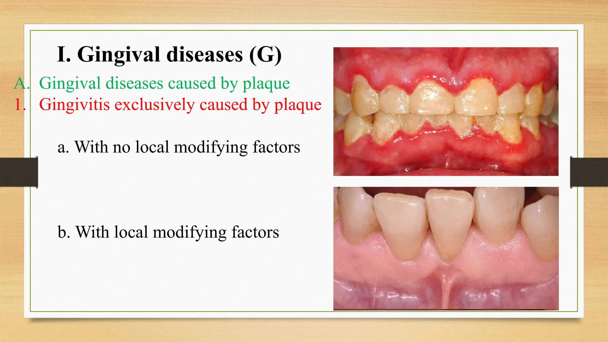
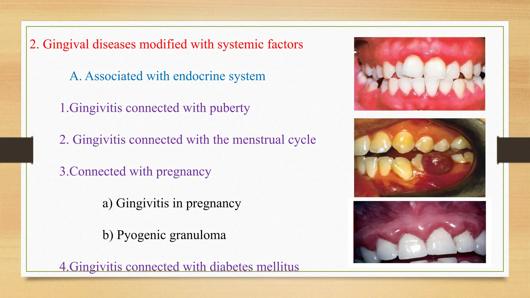
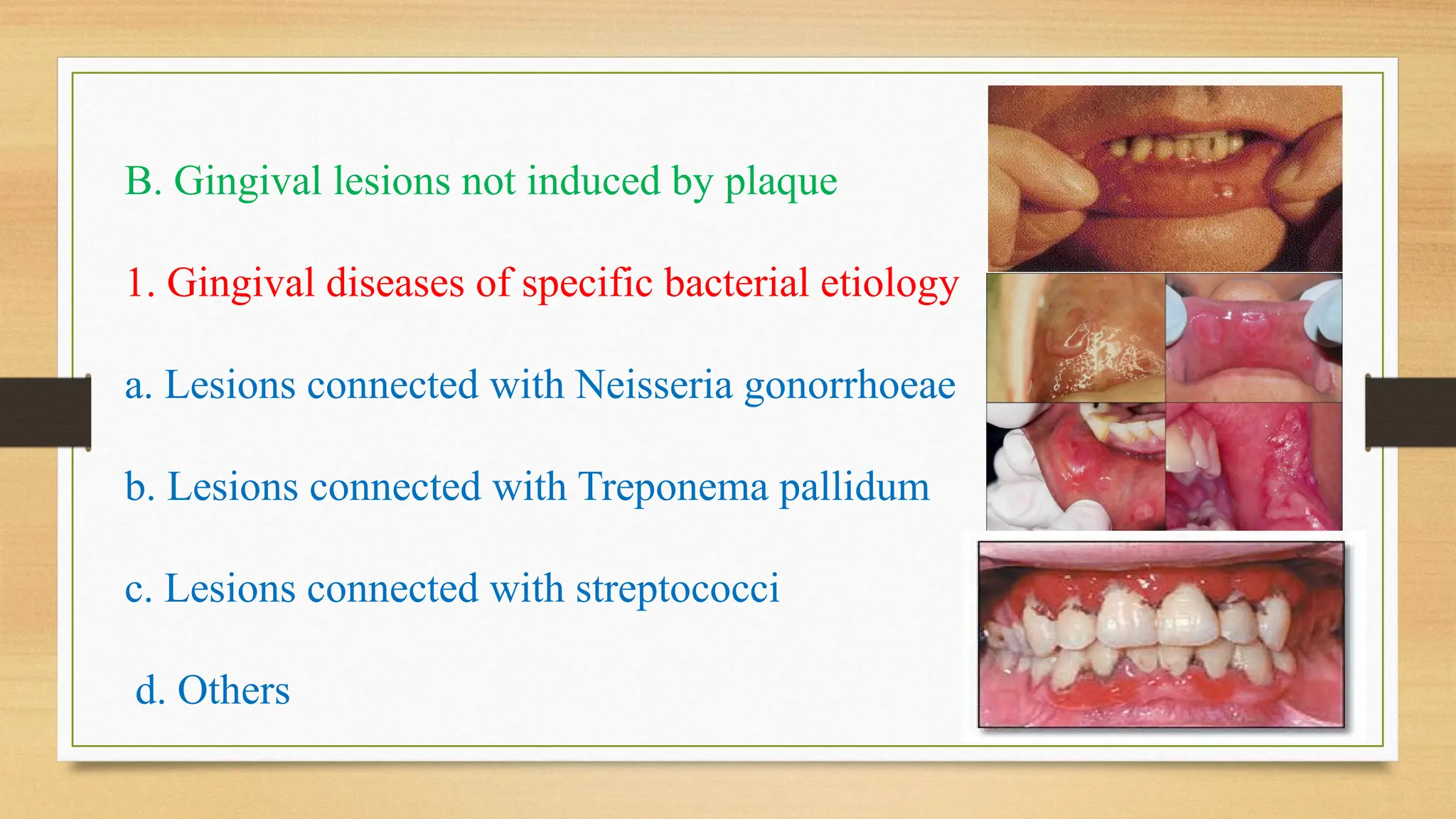
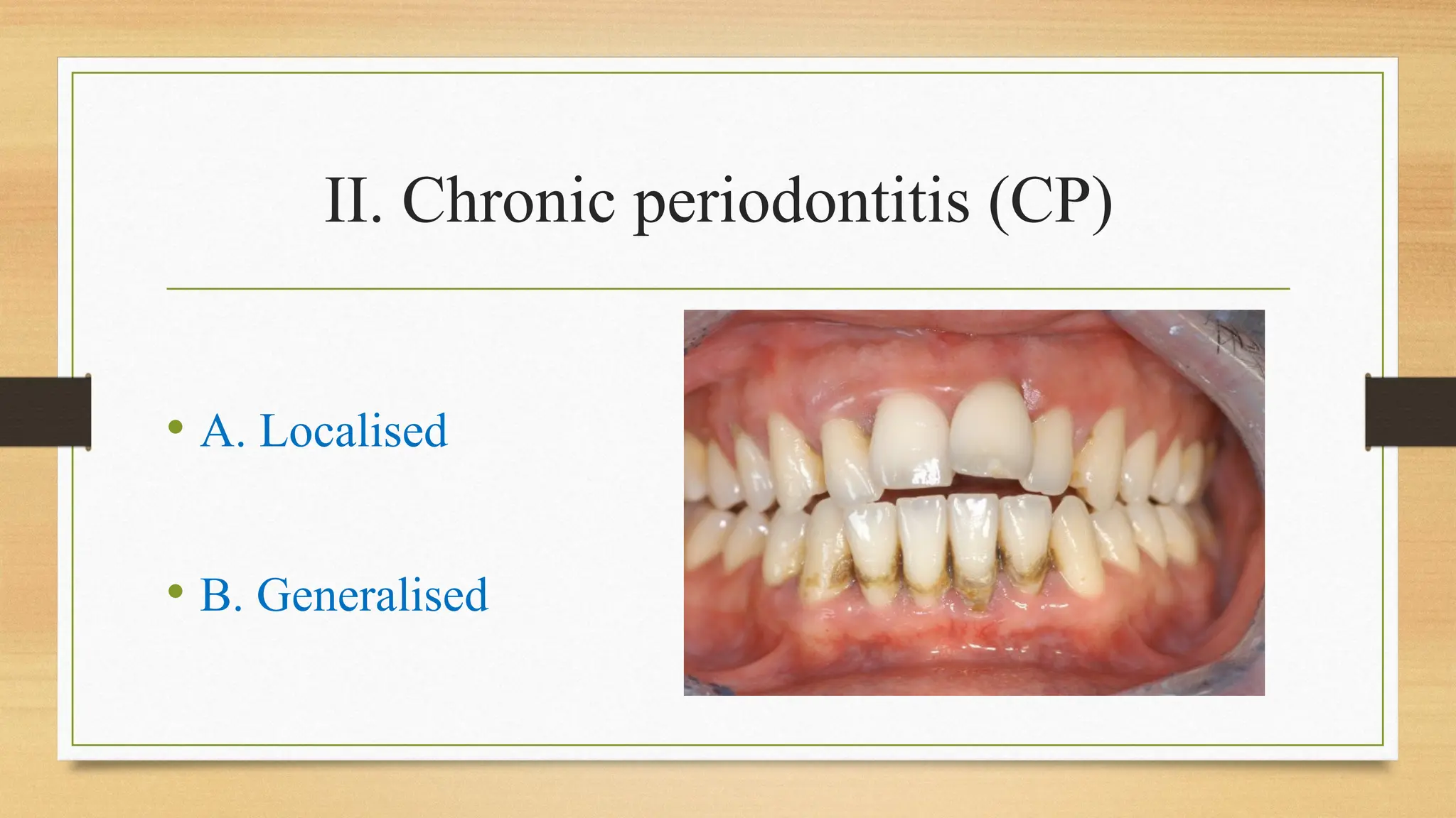
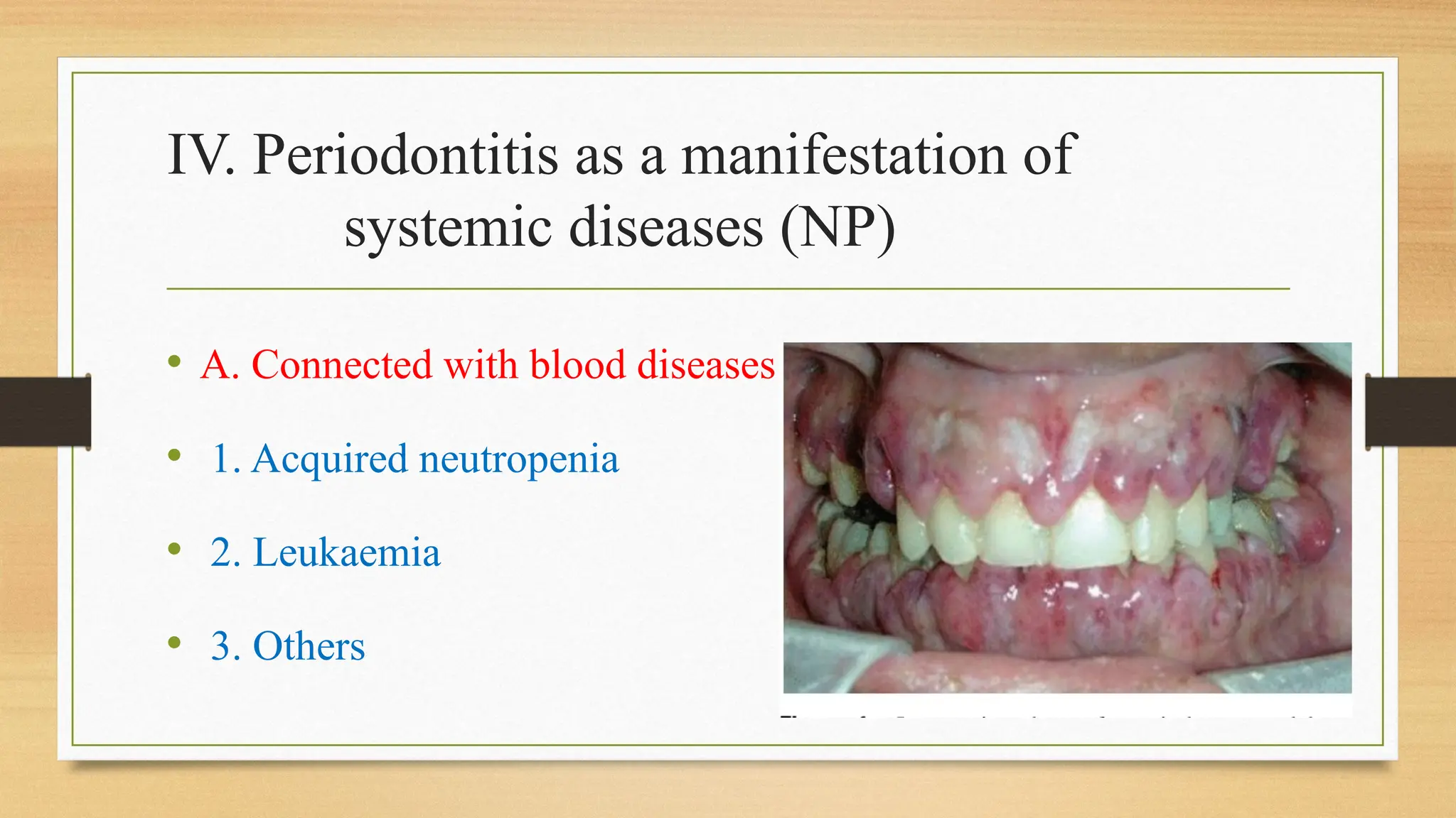
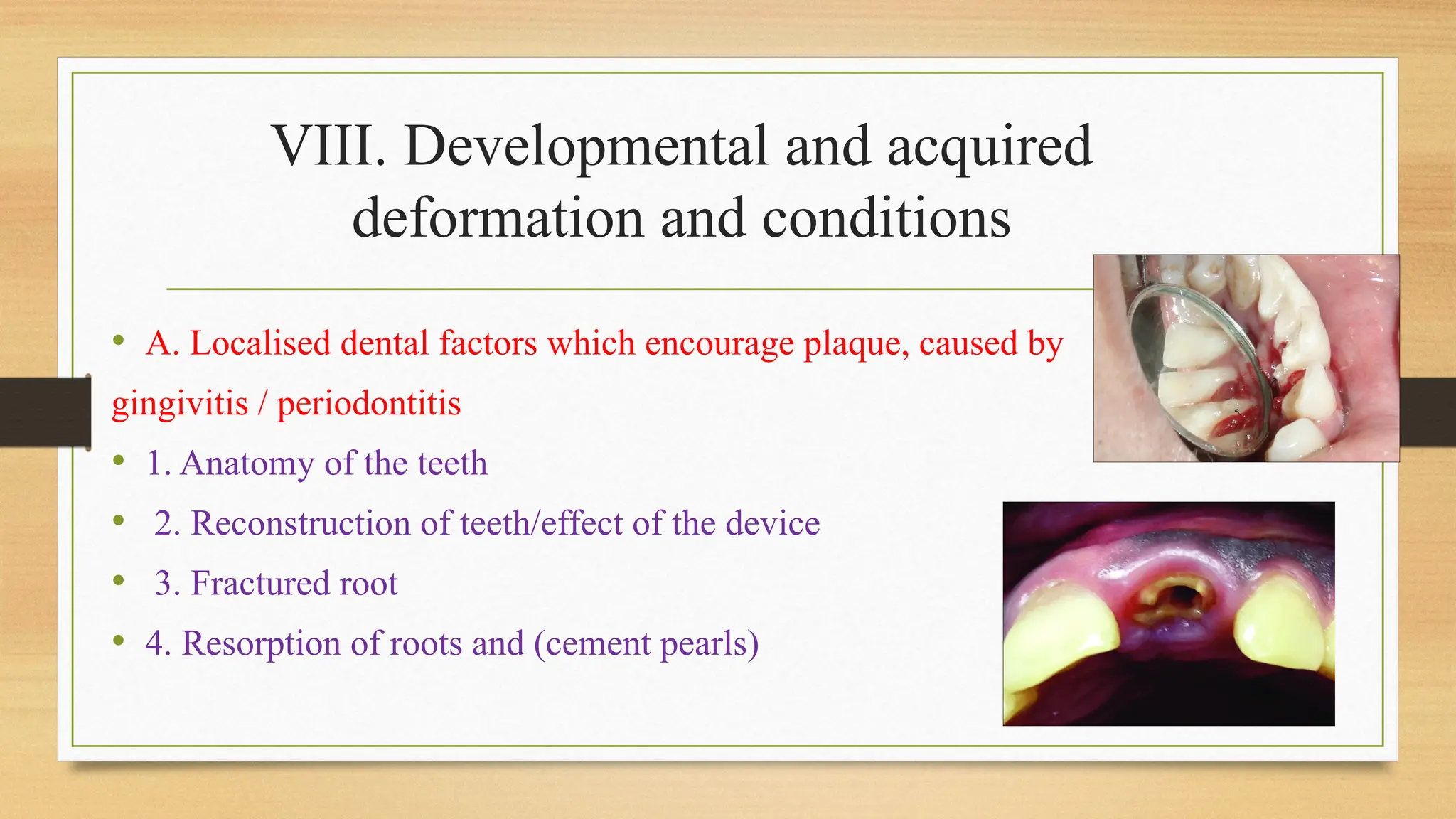
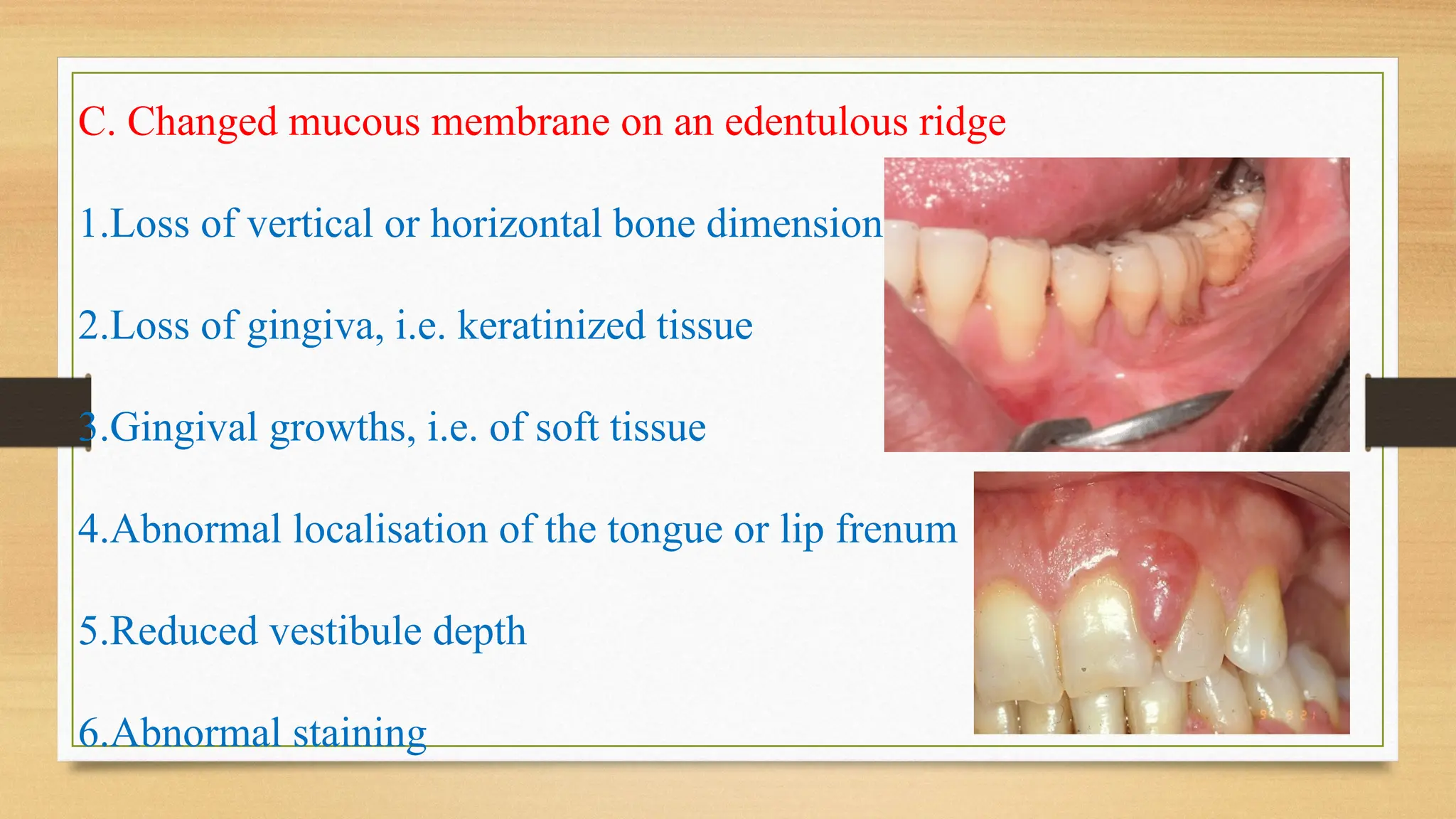
Classification of Periodontal
Diseases
Guided by-
Dr. Monica Mahajani
Dr. Anup Shelke
Dr. Subodh Gaikwad
Dr. Kuldeep Patil
Dr. Chitrika Subhadarshanee
Presented by-
Dr. Pranjal Baheti
Content-
• Introduction
• Need of classification
• Ideal Requisite
• Classification
• Advantages & disadvantages
• Conclusion
• Reference
Introduction:
• Periodontal disease are the infectious disease resulting in
inflammation within the supporting tissues of the teeth,
progressive attachment loss and bone loss.
• Depending on updation of knowledge during different times in
the past regarding the etiopathogenesis and clinical
manifestations, various schemes for classifying periodontal
diseases were proposed.
Need of classification
• Foundation to study the etiology, susceptibility traits,
pathogenesis, and treatment of diseases in an organized manner.
• To give clinicians a way to organize the health care needs of
their patients.
• Diagnosis, prognosis, treatment planning; communication.
Ideal Requisite
• Suitable organizing principle matching the nature of the disease
being classified.
• Simple
• Every disease should fall into at least one of the classes.
• No disease should fall into more than one of the classes.
Dominant paradigms in the evolution of
classification systems
(~1870–1920) Clinical characteristics paradigm
There was dispute about the nature of periodontal diseases;
whether they were caused by local or systemic factors.
Many of the advocates for the etiological role of local factors also
acknowledged that in some cases both local and systemic factors
are responsible.
(~1920–1970) Classical pathology paradigm
At that time a new concept developed that periodontal diseases can be of
2 types-inflammatory and non-inflammatory (‘degenerative’ or
‘dystrophic’)
This was based on the observation that certain forms of periodontal
diseases were due to degenerative changes in the periodontium.
(~1970–present) Infection/ host response paradigm
Infection/ host response paradigm.
W.D. Miller stated three factors which were to be taken into
consideration in every case-
(1)predisposing circumstances,
(2)local irritation,
(3)bacteria.
GOTTLIEB (1921)
Pyorrhea alveolaris
Schmutz
pyorrhea /Filth
pyorrhea
Alveolar
Atrophy
Trauma
from
occlusion
Paradontal
Pyorrhea
Fish described pyorrhea simplex and pyorrhea profunda.
Box divided chronic periodontitis into complex and
simplex, and ascribed a prominent role for occlusal trauma
in the aetiology of complex.
ORBAN’S Classification(1942)
Degeneration Atropy Hypertropy Traumatism
Inflammation
Periodontal
atropy
Periodontal
traumatism
Gingival
hypertropy
Periodontosis
Gingivitis
Periodontitis
IRVING GLICKMAN
I. INFLAMMATORY
A . Chronic Inflammatory Gingival Enlargement
1. Generalized or localized
2. Discrete (Tumor-like)
B. Acute Inflammatory Gingival Enlargement (Gingival abscess)
II. NON-INFLAMMATORY HYPERPLASTIC GINGIVAL
ENLARGEMENT (Gingival hyperplasia)
A . Marginal
B. Diffuse
III. COMBINED GINGIVAL ENLARGEMENT
IV. CONDITIONED GINGIVAL ENLARGEMENT
A . Hormonal
1. Gingival Enlargement of Pregnancy
2. Gingival Enlargement of Puberty
B. Leukemic Gingival Enlargement
C. Gingival Enlargement Associated with Vitamin C Deficiency
V . NEOPLASMS
VI . DEVELOPMENTAL GINGIVAL ENLARGEMENT
1957 AAP Classification
IN F L A M M A T IO N D Y ST R O PH Y
Gingivitis
Periodontitis
PRIMARY
(Simplex)
SECONDARY
(Complex)
 Chronic
 Acute
 Chronic papillary
 Necrotizing
 Fibrotic
 Desquamative
 Ulcerative
 Bullous
Occlusal
traumatism
Periodontal
disuse atropy
 Gingivosis
 Periodontosis
Page and Schroeder (1976)
Juvenile
Rapidly
progressive
Pre-
pubertal
Adult
periodontiti
s
Acute
necrotizing
ulcerative
gingivo-
periodontitis
AAP 1977 Classification
I. Juvenile Periodontitis
II. Chronic Marginal Periodontitis
AAP 1986 Classification
• Juvenile Periodontitis
-Prepubertal
-Localized juvenile periodontitis
-Generalized juvenile periodontitis
• Adult Periodontitis
• Necrotizing Ulcerative Gingivo-Periodontitis
• Refractory Periodontitis
AAP World Workshop in Clinical Periodontics
(1989) Classification
I. Early- onset Periodontitis
A.
B.
Prepubertal
Periodontitis
Localized
Generalized
Juvenile
Periodontitis
Localized
Generalized
II. Adult Periodontitis
III. Necrotising Ulcerative Periodontitis
IV. Refractory Periodontitis
V. Periodontitis associated with Systemic diseases
Drawbacks of the 1989 Classification
• Gingival disease category was absent.
• Non-validated age-dependent criteria in other periodontitis
categories.
• Extensive crossover in rates of progression of the different
categories of periodontal disease.
• ‘Rapidly Progressive Periodontitis’, ‘Refractory Periodontitis’
and ‘Prepubertal Periodontitis ‘were heterogeneous category.
• Extensive overlap in the clinical characteristics of the different
categories of periodontitis.
European Workshop in Periodontology (1993)
Classification
• Adult Periodontitis - Begins at the 4th decade of life, slow rate
of progression of disease.
• Early onset Periodontitis - Begins before the 4 th decade of life,
rapid rate of progression of disease, altered host response is seen.
• Necrotizing Periodontitis - Tissue necrosis with clinical
attachment and bone loss is seen.
Drawbacks
• Elaboration of the broad spectrum of periodontal diseases
encountered in clinical practice was absent.
American Academy of Periodontology (1999)
Classification
I. Gingival diseases
II. Chronic periodontitis (CP)
III. Aggressive periodontitis
IV. Periodontitis as a manifestation of
systemic diseases (NP)
V. Necrotizing periodontal diseases
VI. Periodontal abscesses
VII. Periodontitis with endodontic lesions
VIII. Developmental and acquired
deformation and conditions
I. Gingival diseases (G)
A. Gingival diseases caused by plaque
1. Gingivitis exclusively caused by plaque
a. With no local modifying factors
b. With local modifying factors
2. Gingival diseases modified with systemic factors
A. Associated with endocrine system
1.Gingivitis connected with puberty
2. Gingivitis connected with the menstrual cycle
3.Connected with pregnancy
a) Gingivitis in pregnancy
b) Pyogenic granuloma
4.Gingivitis connected with diabetes mellitus
b. Connected with blood disease
1) Gingivitis connected with leukaemia
2) Other diseases
3. Gingival diseases modified by application of
medications
a. Gingival diseases caused by medications
1) Gingival growths caused by medications
2) Gingivitis caused by medications
a) Gingivitis connected with oral contraceptives
b) Other medications
4. Gingival diseases caused by malnutrition
a. Gingivitis due to lack of vitamin C
b. Others
B. Gingival lesions not induced by plaque
1. Gingival diseases of specific bacterial etiology
a. Lesions connected with Neisseria gonorrhoeae
b. Lesions connected with Treponema pallidum
c. Lesions connected with streptococci
d. Others
2. Gingival diseases of viral etiology
A. Infection with the Herpes virus
1) Primary herpetic gingivostomatitis
2) Recurring oral herpes
3) Varicella zoster infection
B. Others
3. Gingival diseases of fungal etiology
A. Infection with candida
1) Generalised gingival candidiasis
B. Linear gingival erythema
C. Histoplasmosis
D. Others
4. Gingival diseases of genetic etiology
a. Inherited fibromatosis of the gingiva
b. Others
5. Systemic diseases which manifest on the gingiva
A. Changed mucous membrane
1) Lichen planus
2) Pemphigoid
3) Pemphigus vulgaris
4) Erythema multiformis
5) Lupus erythematosus
6)Others
B. Allergic reactions
1) Material in restorative dentistry
a) Mercury
b) Nickel
c) Acrylic
d) Others
2) Reaction to:
a) Toothpaste
b) Mouthwashes
c) Additives in chewing gum
d) Nutritive substitutes
3) Others
6. Traumatic lesions (iatrogenic, accidents)
a. Chemical
b. Physical
c. Thermal
7. Reaction to foreign bodies
8. Not otherwise defined
II. Chronic periodontitis (CP)
• A. Localised
• B. Generalised
III. Aggressive periodontitis (AP)
• A. Localised
• B. Generalised
IV. Periodontitis as a manifestation of
systemic diseases (NP)
• A. Connected with blood diseases
• 1. Acquired neutropenia
• 2. Leukaemia
• 3. Others
B. Connected with genetic disorders
1. Family or cyclic neutropenia
2. Down’s syndrome
3. Leucocyte adhesive deficiency syndrome
4. Papillon-Lefevre syndrome
5. Chediak-Higashi syndrome
6. Histiocytosis or Eosinophilic granuloma syndrome
7. Glycogen storage syndrome
8. Infantile genetic agranulocytosis
9. Cohen’s syndrome
10. Ehlers-Danlos syndrome, type IV and VIII AD
11. Hypophosphatasia
12. Others
C. Not otherwise defined
V. Necrotizing periodontal diseases
• A. Necrotizing ulcerous gingivitis (NUG)
• B. Necrotizing ulcerous periodontitis (NUP)
VI. Periodontal abscesses
• A. Gingival abscess
• B. Periodontal abscess
• C. Pericoronal abscess
VII. Periodontitis with endodontal lesions
• A. Combined perio-endo lesion
VIII. Developmental and acquired
deformation and conditions
• A. Localised dental factors which encourage plaque, caused by
gingivitis / periodontitis
• 1. Anatomy of the teeth
• 2. Reconstruction of teeth/effect of the device
• 3. Fractured root
• 4. Resorption of roots and (cement pearls)
B. Mucogingival deformities and relations in the tooth vicinity
1. Recession
a. Facially and orally
b. Approximally
2. Lack of gingival keratinization
3. Shortened gingival attachment
4. Localisation of the tongue or lip frenum
5. Gingival enlargement
a. Pseudo-pockets
b. Irregular development of the gingival edge
c. Excessive gingival presentation
d. Gingival enlargement
6. Abnormal staining
C. Changed mucous membrane on an edentulous ridge
1.Loss of vertical or horizontal bone dimension
2.Loss of gingiva, i.e. keratinized tissue
3.Gingival growths, i.e. of soft tissue
4.Abnormal localisation of the tongue or lip frenum
5.Reduced vestibule depth
6.Abnormal staining
D. Occlusal trauma
1. Primary occlusal trauma
2. Secondary occlusal trauma
Changes in the 1999 Classification for
Periodontal diseases
• Addition of a Section on “Gingival diseases
• Replacement of “Adult Periodontitis” with “Chronic Periodontitis”
• Replacement of “Early-Onset Periodontitis” With “Aggressive Periodontitis”
• Elimination of a Separate Disease Category for “Refractory Periodontitis”
• Clarification of the Designation “Periodontitis as a Manifestation of Systemic
Diseases”
• Replacement of “Necrotizing Ulcerative Periodontitis” With
“Necrotizing Periodontal diseases”
• Addition of a Category for “Periodontal Abscess” and
“Periodontic-Endodontic Lesions”
• Addition of a Category on “Developmental or Acquired
Deformities and Conditions”
• Essentialistic or Nominalistic disease classification
DISADVANTAGE
• It did not serve as therapeutic guide
• Categorizing aggressive & chronic periodontitis –confusing
• Current evidence does not support the distinction between chronic and
aggressive periodontitis as separate clinical entities
• Confusion in diagnosing a case of plaque-induced gingival
inflammation on a reduced but healthy periodontium –
periodontitis or gingivitis?
• Categories of gingival disease modified by medication & diabetes
mellitus exist but no such periodontitis class exists.
• No mention of peri-implant diseases
Classification of Periodontal &Peri-implant diseases and conditions
2017(AAP & EFP)
A) Periodontal Diseases and Conditions
B) Peri- Implant diseases and conditions
 Peri-implant Health
 Peri implant Mucositis
 Peri-implantitis
 Peri-implant soft and Hard tissue deficiency
0.25%
0.25-1%
>1%
Advantages
• Evidence-based and clinically relevant
• Helps in accurate case selection
• Disease risk and complexity factors
• Reflect on tooth loss
• Periodontal & gingival health have been defined.
• Endo-Perio lesion- classification based on clinical findings
Disadvantages
• very extensive and more complicated
• Some degree of overlap
• Necrotizing gingivitis is included in the periodontitis
• Periodontal abscess is a clinical manifestation and not a disease yet
is considered as a diagnosis
• There is no distinction between periodontal and gingival abscesses.
2018 New Periodontal Classification (By EFP & AAP)
AAP)
Mucogingival deformities & conditions around teeth
1.Periodontal Biotype
• Thin scalloped
• Thick scalloped
• Thick flat
2.Gingival soft tissue recession
• Facial & lingual surface
• Interproximal (papillary)
• Severity of recession(cairo RT1,2,3)
• Gingival thickness
• Gingival width
• Presence of NCCL /Cervical caries
• Presence of hypersensitivity
3.Lack of keratinised gingiva
4.Decrease vestibular depth
5.Aberrunt frenum /muscle position
6.Gingival excess
• Psuedo pocket
• Inconsistent gingival margin
• Excessive gingival display
• Gingival enlargement
7.Abnormal colour
Conclusion
• It is just a guideline that certainly may possess some limitations
in some cases. Therefore, judgement of clinicians is essential to
make a definitive diagnosis. Clinical efficiency of the
classification’s should be evaluated in a future studys.
References
• Lindhe, J. (2008, January 1). Clinical periodontology and implant dentistry. 1. [Basic
concepts]. 5th
Ed
• Newman, M. G., Takei, H., Klokkevold, P. R., & Carranza, F. A. (2011, February 14).
Carranza’s Clinical Periodontology. 10th
Ed
• Sutthiboonyapan, P., Wang, H., & Charatkulangkun, O. (2020, April 8). Flowcharts for
Easy Periodontal Diagnosis Based on the 2018 New Periodontal Classification. Clinical
Advances in Periodontics, 10(3), 155–160. https://doi.org/10.1002/cap.10095
• Caton, J. et al. (2018, June). A new classification scheme for periodontal and
peri implant diseases and conditions – Introduction and key changes from the
‐
1999 classification. Journal of Periodontology, 89(S1).
• Salme E. Lavigne. The 2018 AAP/EFP Classification of Periodontal & Peri-
implant Diseases. Crest® + Oral-B® at dentalcare.com
• Mahajan A.,Kolte R. , Kolte A., Development and Evolution of Classification
of Periodontal Diseases: An Insight. International Dental Journal of Student’s
Research, January – March 2015;3(1):3-11
• Papapanou PN, Sanz M, et al. Periodontitis: Consensus report of Workgroup 2 of
the 2017 World Workshop on the Classification of Periodontal and Peri-Implant
Diseases and Con ditions. J Periodontol. 2018;89(Suppl 1):S173–S182.
• Mittal V, Bhullar RP, Bansal R, Singh K, Bhalodi A, Khinda PK. A practicable
approach for periodontal classification. Dent Res J (Isfahan). 2013 Nov;10(6):697-
703. PMID: 24379855; PMCID: PMC3872618.
• GLICKMAN I. A basic classification of "gingival enlargement". J Periodontol
(1930). 1950 Jul;21(3):131-9. doi: 10.1902/jop.1950.21.3.131. PMID: 15428964.
Periodontal diseases classification.pptx

Periodontal diseases classification.pptx

  • 1.
    Classification of Periodontal Diseases Guidedby- Dr. Monica Mahajani Dr. Anup Shelke Dr. Subodh Gaikwad Dr. Kuldeep Patil Dr. Chitrika Subhadarshanee Presented by- Dr. Pranjal Baheti
  • 2.
    Content- • Introduction • Needof classification • Ideal Requisite • Classification • Advantages & disadvantages • Conclusion • Reference
  • 3.
    Introduction: • Periodontal diseaseare the infectious disease resulting in inflammation within the supporting tissues of the teeth, progressive attachment loss and bone loss. • Depending on updation of knowledge during different times in the past regarding the etiopathogenesis and clinical manifestations, various schemes for classifying periodontal diseases were proposed.
  • 4.
    Need of classification •Foundation to study the etiology, susceptibility traits, pathogenesis, and treatment of diseases in an organized manner. • To give clinicians a way to organize the health care needs of their patients. • Diagnosis, prognosis, treatment planning; communication.
  • 5.
    Ideal Requisite • Suitableorganizing principle matching the nature of the disease being classified. • Simple • Every disease should fall into at least one of the classes. • No disease should fall into more than one of the classes.
  • 6.
    Dominant paradigms inthe evolution of classification systems (~1870–1920) Clinical characteristics paradigm There was dispute about the nature of periodontal diseases; whether they were caused by local or systemic factors. Many of the advocates for the etiological role of local factors also acknowledged that in some cases both local and systemic factors are responsible.
  • 7.
    (~1920–1970) Classical pathologyparadigm At that time a new concept developed that periodontal diseases can be of 2 types-inflammatory and non-inflammatory (‘degenerative’ or ‘dystrophic’) This was based on the observation that certain forms of periodontal diseases were due to degenerative changes in the periodontium.
  • 8.
    (~1970–present) Infection/ hostresponse paradigm Infection/ host response paradigm. W.D. Miller stated three factors which were to be taken into consideration in every case- (1)predisposing circumstances, (2)local irritation, (3)bacteria.
  • 9.
    GOTTLIEB (1921) Pyorrhea alveolaris Schmutz pyorrhea/Filth pyorrhea Alveolar Atrophy Trauma from occlusion Paradontal Pyorrhea
  • 10.
    Fish described pyorrheasimplex and pyorrhea profunda. Box divided chronic periodontitis into complex and simplex, and ascribed a prominent role for occlusal trauma in the aetiology of complex.
  • 11.
    ORBAN’S Classification(1942) Degeneration AtropyHypertropy Traumatism Inflammation Periodontal atropy Periodontal traumatism Gingival hypertropy Periodontosis Gingivitis Periodontitis
  • 12.
    IRVING GLICKMAN I. INFLAMMATORY A. Chronic Inflammatory Gingival Enlargement 1. Generalized or localized 2. Discrete (Tumor-like) B. Acute Inflammatory Gingival Enlargement (Gingival abscess) II. NON-INFLAMMATORY HYPERPLASTIC GINGIVAL ENLARGEMENT (Gingival hyperplasia) A . Marginal B. Diffuse
  • 13.
    III. COMBINED GINGIVALENLARGEMENT IV. CONDITIONED GINGIVAL ENLARGEMENT A . Hormonal 1. Gingival Enlargement of Pregnancy 2. Gingival Enlargement of Puberty B. Leukemic Gingival Enlargement C. Gingival Enlargement Associated with Vitamin C Deficiency V . NEOPLASMS VI . DEVELOPMENTAL GINGIVAL ENLARGEMENT
  • 14.
    1957 AAP Classification INF L A M M A T IO N D Y ST R O PH Y Gingivitis Periodontitis PRIMARY (Simplex) SECONDARY (Complex)  Chronic  Acute  Chronic papillary  Necrotizing  Fibrotic  Desquamative  Ulcerative  Bullous Occlusal traumatism Periodontal disuse atropy  Gingivosis  Periodontosis
  • 15.
    Page and Schroeder(1976) Juvenile Rapidly progressive Pre- pubertal Adult periodontiti s Acute necrotizing ulcerative gingivo- periodontitis
  • 16.
    AAP 1977 Classification I.Juvenile Periodontitis II. Chronic Marginal Periodontitis
  • 17.
    AAP 1986 Classification •Juvenile Periodontitis -Prepubertal -Localized juvenile periodontitis -Generalized juvenile periodontitis • Adult Periodontitis • Necrotizing Ulcerative Gingivo-Periodontitis • Refractory Periodontitis
  • 18.
    AAP World Workshopin Clinical Periodontics (1989) Classification I. Early- onset Periodontitis A. B. Prepubertal Periodontitis Localized Generalized Juvenile Periodontitis Localized Generalized
  • 19.
    II. Adult Periodontitis III.Necrotising Ulcerative Periodontitis IV. Refractory Periodontitis V. Periodontitis associated with Systemic diseases
  • 20.
    Drawbacks of the1989 Classification • Gingival disease category was absent. • Non-validated age-dependent criteria in other periodontitis categories. • Extensive crossover in rates of progression of the different categories of periodontal disease.
  • 21.
    • ‘Rapidly ProgressivePeriodontitis’, ‘Refractory Periodontitis’ and ‘Prepubertal Periodontitis ‘were heterogeneous category. • Extensive overlap in the clinical characteristics of the different categories of periodontitis.
  • 22.
    European Workshop inPeriodontology (1993) Classification • Adult Periodontitis - Begins at the 4th decade of life, slow rate of progression of disease. • Early onset Periodontitis - Begins before the 4 th decade of life, rapid rate of progression of disease, altered host response is seen. • Necrotizing Periodontitis - Tissue necrosis with clinical attachment and bone loss is seen.
  • 23.
    Drawbacks • Elaboration ofthe broad spectrum of periodontal diseases encountered in clinical practice was absent.
  • 24.
    American Academy ofPeriodontology (1999) Classification I. Gingival diseases II. Chronic periodontitis (CP) III. Aggressive periodontitis IV. Periodontitis as a manifestation of systemic diseases (NP) V. Necrotizing periodontal diseases VI. Periodontal abscesses VII. Periodontitis with endodontic lesions VIII. Developmental and acquired deformation and conditions
  • 25.
    I. Gingival diseases(G) A. Gingival diseases caused by plaque 1. Gingivitis exclusively caused by plaque a. With no local modifying factors b. With local modifying factors
  • 26.
    2. Gingival diseasesmodified with systemic factors A. Associated with endocrine system 1.Gingivitis connected with puberty 2. Gingivitis connected with the menstrual cycle 3.Connected with pregnancy a) Gingivitis in pregnancy b) Pyogenic granuloma 4.Gingivitis connected with diabetes mellitus
  • 27.
    b. Connected withblood disease 1) Gingivitis connected with leukaemia 2) Other diseases
  • 28.
    3. Gingival diseasesmodified by application of medications a. Gingival diseases caused by medications 1) Gingival growths caused by medications 2) Gingivitis caused by medications a) Gingivitis connected with oral contraceptives b) Other medications
  • 29.
    4. Gingival diseasescaused by malnutrition a. Gingivitis due to lack of vitamin C b. Others
  • 30.
    B. Gingival lesionsnot induced by plaque 1. Gingival diseases of specific bacterial etiology a. Lesions connected with Neisseria gonorrhoeae b. Lesions connected with Treponema pallidum c. Lesions connected with streptococci d. Others
  • 31.
    2. Gingival diseasesof viral etiology A. Infection with the Herpes virus 1) Primary herpetic gingivostomatitis 2) Recurring oral herpes 3) Varicella zoster infection B. Others
  • 32.
    3. Gingival diseasesof fungal etiology A. Infection with candida 1) Generalised gingival candidiasis B. Linear gingival erythema C. Histoplasmosis D. Others
  • 33.
    4. Gingival diseasesof genetic etiology a. Inherited fibromatosis of the gingiva b. Others
  • 34.
    5. Systemic diseaseswhich manifest on the gingiva A. Changed mucous membrane 1) Lichen planus 2) Pemphigoid 3) Pemphigus vulgaris 4) Erythema multiformis 5) Lupus erythematosus 6)Others
  • 35.
    B. Allergic reactions 1)Material in restorative dentistry a) Mercury b) Nickel c) Acrylic d) Others
  • 36.
    2) Reaction to: a)Toothpaste b) Mouthwashes c) Additives in chewing gum d) Nutritive substitutes 3) Others
  • 37.
    6. Traumatic lesions(iatrogenic, accidents) a. Chemical b. Physical c. Thermal 7. Reaction to foreign bodies 8. Not otherwise defined
  • 38.
    II. Chronic periodontitis(CP) • A. Localised • B. Generalised
  • 39.
    III. Aggressive periodontitis(AP) • A. Localised • B. Generalised
  • 40.
    IV. Periodontitis asa manifestation of systemic diseases (NP) • A. Connected with blood diseases • 1. Acquired neutropenia • 2. Leukaemia • 3. Others
  • 41.
    B. Connected withgenetic disorders 1. Family or cyclic neutropenia 2. Down’s syndrome 3. Leucocyte adhesive deficiency syndrome 4. Papillon-Lefevre syndrome 5. Chediak-Higashi syndrome 6. Histiocytosis or Eosinophilic granuloma syndrome
  • 42.
    7. Glycogen storagesyndrome 8. Infantile genetic agranulocytosis 9. Cohen’s syndrome 10. Ehlers-Danlos syndrome, type IV and VIII AD 11. Hypophosphatasia 12. Others C. Not otherwise defined
  • 43.
    V. Necrotizing periodontaldiseases • A. Necrotizing ulcerous gingivitis (NUG) • B. Necrotizing ulcerous periodontitis (NUP)
  • 44.
    VI. Periodontal abscesses •A. Gingival abscess • B. Periodontal abscess • C. Pericoronal abscess
  • 45.
    VII. Periodontitis withendodontal lesions • A. Combined perio-endo lesion
  • 46.
    VIII. Developmental andacquired deformation and conditions • A. Localised dental factors which encourage plaque, caused by gingivitis / periodontitis • 1. Anatomy of the teeth • 2. Reconstruction of teeth/effect of the device • 3. Fractured root • 4. Resorption of roots and (cement pearls)
  • 47.
    B. Mucogingival deformitiesand relations in the tooth vicinity 1. Recession a. Facially and orally b. Approximally 2. Lack of gingival keratinization 3. Shortened gingival attachment 4. Localisation of the tongue or lip frenum
  • 48.
    5. Gingival enlargement a.Pseudo-pockets b. Irregular development of the gingival edge c. Excessive gingival presentation d. Gingival enlargement 6. Abnormal staining
  • 49.
    C. Changed mucousmembrane on an edentulous ridge 1.Loss of vertical or horizontal bone dimension 2.Loss of gingiva, i.e. keratinized tissue 3.Gingival growths, i.e. of soft tissue 4.Abnormal localisation of the tongue or lip frenum 5.Reduced vestibule depth 6.Abnormal staining
  • 50.
    D. Occlusal trauma 1.Primary occlusal trauma 2. Secondary occlusal trauma
  • 51.
    Changes in the1999 Classification for Periodontal diseases • Addition of a Section on “Gingival diseases • Replacement of “Adult Periodontitis” with “Chronic Periodontitis” • Replacement of “Early-Onset Periodontitis” With “Aggressive Periodontitis” • Elimination of a Separate Disease Category for “Refractory Periodontitis” • Clarification of the Designation “Periodontitis as a Manifestation of Systemic Diseases”
  • 52.
    • Replacement of“Necrotizing Ulcerative Periodontitis” With “Necrotizing Periodontal diseases” • Addition of a Category for “Periodontal Abscess” and “Periodontic-Endodontic Lesions” • Addition of a Category on “Developmental or Acquired Deformities and Conditions” • Essentialistic or Nominalistic disease classification
  • 53.
    DISADVANTAGE • It didnot serve as therapeutic guide • Categorizing aggressive & chronic periodontitis –confusing • Current evidence does not support the distinction between chronic and aggressive periodontitis as separate clinical entities
  • 54.
    • Confusion indiagnosing a case of plaque-induced gingival inflammation on a reduced but healthy periodontium – periodontitis or gingivitis? • Categories of gingival disease modified by medication & diabetes mellitus exist but no such periodontitis class exists. • No mention of peri-implant diseases
  • 55.
    Classification of Periodontal&Peri-implant diseases and conditions 2017(AAP & EFP) A) Periodontal Diseases and Conditions
  • 56.
    B) Peri- Implantdiseases and conditions  Peri-implant Health  Peri implant Mucositis  Peri-implantitis  Peri-implant soft and Hard tissue deficiency
  • 59.
  • 62.
    Advantages • Evidence-based andclinically relevant • Helps in accurate case selection • Disease risk and complexity factors • Reflect on tooth loss • Periodontal & gingival health have been defined. • Endo-Perio lesion- classification based on clinical findings
  • 63.
    Disadvantages • very extensiveand more complicated • Some degree of overlap • Necrotizing gingivitis is included in the periodontitis • Periodontal abscess is a clinical manifestation and not a disease yet is considered as a diagnosis • There is no distinction between periodontal and gingival abscesses.
  • 64.
    2018 New PeriodontalClassification (By EFP & AAP) AAP)
  • 67.
    Mucogingival deformities &conditions around teeth 1.Periodontal Biotype • Thin scalloped • Thick scalloped • Thick flat 2.Gingival soft tissue recession • Facial & lingual surface • Interproximal (papillary) • Severity of recession(cairo RT1,2,3) • Gingival thickness • Gingival width • Presence of NCCL /Cervical caries • Presence of hypersensitivity
  • 68.
    3.Lack of keratinisedgingiva 4.Decrease vestibular depth 5.Aberrunt frenum /muscle position 6.Gingival excess • Psuedo pocket • Inconsistent gingival margin • Excessive gingival display • Gingival enlargement 7.Abnormal colour
  • 69.
    Conclusion • It isjust a guideline that certainly may possess some limitations in some cases. Therefore, judgement of clinicians is essential to make a definitive diagnosis. Clinical efficiency of the classification’s should be evaluated in a future studys.
  • 70.
    References • Lindhe, J.(2008, January 1). Clinical periodontology and implant dentistry. 1. [Basic concepts]. 5th Ed • Newman, M. G., Takei, H., Klokkevold, P. R., & Carranza, F. A. (2011, February 14). Carranza’s Clinical Periodontology. 10th Ed • Sutthiboonyapan, P., Wang, H., & Charatkulangkun, O. (2020, April 8). Flowcharts for Easy Periodontal Diagnosis Based on the 2018 New Periodontal Classification. Clinical Advances in Periodontics, 10(3), 155–160. https://doi.org/10.1002/cap.10095
  • 71.
    • Caton, J.et al. (2018, June). A new classification scheme for periodontal and peri implant diseases and conditions – Introduction and key changes from the ‐ 1999 classification. Journal of Periodontology, 89(S1). • Salme E. Lavigne. The 2018 AAP/EFP Classification of Periodontal & Peri- implant Diseases. Crest® + Oral-B® at dentalcare.com • Mahajan A.,Kolte R. , Kolte A., Development and Evolution of Classification of Periodontal Diseases: An Insight. International Dental Journal of Student’s Research, January – March 2015;3(1):3-11
  • 72.
    • Papapanou PN,Sanz M, et al. Periodontitis: Consensus report of Workgroup 2 of the 2017 World Workshop on the Classification of Periodontal and Peri-Implant Diseases and Con ditions. J Periodontol. 2018;89(Suppl 1):S173–S182. • Mittal V, Bhullar RP, Bansal R, Singh K, Bhalodi A, Khinda PK. A practicable approach for periodontal classification. Dent Res J (Isfahan). 2013 Nov;10(6):697- 703. PMID: 24379855; PMCID: PMC3872618. • GLICKMAN I. A basic classification of "gingival enlargement". J Periodontol (1930). 1950 Jul;21(3):131-9. doi: 10.1902/jop.1950.21.3.131. PMID: 15428964.