REVIEW
published: 08 March2021
doi: 10.3389/fimmu.2021.620510
Frontiers in Immunology | www.frontiersin.org 1 March 2021 | Volume 12 | Article 620510
Edited by:
Liwu Li,
Virginia Tech, United States
Reviewed by:
In Su Cheon,
Mayo Clinic, United States
Anna Ohradanova-Repic,
Medical University of Vienna, Austria
*Correspondence:
Aaron C. Ericsson
ericssona@missouri.edu
Ming Yang
yangmin@health.missouri.edu
Specialty section:
This article was submitted to
Molecular Innate Immunity,
a section of the journal
Frontiers in Immunology
Received: 23 October 2020
Accepted: 17 February 2021
Published: 08 March 2021
Citation:
Zhang C, Yang M and Ericsson AC
(2021) Function of Macrophages in
Disease: Current Understanding on
Molecular Mechanisms.
Front. Immunol. 12:620510.
doi: 10.3389/fimmu.2021.620510
Function of Macrophages in Disease:
Current Understanding on Molecular
Mechanisms
Chunye Zhang1
, Ming Yang2
* and Aaron C. Ericsson1,3,4
*
1
Department of Veterinary Pathobiology, University of Missouri, Columbia, MO, United States, 2
Department of Surgery,
University of Missouri, Columbia, MO, United States, 3
Department of Veterinary Pathobiology, University of Missouri
Metagenomics Center, University of Missouri, Columbia, MO, United States, 4
Department of Veterinary Pathobiology,
University of Missouri Mutant Mouse Resource and Research Center, Columbia, MO, United States
Tissue-resident macrophages (TRMs) are heterogeneous populations originating either
from monocytes or embryonic progenitors, and distribute in lymphoid and non-lymphoid
tissues. TRMs play diverse roles in many physiological processes, including metabolic
function, clearance of cellular debris, and tissue remodeling and defense. Macrophages
can be polarized to different functional phenotypes depending on their origin and tissue
microenvironment. Specific macrophage subpopulations are associated with disease
progression. In studies of fate-mapping and single-cell RNA sequencing methodologies,
several critical molecules have been identified to induce the change of macrophage
function. These molecules are potential markers for diagnosis and selective targets for
novel macrophage-mediated treatment. In this review, we discuss some of the recent
findings regarding less-known molecules and new functions of well-known molecules.
Understanding the mechanisms of these molecules in macrophages has the potential to
yield new macrophage-mediated treatments or diagnostic approaches to disease.
Keywords: macrophage, phenotype, molecules, diagnostic marker, therapy
INTRODUCTION
Macrophages are an essential component of the innate immune system, with a wide distribution
in lymphoid and non-lymphoid tissues throughout the body. Macrophages were initially known
to arise from circulating blood monocytes that continuously migrate to different tissues and
differentiate into macrophages (1). Although the origin of adult tissue-resident macrophages
(TRMs) is still not totally understood, it is now recognized that TRMs are derived from diverse
progenitors, including embryonic origin and monocyte progenitors. Some fate-mapping studies
have demonstrated that major TRMs in mice, such as liver resident Kupffer cells (KCs) and
lung alveolar, splenic, and peritoneal macrophages, are developed before birth and can maintain
themselves independent of circulating blood monocytes (2, 3). Further study also demonstrated
that those TRMs such as KCs in mouse fetal liver originate from yolk sac erythro-myeloid
progenitors (EMPs), which are distinct from adult hematopoietic stem cells (HSCs) (4). In contrast,
other studies showed that some TRMs such as adult cardiac and skeletal muscle macrophages are
derived both from yolk-sac and fetal monocyte progenitors (5, 6), and embryonically developed
TRMs can be replaced by blood monocytes.
Macrophages have pivotal functions in homeostasis and many physiological processes beyond
innate immunity, including metabolic function (7), clearance of cellular debris (8), tissue repair
2.
Zhang et al.Molecular Mechanisms of Macrophage Function
and remodeling (9). In pathogenic conditions, TRMs can be
replaced and joined by recruited monocyte-derived macrophages
to orchestrate an immune response (10). Current studies
using single-cell RNA-sequencing (scRNA-seq) have revealed the
presence of multiple macrophage subsets with distinct functions
in various tissues (11). For example, Remmerie et al. (12)
reported that in fatty liver, hepatic resident KCs were destroyed
and replaced by bone marrow-derived macrophages, and those
recruited macrophages comprise two subsets resembling either
KCs or lipid-associated macrophages expressing Osteopontin.
Moreover, macrophage function and phenotype are impacted by
various factors in physiological and pathological conditions, such
as diet (13) and cytokines (14). The alteration of macrophage
phenotype or polarization is associated with distinct gene
expression profiles (15).
Herein, we summarize the latest findings of new macrophage
markers in different diseases. Firstly, the phenotypes of
macrophages are briefly discussed. Secondly, some macrophage-
associated molecules are selected to discuss their important
roles in diseases, especially with regard to newly identified
functions. Then, the potential application of some molecules
as diagnostic markers or therapeutic targets for treatment is
reviewed. Finally, the methods that are applied to deplete
macrophages are discussed.
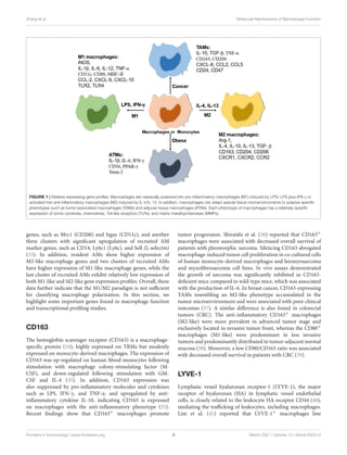
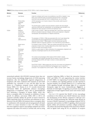
MACROPHAGE PHENOTYPES
Macrophages are very plastic cells with different phenotypes and
functions (Figure 1), which are impacted both by their origin
and resident tissue microenvironment. Broadly, macrophages
can be activated into two distinct subsets based on the
M1/M2 paradigm, classically activated or M1 macrophages and
alternatively activated or M2 macrophages (16). M1 macrophages
are polarized in vitro by Th1 cytokines such as colony-stimulating
factor (GM-CSF), tumor necrosis factor α (TNF-α), and
interferon-γ (IFN-γ) alone or together with lipopolysaccharide
(LPS) from bacteria. M1 macrophages express pro-inflammatory
cytokines such as interleukin-1β (IL-1β), IL-6, IL-12, IL-23, and
TNF-α (17). In contrast, M2 macrophages are polarized by Th2
cytokines such as IL-4 and IL-13 and produce anti-inflammatory
cytokines such as IL-10 and transforming growth factor beta
(TGF-β) (18).
Recent studies have revealed that the M1/M2 paradigm is
not sufficient to encompass all states of macrophage activation.
Macrophages have the ability to change their polarization
in response to different stimuli. For example, macrophage
phenotype changes during tissue repair, switching from pro-
inflammatory phenotype (M1-like) to an anti-inflammatory
phenotype (M2-like) (19). In addition, different polarization and
activation markers of M1- and M2-like macrophages can coexist
in tissues (20). For instance, a high percentage of circulating
macrophages expressing both M1 (CD80, CD86, and TLR4)
and M2 surface markers (CD204, CD163, and CD206) was
shown in human patients with interstitial lung disease (21). With
the analysis of the transcriptomic profiles of macrophages, Liu
et al. (22) reported that polarized M1- or M2-like macrophages
driven by cytokines can be subsequently repolarized to another
phenotype with little or no memory of polarization history.
Therefore, the in vivo phenotype and function of macrophages
remains to be defined under specific tissue microenvironments.
Phenotypic change and functional polarization of
macrophages are accompanied by a change in cellular
metabolism, as M1-like macrophages primarily rely on
glycolysis, whereas M2-like macrophages rely on oxidative
phosphorylation (23). Parallel analysis of macrophage
metabolic and transcriptional profiles also indicates that
metabolic reprogramming impacts macrophage polarization
or activation (24). For example, inhibition of aspartate-
aminotransferase and N-glycosylation interfere with M1 and
M2 macrophage polarization, respectively. Factors that affect
macrophage metabolism may disrupt M1/M2 homeostasis.
Hill et al. (25) reported that multiple distinct populations of
adipose tissue-associated macrophages (ATMs) are present
in adipose tissues in mice and humans, existing with unique
transcriptomes, chromatin landscapes, and functions. Similarly,
pro-inflammatory ATMs in obese mice or humans can express
additional markers of metabolic activation distinct from the
classical markers of activation (26).
In addition to the abilities of engulfing and digesting foreign
pathogens and cellular debris, macrophages can clear away tumor
cells. Tumor-associated macrophages (TAMs) are particularly
abundant immune cells in cancer and exert strong influences
on tumor initiation, progression, and metastasis (27, 28).
Besides, TAMs can secrete different cytokines such as IL-10
and transforming growth factor-β (TGF-β) to suppress T cell-
dependent antitumor function (29). TAMs can be polarized into
pro-inflammatory (M1-like) phenotype and anti-inflammatory
(M2-like) phenotype under the stimuli of different tumor
microenvironments, with the majority of TAMs functioning
as M2-like macrophages (30). Notably, differentially polarized
TAMs may exert opposite effects on tumor development. For
example, high numbers of infiltrating M2-like macrophages
in patients with gastric cancer (GC) were associated with a
low rate of overall survival (OS), while an elevated number
of M1-like macrophages was associated with better OS (31).
Using scRNA-seq data and cell trajectory analysis, Landry
et al. (32) reported that core TAMs evolve toward a pro-
inflammatory state in human glioblastoma, while peripheral
TAMs develop an anti-inflammatory phenotype. Therefore, an
accurate understanding of the function of TAMs is likely to
advance cancer immunotherapy.
IMPORTANT MOLECULES MEDIATING
MACROPHAGE FUNCTION
Currently, scRNA-seq is a critical tool to investigate macrophage
heterogeneity and function. For example, transcriptional
profiling analysis showed that there are five clusters of alveolar
macrophages (AMs) across homeostasis, acute inflammation,
and resolving inflammation, all expressing macrophage-specific
markers, such as CD68 and Lgals3 (Galectin-3), Among them,
two clusters express tissue-resident airspace macrophage marker
Frontiers in Immunology | www.frontiersin.org 2 March 2021 | Volume 12 | Article 620510
3.
Zhang et al.Molecular Mechanisms of Macrophage Function
FIGURE 1 | Relative expressing gene profiles. Macrophages are classically polarized into pro-inflammatory macrophages (M1) induced by LPS/ LPS plus IFN-γ or
activated into anti-inflammatory macrophages (M2) induced by IL-4/IL-13. In addition, macrophages can adapt special tissue microenvironments to polarize specific
phenotypes such as tumor-associated macrophages (TAMs) and adipose tissue macrophages (ATMs). Each phenotype of macrophages has a relatively specific
expression of some cytokines, chemokines, Toll-like receptors (TLRs), and matrix metalloproteinases (MMPs).
genes, such as Mrc1 (CD206) and Itgax (CD11c), and another
three clusters with significant upregulation of recruited AM
marker genes, such as CD14, Ly6c1 (Ly6c), and Sell (L-selectin)
(33). In addition, resident AMs show higher expression of
M2-like macrophage genes and two clusters of recruited AMs
have higher expression of M1-like macrophage genes, while the
last cluster of recruited AMs exhibit relatively low expression of
both M1-like and M2-like gene expression profiles. Overall, these
data further indicate that the M1/M2 paradigm is not sufficient
for classifying macrophage polarization. In this section, we
highlight some important genes found in macrophage function
and transcriptional profiling studies.
CD163
The hemoglobin scavenger receptor (CD163) is a macrophage-
specific protein (34), highly expressed on TRMs but modestly
expressed on monocyte-derived macrophages. The expression of
CD163 was up-regulated on human blood monocytes following
stimulation with macrophage colony-stimulating factor (M-
CSF), and down-regulated following stimulation with GM-
CSF and IL-4 (35). In addition, CD163 expression was
also suppressed by pro-inflammatory molecules and cytokines
such as LPS, IFN-γ, and TNF-α, and upregulated by anti-
inflammatory cytokine IL-10, indicating CD163 is expressed
on macrophages with the anti-inflammatory phenotype (35).
Recent findings show that CD163+ macrophages promote
tumor progression. Shiraishi et al. (36) reported that CD163+
macrophages were associated with decreased overall survival of
patients with pleomorphic sarcoma. Silencing CD163 abrogated
macrophage-induced tumor cell proliferation in co-cultured cells
of human monocyte-derived macrophages and leiomyosarcoma
and myxofibrosarcoma cell lines. In vivo assays demonstrated
the growth of sarcoma was significantly inhibited in CD163-
deficient mice compared to wild-type mice, which was associated
with the production of IL-6. In breast cancer, CD163-expressing
TAMs resembling an M2-like phenotype accumulated in the
tumor microenvironment and were associated with poor clinical
outcomes (37). A similar difference is also found in colorectal
tumors (CRC). The anti-inflammatory CD163+ macrophages
(M2-like) were more prevalent in advanced tumor stage and
exclusively located in invasive tumor front, whereas the CD80+
macrophages (M1-like) were predominant in less invasive
tumors and predominantly distributed in tumor-adjacent normal
mucosa (38). Moreover, a low CD80/CD163 ratio was associated
with decreased overall survival in patients with CRC (39).
LYVE-1
Lymphatic vessel hyaluronan receptor-1 (LYVE-1), the major
receptor of hyaluronan (HA) in lymphatic vessel endothelial
cells, is closely related to the leukocyte HA receptor CD44 (40),
mediating the trafficking of leukocytes, including macrophages.
Lim et al. (41) reported that LYVE-1+ macrophages line
Frontiers in Immunology | www.frontiersin.org 3 March 2021 | Volume 12 | Article 620510
4.
Zhang et al.Molecular Mechanisms of Macrophage Function
murine and human blood vessels, modulated collagen
production in smooth muscle cells to maintain arterial wall
homeostasis. Another study also showed LYVE-1+ cells
expressing macrophage markers CD68 or CD169 that co-
localized with collagen fibers in rat meninges, and some
LYVE-1+ cells had intracellular collagen (42). Dollt et al. (43)
also reported that extracellular domains of LYVE protein from
M2-like macrophages significantly inhibited human and murine
melanoma cell proliferation by acting as a receptor for low-
molecular weight HA. In addition, the LYVE-1+ macrophages
have been shown to play critical roles in tissue remodeling (44)
and murine eyes (45).
MerTK
Synovial tissue macrophages (STMs) play critical roles in
autoimmune diseases including rheumatoid arthritis (RA) (46).
With the analysis of integrated scRNA-seq, deep-phenotypic,
spatial and functional data, Alivernini et al. (47) found
that synovial tissue macrophages (STMs) can be broadly
classified into two populations, MerTK−CD206− STMs and
MerTK+CD206+ STMs. MerTK−CD206− STMs produce pro-
inflammatory cytokines and alarmins and induce inflammatory
responses in synovial fibroblasts, while MerTK+CD206+ STMs
from patients with RA in sustained disease remission produce
lipid mediators that resolve inflammation and induce a
repair phenotype of fibroblast-like synoviocytes. Also, they
found that two STM subpopulations (MerTK+TREM2high
and MerTK+LYVE1+) with unique remission transcriptomic
signatures, enriched in negative regulators of inflammation
(47). Kuo et al. (48) also identified a subset of inflammatory
macrophages expressing heparin-binding EGF-like growth factor
in RA joints, which altered synovial fibroblast gene expression
profile (e.g., IL-33) via up-regulation of epidermal growth factor
receptor (EGFR) response and increased their invasiveness (47,
48). MerTK as a key efferocytosis receptor, is primarily expressed
by CD11b+ F4/80+ large peritoneal macrophages (LPMs).
At steady-state, MerTK-deficient LPMs exhibit significantly
increased pro-inflammatory cytokine expression, when under
stimulation of apoptotic cells, MerTK−/− LPMs increased gene
expression of cell death and apoptosis (49).
SIGLECS
Siglecs (sialic acid-binding immunoglobulin-type lectins)
are transmembrane surface proteins found primarily on
hematopoietic cells, consisting of intracellular tyrosine motifs
involved in cell signaling (50). Siglecs predominantly recognize
sialic acid residues of glycoproteins on the cell membrane.
Fourteen of 15 known Siglecs are present in humans, namely
Siglec-1 to 15 except Siglec-13 which is present in nonhuman
primates. Siglecs on macrophages or monocytes play critical
roles in many different diseases, as described in Table 1. Siglecs
appear to have predominantly pro- or anti-inflammatory
functions in macrophages. For example, Siglec-1, a receptor on
monocytes/macrophages, plays a central role in the pathogenesis
of congenital heart block and contributes to IFN stimulation
(51). Plasma concentrations of Siglec-1 are also strongly
correlated with type I interferon-regulated gene expression in
systemic lupus erythematosus (SLE) patients (75), suggesting a
pro-inflammatory influence.
In the context of nonalcoholic fatty liver disease (NAFLD)
and nonalcoholic steatohepatitis (NASH), liver Kupffer cells and
recruited monocyte-derived macrophages play pivotal roles in
the pathophysiology of NAFLD and NASH (76). In NAFLD
patients, Siglec-7 was mostly expressed in hepatic CCR2+
macrophages, in contrast, its expression was much weaker in
resident macrophages (58). In addition, soluble Siglec-7 was
shown highly produced in monocyte-derived macrophages and
serum sSiglec-7 can be used as an independent marker for
NAFLD in patients with advanced liver fibrosis (58).
Siglec-9, as a mediator of inflammation, is a potential
target for the treatment of sepsis. Pretreatment with a
human anti-Siglec-9 Fab fragment attenuated LPS-induced pro-
inflammatory cytokines TNF-α, IL-6, and IL-1β production
in human peripheral blood mononuclear cell (PBMC)-derived
macrophages and human THP-1-differentiated macrophages
(60). Following exposure to the sialic acid-expressing human
bacterial pathogen group B Streptococcus (GBS), Siglec-14, as
a positive regulator of NLRP3 inflammasome activation on
macrophages, resulted in the release of the pro-inflammatory
cytokine IL-1β. Mononuclear phagocytes are also attractive drug
delivery vehicles for novel cancer treatment owing to their
cancerous tissue-accumulating nature. Given Siglecs are highly
expressed in TAMs and peripheral blood monocytes, sialic
acid-conjugated liposomes have been applied to deliver tumor-
targeting drugs (77). Moreover, other Siglecs including Siglec-2
(52), Siglec-3 (53), Siglec-4 (54), Siglec-5 (55, 56), Siglec-6 (57),
Siglec-8 (59), Siglec-10 (61), Siglec-11 (62), Siglec-12 (63), Siglec-
15 (64) also play important roles in diseases as listed in Table 1.
SIRPα
CD47, known as the “don’t-eat-me” signal, is commonly
overexpressed on the surface of tumor cells (78, 79). Signal
regulatory protein α (SIRPα) on macrophages serves as a
receptor of CD47 to transduce CD47/SIRPα axis mediated
inhibitory function of macrophage phagocytosis of tumor
cells (80). Blockade of CD47-SIRPα signaling is a strategy to
promote macrophage phagocytosis of many cancer cells, which is
discussed in the therapeutic section. A number of preclinical and
clinical investigations are underway to target the CD47/SIRPα
axis for cancer therapy, including examination of the synergistic
effect with other anti-tumor agents such as rituximab (anti-CD20
antibody), cetuximab (an inhibitor of EGFR), and trastuzumab
(an inhibitor of human epidermal growth factor receptor 2,
HER2) (81).
TREM2
The prevalence of obesity has reached epidemic levels, and over
44% of adults are overweight worldwide. Obesity substantially
Frontiers in Immunology | www.frontiersin.org 4 March 2021 | Volume 12 | Article 620510
5.
Zhang et al.Molecular Mechanisms of Macrophage Function
TABLE 1 | The role of Siglecs in macrophage function.
Siglecs Diseases Function References
Siglec-1 (CD169) Congenital heart
block (CHB)
The expression of IFN and type I IFN-stimulated genes, including Siglec-1, a receptor
on monocytes/macrophages, play an important role in the pathogenesis of congenital
heart block.
(51)
Siglec-2 (CD22) Aging brain CD22 (siglec-2) mediated the anti-phagocytic effect and inhibition of CD22 promoted
the clearance of myelin debris, amyloid-β oligomers, and α-synuclein fibrils in vivo. A
long-term CNS-delivery of a CD22 blocking antibody activated microglia, resulting in
the improvement of cognitive function in aged mice.
(52)
Siglec-3 (CD33) Alzheimer’s
disease
Deletion of hCD33 in macrophage cell lines U937 and THP-1 increased cargo uptake
in vitro. In addition, transgenic mice expressing hCD33 in the microglial cell lineage
showed inhibited cargo uptake in primary microglia.
(53)
Siglec-4
(Myelin-associated
Glycoprotein, or
MAG)
CNS pathology Myelin-associated glycoprotein (MAG), a minor constituent of central and peripheral
nervous system myelin, binds to gangliosides GD1a and GT1b, prominent molecules
on the axon surface, mediating axon stability in the central nervous system and
peripheral nervous system.
(54)
Siglec-5 (CD170) Asthma Siglec-5 expression was significantly increased in patients receiving inhaled
corticosteroids, exerting a beneficial effect. Double staining of cells indicated that
Siglec-5 was expressed in monocyte/macrophages and neutrophils, but not in
lymphocytes. Exposure to the sialic acid-expressing human bacterial pathogen group
B Streptococcus (GBS), Siglec-5 negatively regulates inflammation.
(55, 56)
Siglec-6 (CDw327) Colorectal cancer
(CRC)
The siglec-6 expression on mast cells is involved in their function in the tumor
microenvironment of CRC, but no evidence shown in macrophages.
(57)
Siglec-7 (CDw328) Non-alcoholic fatty
liver disease
(NAFLD)
Serum Siglec-7 could serve as an independent marker for advanced liver fibrosis in
patients with NAFLD.
(58)
Siglec-8 (Siglec-F
in mouse)
Lung disease Therapeutic targeting of Siglec-8 has the potential to impact blood as well as lung
eosinophils, which may be associated with an improved outcome in eosinophilic lung
diseases.
(59)
Siglec-9 (CD329
and Siglec-E in
mouse)
Sepsis Blockade Siglec-9 induced inflammation by anti-Siglec-9 Fab fragment (hS9-Fab03)
is a potential therapeutic agent for sepsis.
(60)
Siglec-10
(Siglec-G in
mouse)
Ovarian and breast
cancers
Blockade of CD24-Siglec-10 signaling is a potential therapeutic strategy for breast
and ovarian cancer immunotherapy.
(61)
Siglec-11 Neural disease The polysialic acid (polySia) with an average degree of polymerization 20 (avDP20)
neutralized the LPS-triggered increase in macrophage phagocytosis, by interacting
with SIGLEC-11.
(62)
Siglec-12
(Siglec-like
molecule-1)
Prostate cancer The stable expression of Siglec-12 enhanced prostate cancer cell growth in nude
mice. Anti-Siglec-12 monoclonal antibodies were internalized by
Siglec-12-expressing prostate carcinoma cells, providing a target.
(63)
Siglec-14 Bacteria Siglec-14 enhances NLRP3 inflammasome activation in macrophage, in response to
known inflammasome activators or the sialic acid-expressing human bacterial
pathogen GBS.
(56)
Siglec-15(Siglec-H
in mouse)
Cancer Siglec-15 on macrophages may contribute to tumor progression by targeting the
sialyl-Tn (sTn) antigen, a tumor-associated glycan structure, which modulating TGF-β
secretion in tumor microenvironments.
(64)
increases the risk of many diseases, including type 2 diabetes
mellitus, NAFLD, hypertension, myocardial infarction, stroke,
osteoarthritis, and cancer (82). Recently, scRNA-seq analysis
of the immune cells in murine lipid-associated macrophages
(LAMs) (83), NASH-associated macrophages (84), and aortic
macrophages (85) indicated that a subset of macrophages
expressing triggering receptor expressed on myeloid cells 2
(TREM2), which drives a gene expression program involved
in phagocytosis, lipid catabolism, and energy metabolism.
Further study showed that TREM2+ macrophages arose
from circulating monocytes and positioned around enlarged
adipocytes and scRNA-seq data of human adipose tissue
indicated that LAM cells, as well as the TREM2 pathway,
were highly conserved (83). TREM2 deletion abrogated
macrophage recruitment to enlarged adipocytes and caused
massive adipocyte hypertrophy, systemic hypercholesterolemia,
inflammation, and glucose intolerance, indicating the protective
role of TREM2+ macrophages (83). TREM2−/− mice fed
a high-fat diet (HFD) exhibited a reduction of infiltrating
F4/80+CD11c+ macrophages in adipose tissue but displayed
augmentation of pro-inflammatory cytokines IL-1β, IL-6, and
inducible nitric oxide synthase (iNOS), adipocyte hypertrophy,
Frontiers in Immunology | www.frontiersin.org 5 March 2021 | Volume 12 | Article 620510
6.
Zhang et al.Molecular Mechanisms of Macrophage Function
hepatic steatosis, and insulin resistance compared with WT
controls (86).
TREM2 expression promotes the transition of liver
macrophages from the pro-inflammatory to the tissue repair
phase and impacts endothelial cell differentiation during tissue
recovery in mouse models of acetaminophen (acute) or CCl4
(chronic)-induced hepatotoxic injury (87). In addition, TREM2
expression prevents lung macrophage apoptosis during acute
parainfluenza virus infections. A more active soluble form of
TREM2 (sTREM2) was found after clearance of infection, which
is unexpectedly active in preventing macrophage apoptosis (88).
However, the function of TREM2 may change according
to disease stage and location, with several studies suggesting
CNS-specific functions of TREM2. Recent genetic studies show
that TREM2 mutation is also associated with a higher risk
of Alzheimer’s disease (AD) and multiple neurodegenerative
disorders (89). In mice, TREM2 deficiency enhanced macrophage
activation near the lesion of traumatic brain injury (TBI) but
significantly reduced macrophage activation distant from the
lesion compared to control groups in the acute stage (90). In
addition, TREM2 deficiency resulted in a protective function at
later time points, as the TREM2−/− mice showed a reduction
of hippocampal atrophy and rescue of TBI-induced behavioral
changes compared to wild-type mice.
OTHERS
In addition to the molecules as described above, there are several
less well-studied molecules expression in macrophages, including
osteopontin (encoded by gene Spp1) (12), CLEC2 (C-type lectin
domain family 1 member B, encoded by gene Clec1b) (91), and
CD48 (92). Overall, understanding the molecular mechanism of
macrophages in tissue microenvironment is critical to design
macrophage-mediated therapy in diseases.
PROGNOSTIC MARKERS
Two of the best studies on macrophage-associated prognostic
markers include soluble CD163 (sCD163)+ and CD204+ TAMs,
with particular utility in the context of liver disease and various
cancers, respectively. Soluble CD163 (sCD163) is upregulated
during macrophage proliferation and activation and is a marker
for diagnosis of the severity and progression of liver disease. For
instance, Lidofsky et al. (65) reported that serum sCD163 from
macrophages in patients infected with human immunodeficiency
virus (HIV) and hepatitis C virus (HCV) was positively associated
with the severity of liver fibrosis from mild to moderate stage,
with an Ishak fibrosis score up to 4, but not in established
cirrhosis. These results suggest that sCD163 is a dynamic
biomarker of hepatic fibrogenesis rather than cirrhosis in patients
with viral infections (65). Another study found that sCD163
was associated with primary biliary cholangitis disease severity
and long-term (a median of 8.6 years) prognosis, following a
study in 201 patients (66). Besides, using sCD163 as a marker
increased the prediction accuracy of poor outcomes of PBC.
The sCD163 levels were also elevated in patients with early
allograft dysfunction after liver transplantation, as macrophages
were activated in the implanted liver when exposed to ischemia
and reperfusion injury (67).
Accumulating evidence reveals that CD204+ TAMs promote
cancer cell proliferation, invasion, and metastasis, resulting in
a poor survival rate. Kawajiri and colleagues reported that a
high number of CD204+ TAMs was associated with poorer
3-year overall survival (OS) and cumulative incidences of
relapse, and a poorer prognosis in allogeneic hematopoietic
cell transplantation for malignant lymphomas, such as T-cell
lymphoma and leukemia (68). In lung adenocarcinoma, the
expression of CD204 in TAMs was associated with a low 5-
year disease-free survival rate and the aggressiveness of lung
adenocarcinoma (69). CD204+ TAMs located in the tumor
stroma area were also identified as useful prognostic markers
in non-small-cell lung cancer (NSCLC) (70) and an increased
number of CD204+ TAMs was positively associated with
worse clinical prognoses in breast cancer, including relapse-
free survival, distant relapse-free survival and breast cancer-
specific survival (71). CD204 is also a useful marker for TAMs
contributing to the angiogenesis, progression and prognosis of
esophageal squamous cell carcinomas (72).
Dual markers CD163 and CD204 can also be used in diagnosis
(Table 2). For instance, the numbers and percentages of M2-
polarized alveolar macrophages expressing markers CD163+,
CD204+, and CD206+ increases with the severity of chronic
obstructive pulmonary disease, with higher numbers in smoker
patients than non-smokers (73). In clinical findings, the number
of CD163+CD204+ TAMs is negatively correlated with that
of CD25+ cells (presumably activated lymphocytes) and 5-
year progression-free survival. Dual CD163+CD204+ TAMs
possibly play a vital role in the invasion and metastasis of
oral squamous cell carcinoma by T-cell regulation via IL-10
and PD-L1 production, relative to CD163+CD204− TAMs or
CD163−CD204+ TAMs (74).
THERAPEUTIC TARGETS FOR DISEASES
While M2-like pro-tumoral phenotype is dominant in tumor
microenvironment, targeting TAMs or conversion of TAMs to
an M1-like anti-tumoral phenotype is an emerging strategy
for targeting TAMs-mediated cancer therapy (93). An in vivo
assay showed that a hybrid peptide composed of melittin and
the pro-apoptotic peptide, selectively targeted TAMs without
impacting affecting other leukocytes, such as T cells and dendritic
cells. Treatment of the hybrid peptide resulted in apoptosis
of CD206+ M2-like TAMs and reduction of tumor growth
and angiogenesis, showing as a promising cancer therapeutic
agent (94).
Targeting “Don’t eat me” signaling pathway such as blocking
the CD47-SIRPα interaction can improve macrophage
phagocytosis of tumor cells. Monotherapy of TTI-621, a
fusion protein consisting of CD47 binding domain of SIRPα,
is well-tolerated and shows rapidly active responses in adult
patients with relapsed/refractory percutaneously accessible solid
tumors (95). Another study showed that anti-human SIRPα
Frontiers in Immunology | www.frontiersin.org 6 March 2021 | Volume 12 | Article 620510
7.
Zhang et al.Molecular Mechanisms of Macrophage Function
TABLE 2 | Macrophage expressing markers CD163, CD204, or both in disease diagnosis.
Markers Diseases Function References
CD163 Liver fibrosis Under the coinfection with human immunodeficiency virus (HIV) or hepatitis C virus
(HCV), serum (s)CD163 levels accompanied periportal CD163+ macrophage
enrichment was associated with mild to moderate fibrosis, but not cirrhosis.
(65)
Primary biliary
cholangitis
(PBC)
sCD163 represents a non-invasive measure of that provides useful long-term
prognostic for PBC.
(66)
Early allograft
dysfunction
(EAD)
The sCD163 levels in patients were also elevated in patients with early allograft
dysfunction (EAD) after liver transplantation, as macrophages were activated in the
implanted liver when exposed to ischemia and reperfusion injury.
(67)
CD204 Malignant
lymphomas,
such as T-cell
lymphoma
and leukemia
A high number of CD204+
TAMs was associated with 3-year poorer overall survival
(OS) and cumulative incidences of relapse, and the poorer prognosis in allogeneic
hematopoietic cell transplantation for malignant lymphomas.
(68)
Lung
adenocarcinoma
The expression of CD204 in TAMs was associated with a low 5-year disease-free
survival (DFS) rate and the aggressiveness of lung adenocarcinoma. In lung
adenocarcinoma, CD204+
TAMs located in the tumor stroma area were the
preferable marker for prognostic prediction in non-small-cell lung cancer (NSCLC).
(69, 70)
Breast cancer
(BC)
An increased number of CD204+
TAMs was positively associated with worse clinical
prognoses in breast cancer, including relapse-free survival, distant relapse-free
survival and breast cancer-specific survival.
(71)
Esophageal
squamous
cell
carcinomas
(ESCCs)
CD204 is a useful marker for TAMs contributing to the angiogenesis, progression and
prognosis of esophageal squamous cell carcinomas (ESCCs).
(72)
CD163
and
CD204
Chronic
obstructive
pulmonary
disease
(COPD)
The numbers and percentages of M2 markers CD163+
CD204+
alveolar
macrophages were positively associated with the severity of chronic obstructive
pulmonary disease (COPD), with higher numbers in smoker patients than
non-smokers.
(73)
Oral
squamous
cell
carcinoma
(OSCC)
Dual CD163+
CD204+
TAMs possibly play a key role in the invasion and metastasis of
oral squamous cell carcinoma (OSCC) by T-cell regulation via IL-10 and PD-L1
production, more valuable than CD163+
CD204−
TAMs or CD163−
CD204+
TAMs.
(74)
monoclonal antibody (Ab) KWAR23 treatment alone does not
increase human macrophage phagocytosis of CD20-expressing
lymphoma cells, but significantly augments phagocytosis of
lymphoma cells when combined with treatment of anti-CD20
antibodies (e.g., rituximab) in vitro, and this effect was also
shown in a lymphoma xenograft mouse model expressing
human SIRPα (96). Murata et al. (97) recently showed that
anti-human SIRPα Ab significantly enhances macrophage
phagocytosis of lymphoma cancer cells in immunodeficient
mice harboring a transgene of human SIRPα, which also
shows a synergistic effect of rituximab on inhibition of tumor
growth. In addition, anti-mouse SIRPα (P84 or MY-1) Ab
treatment markedly inhibits the growth of tumors formed by
SIRPα-expressing renal cell carcinoma and melanoma in mice.
Moreover, the anti-SIRPα Ab treatment shows a synergistic effect
in combination with rituximab and anti-programmed cell death
1 (PD-1) on tumor formation in mice (98). Other strategies
have been applied to improve macrophage-mediated anti-tumor
responses and reduce the toxicity to normal cells, such as using
exosomes harboring SIRPα to block the interaction between
CD47 and SIRPα (99) and augmenting the contact interface
of CD47-SIRPα via engineering N-terminal peptide extension
(80). In addition, new signaling pathways are also reported to
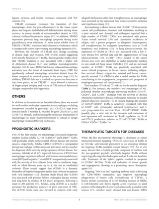
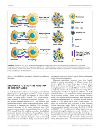
function similarly. For example, Barkal et al. (61) reported that
CD24 signaling through macrophage Siglec-10 is a potential
therapeutic target for cancer immunotherapy (Figure 2), as
blockade of CD24-Siglec-10 signaling can promote macrophage
phagocytosis of triple-negative breast cancer and ovarian cancer
cells (61).
Cai et al. (100) reported that MerKT in liver macrophage
contributes to liver fibrosis since MerKT-deficient mice slowed
the development of liver fibrosis compared to wild-type mice
when fed a NASH-promoting diet. Molecular study showed an
increase of MerKT expression on macrophages induced TGF-b1
expression and secretion via ERK1/2 signaling, which promoted
hepatic stellate cell activation to produce collagen production.
The authors also found that blockade of the interaction of MerKT
with its ligand Gas6 using RU-30, an inhibitor of receptors
Frontiers in Immunology | www.frontiersin.org 7 March 2021 | Volume 12 | Article 620510
8.
Zhang et al.Molecular Mechanisms of Macrophage Function
FIGURE 2 | Blockade of “Don’t eat me” signalings increases macrophage phagocytosis to tumor cells. Cancer cells are capable of evading macrophage
phagocytosis by expressing “don’t eat me” signals, including CD47, PD-L1 (CD274), the beta-microglobulin subunit of the major histocompatibility class I complex
(B2M), and CD24, resulting in tumor growth (top panel). Partly blockade (middle panel) and fully blockade (low panel) of these “Don’t eat me” signalings may induce
tumor shrink and tumor damage, respectively.
(Tyro-3, Axl, and Mertk), significantly inhibited the production
of collagen.
STRATEGIES TO STUDY THE FUNCTION
OF MACROPHAGES
To study the precise functions of macrophages under different
physiological and pathological conditions, several strategies
have been used to deplete macrophages in various mouse
models, including genetic ablation (101), nanotechnology-
based depletion by clodronate-encapsulated liposomes (102),
and antibody-mediated depletion (103). These strategies have
produced encouraging results in experimental murine models.
For example, in transgenic mice based on CD11b promoter-
mediated expression of the human diphtheria toxin (DT)
receptor, transient depletion of macrophages occurs in vivo when
DT is injected. Using this model, the researchers identified that
functionally distinct subpopulations of macrophages exist in
liver injury and recovery, inducing liver fibrosis by promoting
extracellular matrix deposition during liver injury or promoting
the resolution of liver fibrosis during recovery (104). These
methods are however not perfectly specific for macrophages and
induce only temporary ablation.
Clodronate-encapsulated liposomes have been broadly
applied to deplete macrophages in different tissues (bone
marrow, spleen, liver, lungs, brain, gut, peritoneal cavity,
lymph nodes/vessels) and blood. A protocol used to deplete
macrophages in mice was described before (102). For instance,
a single i.p. injection of clodronate liposome during chronic
cholangiopathy resulted in significant inhibition of ECM
deposition in mice (105). In a murine model of Azoxymethane
(AOM)/Dextran Sodium Sulfate (DSS)-induced colorectal
cancer, mice received multiple treatments with clodronate to
deplete macrophages resulted in a significant decrease of tumor
number by ∼35%, specifically for large (≥1 mm) tumors. This
effect was accompanied with a decrease in gene expression
of pan macrophage marker F4/80 as well as expression of
markers associated with M2 macrophages (IL-13, IL-10, TGF-β,
and CCL17) (106). In addition, macrophage depletion was also
associated with a significantly increased relative abundance of the
Firmicutes phylum in stool, demonstrating that macrophages are
important mediators of tumor growth and directly or indirectly
capable of influencing the gut microbiota during colorectal
cancer development (106). TAMs increased as urethane-induced
Frontiers in Immunology | www.frontiersin.org 8 March 2021 | Volume 12 | Article 620510
9.
Zhang et al.Molecular Mechanisms of Macrophage Function
lung tumor grew in mice, which exhibited a mixed M1/M2-
like macrophage phenotype. Liposomal clodronate treatment
(one time of intrathecal administration plus five times of i.v.
administration) significantly decreased the alveolar macrophage
population (> 50%) and resulted in a 50% reduction in tumor
burden compared to vehicle liposome-treated mice (107). A
subsequent study demonstrated that inhibition of recruited
macrophages by blockade of CCL2/CCR2 signaling did not
attenuate lung cancer progression, as CCR2-deficient mice
showed similar tumor growth rate compared to wild-type mice.
However, depletion of the entire macrophages in a specific
tissue or whole body may result in unwanted side effects. For
instance, depletion of peritoneal macrophages with clodronate
injection induced a dramatic decrease in neutrophil recruitment
in experimental peritonitis (108). Another study showed
transient depletion of macrophages may exacerbate liver injury
in NASH patients since monocytes-derived KCs show more
pro-inflammatory activity and are less efficient in hepatic
triglyceride storage (91). As we discussed above molecular
markers, some subpopulations of macrophages also benefit the
resolution of diseases, such as LYVE-1+ macrophages. Thus,
deletion of specific macrophage subpopulation or specific gene
in macrophages under investigation may represent a more
reasonable approach.
SUMMARY
Macrophages are a key population of innate immunity,
with powerful influences on homeostasis, tissue repair,
obesity, and cancer. Macrophages consist of two populations,
tissue-resident macrophages with a prenatal origin and
postnatal monocyte-derived macrophages. Independent of
their origin, macrophages are very plastic cells, being able
to change their phenotype according to local environmental
stimuli. They are usually polarized into M1-like or M2-like
phenotype; however, recent studies suggest that the phenotype of
macrophages cannot be simply divided into M1/M2 dichotomy,
and additional classifications, such as TAMs and ATMs, are
applied to define the tissue-specific macrophages. In this review,
promising candidate macrophage markers are highlighted
due to their potential application in diagnosis and treatment
against diseases.
Several methods have been applied to target TAMs or ATMs
to prevent tumor growth or inflammation, such as macrophage
depletion, blockade of anti-phagocytic signaling (e.g., Siglec-10
or SIRPα). Therefore, understanding the molecular mechanisms
of macrophage function in disease provides potent diagnostic
markers and/or therapeutic strategies.
AUTHOR CONTRIBUTIONS
CZ and MY conceived and wrote the manuscript. AE provided
guidance for part of manuscript, critically reviewed and
significantly edited the manuscript. All authors contributed to the
article and approved the submitted version.
FUNDING
This manuscript was supported by University of Missouri
Postdoctoral Research Award.
REFERENCES
1. Epelman S, Lavine KJ, Randolph GJ. Origin and
functions of tissue macrophages. Immunity. (2014) 41:21–
35. doi: 10.1016/j.immuni.2014.06.013
2. Yona S, Kim KW, Wolf Y, Mildner A, Varol D, Breker M, et al. Fate mapping
reveals origins and dynamics of monocytes and tissue macrophages under
homeostasis. Immunity. (2013) 38:79–91. doi: 10.1016/j.immuni.2012.12.001
3. Hashimoto D, Chow A, Noizat C, Teo P, Beasley MB, Leboeuf M, et al.
Tissue-resident macrophages self-maintain locally throughout adult life
with minimal contribution from circulating monocytes. Immunity. (2013)
38:792–804. doi: 10.1016/j.immuni.2013.04.004
4. Gomez Perdiguero Klapproth EK, Schulz C, Busch K, Azzoni E,
Crozet L, Garner HC, et al. Tissue-resident macrophages originate from
yolk-sac-derived erythro-myeloid progenitors. Nature. (2015) 518:547–
51. doi: 10.1038/nature13989
5. Epelman S, Lavine KJ, Beaudin AE, Sojka DK, Carrero JA, Calderon B, et al.
Embryonic and adult-derived resident cardiac macrophages are maintained
through distinct mechanisms at steady state and during inflammation.
Immunity. (2014) 40:91–104. doi: 10.1016/j.immuni.2013.11.019
6. Wang X, Sathe AA, Smith GR, Ruf-Zamojski F, Nair V, Lavine KJ,
et al. Heterogeneous origins and functions of mouse skeletal muscle-
resident macrophages. Proc Natl Acad Sci U S A. (2020) 117:20729–
40. doi: 10.1073/pnas.1915950117
7. Saeki N, Imai Y. Reprogramming of synovial macrophage metabolism by
synovial fibroblasts under inflammatory conditions. Cell Commun Signal.
(2020) 18:188. doi: 10.1186/s12964-020-00678-8
8. Herzog C, Pons Garcia L, Keatinge M, Greenald D, Moritz C, Peri F,
et al. Rapid clearance of cellular debris by microglia limits secondary
neuronal cell death after brain injury in vivo. Development. 146:9.
(2019) doi: 10.1242/dev.174698
9. Bosurgi L, Cao YG, Cabeza-Cabrerizo M, Tucci A, Hughes LD, Kong Y, et al.
Macrophage function in tissue repair and remodeling requires IL-4 or IL-13
with apoptotic cells. Science. (2017) 356:1072–6. doi: 10.1126/science.aai8132
10. Greenhalgh AD, Zarruk JG, Healy LM, Baskar Jesudasan SJ, Jhelum P,
Salmon CK, et al. Peripherally derived macrophages modulate microglial
function to reduce inflammation after CNS injury. PLoS Biol. (2018)
16:e2005264. doi: 10.1371/journal.pbio.2005264
11. Fastrès A, Pirottin D, Fievez L, Tutunaru A-C, Bolen G, Merveille A-
C, et al. Identification of pro-fibrotic macrophage populations by single-
cell transcriptomic analysis in west highland white terriers affected
with canine idiopathic pulmonary fibrosis. Front Immunol. (2020)
11:611749. doi: 10.3389/fimmu.2020.611749
12. Remmerie A, Martens L, Thoné T, Castoldi A, Seurinck R, Pavie B,
et al. Osteopontin expression identifies a subset of recruited macrophages
distinct from Kupffer cells in the fatty liver. Immunity. (2020) 53:641–57.
doi: 10.1016/j.immuni.2020.08.004
13. Sárvári AK, Van Hauwaert EL, Markussen LK, Gammelmark E, Marcher
AB, Ebbesen MF, et al. Plasticity of epididymal adipose tissue in
response to diet-induced obesity at single-nucleus resolution. Cell Metab.
(2020) doi: 10.1016/j.cmet.2020.12.004
14. Liu D, Guo M, Zhou P, Xiao J, Ji X. TSLP promote M2 macrophages
polarization and cardiac healing after myocardial infarction. Biochem
Biophys Res Commun. (2019) 516:437–44. doi: 10.1016/j.bbrc.2019.0
6.041
15. Van Hove Martens L, Scheyltjens I, De Vlaminck K, Pombo
Antunes AR, De Prijck S, Vandamme N, et al. A single-cell atlas of
mouse brain macrophages reveals unique transcriptional identities
Frontiers in Immunology | www.frontiersin.org 9 March 2021 | Volume 12 | Article 620510
10.
Zhang et al.Molecular Mechanisms of Macrophage Function
shaped by ontogeny and tissue environment. Nat Neurosci. (2019)
22:1021–35. doi: 10.1038/s41593-019-0393-4
16. Mills CD, Kincaid K, Alt JM, Heilman MJ, Hill AM. M-1/M-2
macrophages and the Th1/Th2 paradigm. J Immunol. (2000) 164:6166–
73. doi: 10.4049/jimmunol.164.12.6166
17. Chen Y, Zhang X. Pivotal regulators of tissue homeostasis
and cancer: macrophages. Exp Hematol Oncol. (2017)
6:23. doi: 10.1186/s40164-017-0083-4
18. Gordon S, Martinez FO. Alternative activation of macrophages:
mechanism and functions. Immunity. (2010) 32:593–
604. doi: 10.1016/j.immuni.2010.05.007
19. Novak ML, Koh TJ. Macrophage phenotypes during tissue repair. J Leukocyte
Biol. (2013) 93:875–81. doi: 10.1189/jlb.1012512
20. Orecchioni M, Ghosheh Y, Pramod AB, Ley K. Macrophage polarization:
different gene signatures in M1(LPS+) vs. classically and M2(LPS-
) vs. alternatively activated macrophages. Front Immunol. (2019)
10:1084. doi: 10.3389/fimmu.2019.01084
21. Trombetta AC, Soldano S, Contini P, Tomatis V, Ruaro B, Paolino S, et al. A
circulating cell population showing both M1 and M2 monocyte/macrophage
surface markers characterizes systemic sclerosis patients with lung
involvement. Resp Res. (2018) 19:186. doi: 10.1186/s12931-018-0891-z
22. Liu SX, Gustafson HH, Jackson DL, Pun SH, Trapnell C. Trajectory analysis
quantifies transcriptional plasticity during macrophage polarization. Sci Rep.
(2020) 10:12273. doi: 10.1038/s41598-020-68766-w
23. Viola A, Munari F, Sánchez-Rodríguez R, Scolaro T, Castegna A. The
metabolic signature of macrophage responses. Front Immunol. (2019)
10:1462. doi: 10.3389/fimmu.2019.01462
24. Jha AK, Huang SC, Sergushichev A, Lampropoulou V, Ivanova
Y, Loginicheva E, et al. Network integration of parallel
metabolic and transcriptional data reveals metabolic modules
that regulate macrophage polarization. Immunity. (2015)
42:419–30. doi: 10.1016/j.immuni.2015.02.005
25. Hill DA, Lim H-W, Kim YH, Ho WY, Foong YH, Nelson VL,
et al. Distinct macrophage populations direct inflammatory versus
physiological changes in adipose tissue. Proc Natl Acad Sci USA. (2018)
115:E5096. doi: 10.1073/pnas.1802611115
26. Kratz M, Coats BR, Hisert KB, Hagman D, Mutskov V, Peris E, et al.
Metabolic dysfunction drives a mechanistically distinct proinflammatory
phenotype in adipose tissue macrophages. Cell Metab. (2014) 20:614–
25. doi: 10.1016/j.cmet.2014.08.010
27. Wenes M, Shang M, Di Matteo M, Goveia J, Martín-Pérez R, Serneels J, et al.
Macrophage metabolism controls tumor blood vessel morphogenesis and
metastasis. Cell Metab. (2016) 24:701–15. doi: 10.1016/j.cmet.2016.09.008
28. Tu D, Dou J, Wang M, Zhuang H, Zhang X. M2 macrophages contribute
to cell proliferation and migration of breast cancer. Cell Biol Int.
(2020). doi: 10.21203/rs.3.rs-39373/v1. [Epub ahead of print].
29. Ruffell B, Chang-Strachan D, Chan V, Rosenbusch A, Ho CM, Pryer
N, et al. Macrophage IL-10 blocks CD8+ T cell-dependent responses to
chemotherapy by suppressing IL-12 expression in intratumoral dendritic
cells. Cancer Cell. (2014) 26:623–37. doi: 10.1016/j.ccell.2014.09.006
30. Yuan H, Lin Z, Liu Y, Jiang Y, Liu K, Tu M, et al. Intrahepatic
cholangiocarcinoma induced M2-polarized tumor-associated macrophages
facilitate tumor growth and invasiveness. Cancer Cell Int. (2020)
20:586. doi: 10.1186/s12935-020-01687-w
31. Wang XL, Jiang JT, Wu CP. Prognostic significance of tumor-associated
macrophage infiltration in gastric cancer: a meta-analysis. Genet Mol Res.
(2016) 15. doi: 10.4238/gmr15049040
32. Landry AP, Balas M, Alli S, Spears J, Zador Z. Distinct regional ontogeny and
activation of tumor associated macrophages in human glioblastoma. Sci Rep.
(2020) 10:19542. doi: 10.1038/s41598-020-76657-3
33. Mould KJ, Jackson ND, Henson PM, Seibold M, Janssen WJ. Single cell RNA
sequencing identifies unique inflammatory airspace macrophage subsets. JCI
Insight. 4:5. (2019) doi: 10.1172/jci.insight.126556
34. Etzerodt A, Moestrup SK. CD163 and inflammation: biological,
diagnostic, therapeutic aspects. Antioxid Redox Sign. (2013)
18:2352–63. doi: 10.1089/ars.2012.4834
35. Buechler C, Ritter M, Orsó E, Langmann T, Klucken J, Schmitz G.
Regulation of scavenger receptor CD163 expression in human monocytes
and macrophages by pro- and antiinflammatory stimuli. J Leukoc Biol. (2000)
67:97–103. doi: 10.1002/jlb.67.1.97
36. Shiraishi D, Fujiwara Y, Horlad H, Saito Y, Iriki T, Tsuboki
J, et al. CD163 is required for protumoral activation of
macrophages in human and murine sarcoma. Cancer Res. (2018)
78:3255–66. doi: 10.1158/0008-5472.CAN-17-2011
37. Ramos RN, Rodriguez C, Hubert M, Ardin M, Treilleux I, Ries CH, et al.
CD163(+) tumor-associated macrophage accumulation in breast cancer
patients reflects both local differentiation signals and systemic skewing of
monocytes. Clin Transl Immunol. (2020) 9:e1108. doi: 10.1002/cti2.1108
38. Pinto ML, Rios E, Durães C, Ribeiro R, Machado JC, Mantovani
A, et al. The two faces of tumor-associated macrophages and their
clinical significance in colorectal cancer. Front Immunol. (2019)
10:1875. doi: 10.3389/fimmu.2019.01875
39. Rozek LS, Schmit SL, Greenson JK, Tomsho LP, Rennert HS, Rennert
G, et al. Tumor-Infiltrating lymphocytes, Crohn’s-like lymphoid
reaction, and survival from colorectal cancer. J Natl Cancer Inst. (2016)
108:27. doi: 10.1093/jnci/djw027
40. Banerji S, Ni J, Wang SX, Clasper S, Su J, Tammi R, et al. LYVE-1, a
new homologue of the CD44 glycoprotein, is a lymph-specific receptor for
hyaluronan. J Cell Biol. (1999) 144:789–801. doi: 10.1083/jcb.144.4.789
41. Lim HY, Lim SY, Tan CK, Thiam CH, Goh CC, Carbajo D, et al. Hyaluronan
receptor LYVE-1-expressing macrophages maintain arterial tone through
hyaluronan-mediated regulation of smooth muscle cell collagen. Immunity.
(2018) 49:326–41.e7. doi: 10.1016/j.immuni.2018.06.008
42. Brezovakova V, Jadhav S. Identification of Lyve-1 positive macrophages
as resident cells in meninges of rats. J Comp Neurol. (2020) 528:2021–
32. doi: 10.1002/cne.24870
43. Dollt C, Becker K, Michel J, Melchers S, Weis C-A, Schledzewski K,
et al. The shedded ectodomain of Lyve-1 expressed on M2-like tumor-
associated macrophages inhibits melanoma cell proliferation. Oncotarget.
(2017) 8:103682. doi: 10.18632/oncotarget.21771
44. Oe S, Masum MA, Ichii O, Nishimura T, Nakamura T, Namba T, et al.
Spatiotemporal histological changes observed in mouse subcutaneous tissues
during the foreign body reaction to silicone. J Biomed Mater Res A.
(2020). doi: 10.1002/jbm.a.37115. [Epub ahead of print].
45. Xu H, Chen M, Reid DM, Forrester JV. LYVE-1-positive macrophages are
present in normal murine eyes. Invest Ophthalmol Vis Sci. (2007) 48:2162–
71. doi: 10.1167/iovs.06-0783
46. Kurowska-Stolarska M, Alivernini S. Synovial tissue macrophages: friend or
foe? RMD Open. (2017) 3:e000527. doi: 10.1136/rmdopen-2017-000527
47. Alivernini S, MacDonald L, Elmesmari A, Finlay S, Tolusso B, Gigante MR,
et al. Distinct synovial tissue macrophage subsets regulate inflammation
and remission in rheumatoid arthritis. Nature Medicine. (2020) 26:1295–
306. doi: 10.1038/s41591-020-0939-8
48. Kuo D, Ding J, Cohn IS, Zhang F, Wei K, Rao DA, et al. HBEGF(+)
macrophages in rheumatoid arthritis induce fibroblast invasiveness. Sci
Transl Med. (2019) 11:eaau8587. doi: 10.1126/scitranslmed.aau8587
49. Lantz C, Radmanesh B, Liu E, Thorp EB, Lin J. Single-cell
RNA sequencing uncovers heterogenous transcriptional signatures
in macrophages during efferocytosis. Scientific Reports. (2020)
10:14333. doi: 10.1038/s41598-020-70353-y
50. von Gunten S, Bochner BS. Basic and clinical immunology of Siglecs. Ann N
Y Acad Sci. (2008) 1143:61–82. doi: 10.1196/annals.1443.011
51. Clancy RM, Halushka M, Rasmussen SE, Lhakhang T, Chang M, Buyon
JP. Siglec-1 macrophages and the contribution of IFN to the development
of autoimmune congenital heart block. J Immunol. (2019) 202:48–
55. doi: 10.4049/jimmunol.1800357
52. Pluvinage JV, Haney MS, Smith BAH, Sun J, Iram T, Bonanno L, et al. CD22
blockade restores homeostatic microglial phagocytosis in ageing brains.
Nature. (2019) 568:187–192. doi: 10.1038/s41586-019-1088-4
53. Bhattacherjee A, Rodrigues E, Jung J, Luzentales-Simpson M, Enterina
JR, Galleguillos DCD, et al. Repression of phagocytosis by human
CD33 is not conserved with mouse CD33. Commun Biol. (2019)
2:450. doi: 10.1038/s42003-019-0698-6
54. Pan B, Fromholt SE, Hess EJ, Crawford TO, Griffin JW, Sheikh KA,
et al. Myelin-associated glycoprotein and complementary axonal ligands,
gangliosides, mediate axon stability in the CNS and PNS: neuropathology
Frontiers in Immunology | www.frontiersin.org 10 March 2021 | Volume 12 | Article 620510
11.
Zhang et al.Molecular Mechanisms of Macrophage Function
and behavioral deficits in single- and double-null mice. Exp Neurol. (2005)
195:208–217. doi: 10.1016/j.expneurol.2005.04.017
55. Wielgat P, Mroz RM, Stasiak-Barmuta A, Szepiel P, Chyczewska E,
Braszko JJ, et al. Inhaled corticosteroids increase siglec-5/14 expression
in sputum cells of COPD patients. Adv Exp Med Biol. (2015) 839:1–
5. doi: 10.1007/5584_2014_51
56. Tsai CM, Riestra AM, Ali SR, Fong JJ, Liu JZ, Hughes G, et al. Siglec-
14 enhances NLRP3-inflammasome activation in macrophages. J Innate
Immun. (2019) 2019:1-11. doi: 10.1159/000504323
57. Yu Y, Blokhuis BRJ, Diks MAP, Keshavarzian A, Garssen J,
Redegeld FA. Functional inhibitory siglec-6 is upregulated in human
colorectal cancer-associated mast cells. Front Immunol. (2018)
9:2138. doi: 10.3389/fimmu.2018.02138
58. Sakamoto Y, Yoshio S, Doi H, Kawai H, Shimagaki T, Mori T, et al. Serum
soluble sialic acid-binding immunoglobulin-like lectin-7 concentration as
an indicator of liver macrophage activation and advanced fibrosis in
patients with non-alcoholic fatty liver disease. Hepatol Res. (2020) 50:466–77.
doi: 10.1111/hepr.13464
59. Johansson MW, Kelly EA, Nguyen CL, Jarjour NN, Bochner BS.
Characterization of Siglec-8 expression on lavage cells after segmental
lung allergen challenge. Int Arch Allergy Immunol. (2018) 177:16–
8. doi: 10.1159/000488951
60. Chu S, Zhu X, You N, Zhang W, Zheng F, Cai B, et al. The fab fragment
of a human anti-siglec-9 monoclonal antibody suppresses LPS-induced
inflammatory responses in human macrophages. Front Immunol. (2016)
7:649. doi: 10.3389/fimmu.2016.00649
61. Barkal AA, Brewer RE, Markovic M, Kowarsky M, Barkal SA, Zaro BW,
et al. CD24 signalling through macrophage Siglec-10 is a target for cancer
immunotherapy. Nature. (2019) 572:392–6. doi: 10.1038/s41586-019-1456-0
62. Shahraz A, Kopatz J, Mathy R, Kappler J, Winter D, Kapoor S, et al. Anti-
inflammatory activity of low molecular weight polysialic acid on human
macrophages. Sci Rep. (2015) 5:16800. doi: 10.1038/srep16800
63. Mitra N, Banda K, Altheide TK, Schaffer L, Johnson-Pais TL, Beuten J,
et al. SIGLEC12, a human-specific segregating (pseudo)gene, encodes a
signaling molecule expressed in prostate carcinomas. J Biol Chem. (2011)
286:23003–11. doi: 10.1074/jbc.M111.244152
64. Takamiya R, Ohtsubo K, Takamatsu S, Taniguchi N, Angata T.
The interaction between Siglec-15 and tumor-associated sialyl-Tn
antigen enhances TGF-β secretion from monocytes/macrophages
through the DAP12-Syk pathway. Glycobiology. (2013) 23:178–
87. doi: 10.1093/glycob/cws139
65. Lidofsky A, Holmes JA, Feeney ER, Kruger AJ, Salloum S, Zheng H, et al.
Macrophage activation marker soluble CD163 is a dynamic marker of liver
fibrogenesis in human immunodeficiency virus/hepatitis C virus coinfection.
J Infect Dis. (2018) 218:1394–403. doi: 10.1093/infdis/jiy331
66. Bossen L, Rebora P, Bernuzzi F, Jepsen P, Gerussi A, Andreone P, et al.
Soluble CD163 and mannose receptor as markers of liver disease severity
and prognosis in patients with primary biliary cholangitis. Liver Int. (2020)
40:1408–14. doi: 10.1111/liv.14466
67. Thomsen KL, Robertson FP, Holland-Fischer P, Davidson BR, Mookerjee
RP, Moller HJ, et al. The macrophage activation marker soluble CD163 is
associated with early allograft dysfunction after liver transplantation. J Clin
Exp Hepatol. (2019) 9:302–11. doi: 10.1016/j.jceh.2018.09.006
68. Kawajiri A, Kitano S, Maeshima AM, Inamoto Y, Tajima K, Takemura T, et al.
Association of CD204(+) macrophages with poor outcomes of malignant
lymphomas not in remission treated by allogeneic HCT. Eur J Haematol.
(2019) 103:578–87. doi: 10.1111/ejh.13324
69. Sun Y, Xu S. Tumor-Associated CD204-positive macrophage is a prognostic
marker in clinical stage i lung adenocarcinoma. Biomed Res Int. (2018)
2018:8459193. doi: 10.1155/2018/8459193
70. Li Z, Maeda D, Yoshida M, Umakoshi M, Nanjo H, Shiraishi K, et al. The
intratumoral distribution influences the prognostic impact of CD68- and
CD204-positive macrophages in non-small cell lung cancer. Lung Cancer.
(2018) 123:127–135. doi: 10.1016/j.lungcan.2018.07.015
71. Miyasato Y, Shiota T, Ohnishi K, Pan C, Yano H, Horlad H, et al.
High density of CD204-positive macrophages predicts worse clinical
prognosis in patients with breast cancer. Cancer science. (2017) 108:1693–
700. doi: 10.1111/cas.13287
72. Shigeoka M, Urakawa N, Nakamura T, Nishio M, Watajima T, Kuroda
D, et al. Tumor associated macrophage expressing CD204 is associated
with tumor aggressiveness of esophageal squamous cell carcinoma. Cancer
Science. (2013) 104:1112–9. doi: 10.1111/cas.12188
73. Kaku Y, Imaoka H, Morimatsu Y, Komohara Y, Ohnishi K, Oda H, et al.
Overexpression of CD163, CD204 and CD206 on alveolar macrophages in
the lungs of patients with severe chronic obstructive pulmonary disease.
PLoS ONE. (2014) 9:e87400. doi: 10.1371/journal.pone.0087400
74. Kubota K, Moriyama M, Furukawa S, Rafiul ASM H, Maruse Y, Jinno T,
et al. CD163+CD204+ tumor-associated macrophages contribute to T cell
regulation via interleukin-10 and PD-L1 production in oral squamous cell
carcinoma. Sci Rep. (2017) 7:1755. doi: 10.1038/s41598-017-01661-z
75. Oliveira JJ, Karrar S, Rainbow DB, Pinder CL, Clarke P, Rubio Garcia
A, et al. The plasma biomarker soluble SIGLEC-1 is associated with
the type I interferon transcriptional signature, ethnic background and
renal disease in systemic lupus erythematosus. Arthritis Res Ther. (2018)
20:152. doi: 10.1186/s13075-018-1649-1
76. Kazankov K, Jorgensen SMD, Thomsen KL, Moller HJ, Vilstrup H, George
J, et al. The role of macrophages in nonalcoholic fatty liver disease and
nonalcoholic steatohepatitis. Nat Rev Gastroenterol Hepatol. (2019) 16:145–
9. doi: 10.1038/s41575-018-0082-x
77. Ding J, Zhao D, Hu Y, Liu M, Liao X, Zhao B, et al. Terminating the
renewal of tumor-associated macrophages: A sialic acid-based targeted
delivery strategy for cancer immunotherapy. Int J Pharm. (2019)
571:118706. doi: 10.1016/j.ijpharm.2019.118706
78. Willingham SB, Volkmer JP, Gentles AJ, Sahoo D, Dalerba P, Mitra SS,
et al. The CD47-signal regulatory protein alpha (SIRPa) interaction is a
therapeutic target for human solid tumors. Proc Natl Acad Sci USA. (2012)
109:6662–7. doi: 10.1073/pnas.1121623109
79. Horrigan SK. Replication study: the CD47-signal regulatory protein alpha
(SIRPa) interaction is a therapeutic target for human solid tumors. Elife.
(2017) 6:18173. doi: 10.7554/eLife.18173
80. Ho CC, Guo N, Sockolosky JT, Ring AM, Weiskopf K, Özkan E,
et al. Garcia: “Velcro” engineering of high affinity CD47 ectodomain
as signal regulatory protein α (SIRPα) antagonists that enhance
antibody-dependent cellular phagocytosis. J Biol Chem. (2015)
290:12650–63. doi: 10.1074/jbc.M115.648220
81. Weiskopf K. Cancer immunotherapy targeting the CD47/SIRPα axis. Eur J
Cancer. (2017) 76:100-9. doi: 10.1016/j.ejca.2017.02.013
82. Blüher M. Obesity: global epidemiology and pathogenesis. Nat Rev
Endocrinol. (2019) 15:288–98. doi: 10.1038/s41574-019-0176-8
83. Jaitin DA, Adlung L, Thaiss CA, Weiner A, Li B, Descamps
H, et al. Lipid-associated macrophages control metabolic
homeostasis in a Trem2-dependent manner. Cell. (2019)
178:686–98.e14. doi: 10.1016/j.cell.2019.05.054
84. Xiong X, Kuang H, Ansari S, Liu T, Gong J, Wang S, et al.
Landscape of intercellular crosstalk in healthy and NASH liver
revealed by single-cell secretome gene analysis. Mol Cell. (2019)
75:644–60.e5. doi: 10.1016/j.molcel.2019.07.028
85. Cochain C, Vafadarnejad E, Arampatzi P, Pelisek J, Winkels H, Ley K, et al.
Single-Cell RNA-Seq reveals the transcriptional landscape and heterogeneity
of aortic macrophages in murine atherosclerosis. Circ Res. (2018) 122:1661–
74. doi: 10.1161/CIRCRESAHA.117.312509
86. Liu C, Li P, Li H, Wang S, Ding L, Wang H, et al. TREM2 regulates obesity-
induced insulin resistance via adipose tissue remodeling in mice of high-fat
feeding. J Trans Med. (2019) 17:300. doi: 10.1186/s12967-019-2050-9
87. Coelho I, Duarte N, Barros A, Macedo MP, Penha-Gonçalves C.
Trem-2 promotes emergence of restorative macrophages and endothelial
cells during recovery from hepatic tissue damage. bioRxiv. (2019)
2019:823773. doi: 10.1101/823773
88. Wu K, Byers DE, Jin X, Agapov E, Alexander-Brett J, Patel AC, et al. TREM-
2 promotes macrophage survival and lung disease after respiratory viral
infection. J Exp Med. (2015) 212:681–97. doi: 10.1084/jem.20141732
89. Gratuze M, Leyns CEG, Holtzman DM. New insights into the
role of TREM2 in Alzheimer’s disease. Mol Neurodegen. (2018)
13:66. doi: 10.1186/s13024-018-0298-9
90. Saber M, Kokiko-Cochran O, Puntambekar SS, Lathia JD, Lamb BT.
Triggering receptor expressed on myeloid cells 2 deficiency alters acute
Frontiers in Immunology | www.frontiersin.org 11 March 2021 | Volume 12 | Article 620510
![Zhang et al. Molecular Mechanisms of Macrophage Function
shaped by ontogeny and tissue environment. Nat Neurosci. (2019)
22:1021–35. doi: 10.1038/s41593-019-0393-4
16. Mills CD, Kincaid K, Alt JM, Heilman MJ, Hill AM. M-1/M-2
macrophages and the Th1/Th2 paradigm. J Immunol. (2000) 164:6166–
73. doi: 10.4049/jimmunol.164.12.6166
17. Chen Y, Zhang X. Pivotal regulators of tissue homeostasis
and cancer: macrophages. Exp Hematol Oncol. (2017)
6:23. doi: 10.1186/s40164-017-0083-4
18. Gordon S, Martinez FO. Alternative activation of macrophages:
mechanism and functions. Immunity. (2010) 32:593–
604. doi: 10.1016/j.immuni.2010.05.007
19. Novak ML, Koh TJ. Macrophage phenotypes during tissue repair. J Leukocyte
Biol. (2013) 93:875–81. doi: 10.1189/jlb.1012512
20. Orecchioni M, Ghosheh Y, Pramod AB, Ley K. Macrophage polarization:
different gene signatures in M1(LPS+) vs. classically and M2(LPS-
) vs. alternatively activated macrophages. Front Immunol. (2019)
10:1084. doi: 10.3389/fimmu.2019.01084
21. Trombetta AC, Soldano S, Contini P, Tomatis V, Ruaro B, Paolino S, et al. A
circulating cell population showing both M1 and M2 monocyte/macrophage
surface markers characterizes systemic sclerosis patients with lung
involvement. Resp Res. (2018) 19:186. doi: 10.1186/s12931-018-0891-z
22. Liu SX, Gustafson HH, Jackson DL, Pun SH, Trapnell C. Trajectory analysis
quantifies transcriptional plasticity during macrophage polarization. Sci Rep.
(2020) 10:12273. doi: 10.1038/s41598-020-68766-w
23. Viola A, Munari F, Sánchez-Rodríguez R, Scolaro T, Castegna A. The
metabolic signature of macrophage responses. Front Immunol. (2019)
10:1462. doi: 10.3389/fimmu.2019.01462
24. Jha AK, Huang SC, Sergushichev A, Lampropoulou V, Ivanova
Y, Loginicheva E, et al. Network integration of parallel
metabolic and transcriptional data reveals metabolic modules
that regulate macrophage polarization. Immunity. (2015)
42:419–30. doi: 10.1016/j.immuni.2015.02.005
25. Hill DA, Lim H-W, Kim YH, Ho WY, Foong YH, Nelson VL,
et al. Distinct macrophage populations direct inflammatory versus
physiological changes in adipose tissue. Proc Natl Acad Sci USA. (2018)
115:E5096. doi: 10.1073/pnas.1802611115
26. Kratz M, Coats BR, Hisert KB, Hagman D, Mutskov V, Peris E, et al.
Metabolic dysfunction drives a mechanistically distinct proinflammatory
phenotype in adipose tissue macrophages. Cell Metab. (2014) 20:614–
25. doi: 10.1016/j.cmet.2014.08.010
27. Wenes M, Shang M, Di Matteo M, Goveia J, Martín-Pérez R, Serneels J, et al.
Macrophage metabolism controls tumor blood vessel morphogenesis and
metastasis. Cell Metab. (2016) 24:701–15. doi: 10.1016/j.cmet.2016.09.008
28. Tu D, Dou J, Wang M, Zhuang H, Zhang X. M2 macrophages contribute
to cell proliferation and migration of breast cancer. Cell Biol Int.
(2020). doi: 10.21203/rs.3.rs-39373/v1. [Epub ahead of print].
29. Ruffell B, Chang-Strachan D, Chan V, Rosenbusch A, Ho CM, Pryer
N, et al. Macrophage IL-10 blocks CD8+ T cell-dependent responses to
chemotherapy by suppressing IL-12 expression in intratumoral dendritic
cells. Cancer Cell. (2014) 26:623–37. doi: 10.1016/j.ccell.2014.09.006
30. Yuan H, Lin Z, Liu Y, Jiang Y, Liu K, Tu M, et al. Intrahepatic
cholangiocarcinoma induced M2-polarized tumor-associated macrophages
facilitate tumor growth and invasiveness. Cancer Cell Int. (2020)
20:586. doi: 10.1186/s12935-020-01687-w
31. Wang XL, Jiang JT, Wu CP. Prognostic significance of tumor-associated
macrophage infiltration in gastric cancer: a meta-analysis. Genet Mol Res.
(2016) 15. doi: 10.4238/gmr15049040
32. Landry AP, Balas M, Alli S, Spears J, Zador Z. Distinct regional ontogeny and
activation of tumor associated macrophages in human glioblastoma. Sci Rep.
(2020) 10:19542. doi: 10.1038/s41598-020-76657-3
33. Mould KJ, Jackson ND, Henson PM, Seibold M, Janssen WJ. Single cell RNA
sequencing identifies unique inflammatory airspace macrophage subsets. JCI
Insight. 4:5. (2019) doi: 10.1172/jci.insight.126556
34. Etzerodt A, Moestrup SK. CD163 and inflammation: biological,
diagnostic, therapeutic aspects. Antioxid Redox Sign. (2013)
18:2352–63. doi: 10.1089/ars.2012.4834
35. Buechler C, Ritter M, Orsó E, Langmann T, Klucken J, Schmitz G.
Regulation of scavenger receptor CD163 expression in human monocytes
and macrophages by pro- and antiinflammatory stimuli. J Leukoc Biol. (2000)
67:97–103. doi: 10.1002/jlb.67.1.97
36. Shiraishi D, Fujiwara Y, Horlad H, Saito Y, Iriki T, Tsuboki
J, et al. CD163 is required for protumoral activation of
macrophages in human and murine sarcoma. Cancer Res. (2018)
78:3255–66. doi: 10.1158/0008-5472.CAN-17-2011
37. Ramos RN, Rodriguez C, Hubert M, Ardin M, Treilleux I, Ries CH, et al.
CD163(+) tumor-associated macrophage accumulation in breast cancer
patients reflects both local differentiation signals and systemic skewing of
monocytes. Clin Transl Immunol. (2020) 9:e1108. doi: 10.1002/cti2.1108
38. Pinto ML, Rios E, Durães C, Ribeiro R, Machado JC, Mantovani
A, et al. The two faces of tumor-associated macrophages and their
clinical significance in colorectal cancer. Front Immunol. (2019)
10:1875. doi: 10.3389/fimmu.2019.01875
39. Rozek LS, Schmit SL, Greenson JK, Tomsho LP, Rennert HS, Rennert
G, et al. Tumor-Infiltrating lymphocytes, Crohn’s-like lymphoid
reaction, and survival from colorectal cancer. J Natl Cancer Inst. (2016)
108:27. doi: 10.1093/jnci/djw027
40. Banerji S, Ni J, Wang SX, Clasper S, Su J, Tammi R, et al. LYVE-1, a
new homologue of the CD44 glycoprotein, is a lymph-specific receptor for
hyaluronan. J Cell Biol. (1999) 144:789–801. doi: 10.1083/jcb.144.4.789
41. Lim HY, Lim SY, Tan CK, Thiam CH, Goh CC, Carbajo D, et al. Hyaluronan
receptor LYVE-1-expressing macrophages maintain arterial tone through
hyaluronan-mediated regulation of smooth muscle cell collagen. Immunity.
(2018) 49:326–41.e7. doi: 10.1016/j.immuni.2018.06.008
42. Brezovakova V, Jadhav S. Identification of Lyve-1 positive macrophages
as resident cells in meninges of rats. J Comp Neurol. (2020) 528:2021–
32. doi: 10.1002/cne.24870
43. Dollt C, Becker K, Michel J, Melchers S, Weis C-A, Schledzewski K,
et al. The shedded ectodomain of Lyve-1 expressed on M2-like tumor-
associated macrophages inhibits melanoma cell proliferation. Oncotarget.
(2017) 8:103682. doi: 10.18632/oncotarget.21771
44. Oe S, Masum MA, Ichii O, Nishimura T, Nakamura T, Namba T, et al.
Spatiotemporal histological changes observed in mouse subcutaneous tissues
during the foreign body reaction to silicone. J Biomed Mater Res A.
(2020). doi: 10.1002/jbm.a.37115. [Epub ahead of print].
45. Xu H, Chen M, Reid DM, Forrester JV. LYVE-1-positive macrophages are
present in normal murine eyes. Invest Ophthalmol Vis Sci. (2007) 48:2162–
71. doi: 10.1167/iovs.06-0783
46. Kurowska-Stolarska M, Alivernini S. Synovial tissue macrophages: friend or
foe? RMD Open. (2017) 3:e000527. doi: 10.1136/rmdopen-2017-000527
47. Alivernini S, MacDonald L, Elmesmari A, Finlay S, Tolusso B, Gigante MR,
et al. Distinct synovial tissue macrophage subsets regulate inflammation
and remission in rheumatoid arthritis. Nature Medicine. (2020) 26:1295–
306. doi: 10.1038/s41591-020-0939-8
48. Kuo D, Ding J, Cohn IS, Zhang F, Wei K, Rao DA, et al. HBEGF(+)
macrophages in rheumatoid arthritis induce fibroblast invasiveness. Sci
Transl Med. (2019) 11:eaau8587. doi: 10.1126/scitranslmed.aau8587
49. Lantz C, Radmanesh B, Liu E, Thorp EB, Lin J. Single-cell
RNA sequencing uncovers heterogenous transcriptional signatures
in macrophages during efferocytosis. Scientific Reports. (2020)
10:14333. doi: 10.1038/s41598-020-70353-y
50. von Gunten S, Bochner BS. Basic and clinical immunology of Siglecs. Ann N
Y Acad Sci. (2008) 1143:61–82. doi: 10.1196/annals.1443.011
51. Clancy RM, Halushka M, Rasmussen SE, Lhakhang T, Chang M, Buyon
JP. Siglec-1 macrophages and the contribution of IFN to the development
of autoimmune congenital heart block. J Immunol. (2019) 202:48–
55. doi: 10.4049/jimmunol.1800357
52. Pluvinage JV, Haney MS, Smith BAH, Sun J, Iram T, Bonanno L, et al. CD22
blockade restores homeostatic microglial phagocytosis in ageing brains.
Nature. (2019) 568:187–192. doi: 10.1038/s41586-019-1088-4
53. Bhattacherjee A, Rodrigues E, Jung J, Luzentales-Simpson M, Enterina
JR, Galleguillos DCD, et al. Repression of phagocytosis by human
CD33 is not conserved with mouse CD33. Commun Biol. (2019)
2:450. doi: 10.1038/s42003-019-0698-6
54. Pan B, Fromholt SE, Hess EJ, Crawford TO, Griffin JW, Sheikh KA,
et al. Myelin-associated glycoprotein and complementary axonal ligands,
gangliosides, mediate axon stability in the CNS and PNS: neuropathology
Frontiers in Immunology | www.frontiersin.org 10 March 2021 | Volume 12 | Article 620510](https://image.slidesharecdn.com/pathologyassignment-250311065413-dcdcde1c/85/pathology-assignment-pdfghjjt-n-fnfdjfdd-10-320.jpg)