Learning Objectives (1of 2)
• Compare benign versus malignant tumors; discuss
naming of tumors and exceptions to standard
terminology
• Summarize principal types of lymphoma
• Differentiate infiltrating versus in situ carcinoma;
role of Pap smear in early diagnoses of neoplasms
• Explain classification and clinical manifestation of
leukemia
• Differentiate leukemia versus multiple myeloma
• Explain mechanism of body’s immunologic
defenses against tumor
3.
Learning Objectives (2of 2)
• Summarize modalities and side effects of cancer
treatment
• Describe applications and limitations of tumor-
associated antigens
• Compare incidence and survival rates for various
malignant tumors
• Explain role of late recurrence and role of adjuvant therapy
• Understand role of oncogenes(a gene which in certain
circumstances can transform a cell into a tumour cell) and
disturbance in suppressor gene function in the pathogenesis
of tumors
4.
Definition
A tumor suppressorgene, or
antioncogene, is a gene that protects
a cell from one step on the path to
cancer. When this gene mutates to
cause a loss or reduction in its
function, the cell can progress to
cancer, usually in combination with
other genetic changes.
5.
Definition
Tumor suppressor genesare normal genes
that slow down cell division, repair DNA
mistakes, or tell cells when to die (a
process known as apoptosis or
programmed cell death). When tumor
suppressor genes don't work properly, cells
can grow out of control, which can lead to
cancer.
6.
Definition
• Neoplasm: NEO= new + PLASM = growth
• Cancer: any type of malignant growth
– Unrestrained growth and spread
– Cells do not respond to control mechanisms
that normally regulate cell growth and
differentiation
– Serves no useful purpose
– Terms neoplasm and tumor may be used
interchangeably
7.
Warning Signs forCancer
• Change in bowel/bladder habits or function
• A sore that does not heal
• Unusual bleeding or discharge
• Thickening or lump in breast or elsewhere
• Indigestion or difficulty swallowing
• Obvious change in growth or infiltrator
• Nagging cough or hoarseness
8.
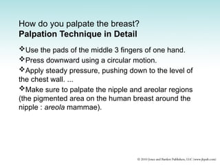
How do youpalpate the breast?
Palpation Technique in Detail
Use the pads of the middle 3 fingers of one hand.
Press downward using a circular motion.
Apply steady pressure, pushing down to the level of
the chest wall. ...
Make sure to palpate the nipple and areolar regions
(the pigmented area on the human breast around the
nipple : areola mammae).
9.
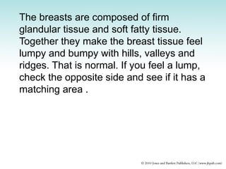
The breasts arecomposed of firm
glandular tissue and soft fatty tissue.
Together they make the breast tissue feel
lumpy and bumpy with hills, valleys and
ridges. That is normal. If you feel a lump,
check the opposite side and see if it has a
matching area .
10.
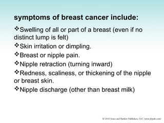
symptoms of breastcancer include:
Swelling of all or part of a breast (even if no
distinct lump is felt)
Skin irritation or dimpling.
Breast or nipple pain.
Nipple retraction (turning inward)
Redness, scaliness, or thickening of the nipple
or breast skin.
Nipple discharge (other than breast milk)
11.
CTD
If you feela lump in your breast, try not to panic
or worry. Most lumps are not breast cancer, but
something less serious, such as a benign
breast condition. Some lumps will go away on
their own. In younger women, lumps are often
related to menstrual periods and will go away
by the end of the cycle.
Benign Tumors
• Namedby adding suffix - oma to the name
of the cells of origin
– Adenoma: from glandular epithelium
– Angioma: from blood vessels
– Chondroma: from cartilage
– Polyps or papilloma: benign tumor on stalk
arising from an epithelial surface
14.
Malignant Tumors (1of 2)
• Start from a single cell that has sustained
damage to its genome, causing it to proliferate
abnormally
• twin of identical cells is formed; if unchecked,
eventually develops into a distinct tumor
• Exhibit behavior different from that of normal
cells
• Do not respond to normal growth regulatory
signals
• Proliferate unnecessarily
15.
Malignant Tumors (2of 2)
• May secrete growth factors to stimulate their own
growth, allowing tumors to flourish at the expense
of surrounding normal cells
• Secrete enzymes that break down normal cell and
tissue barriers, allowing them to
– Infiltrate into adjacent tissues
– Invade lymphatic channels and blood vessels
– Spread throughout the body
• Tumor cells do not normally “wear out” as normal
cells, but become “immortal” and can proliferate
indefinitely
16.
Tumor Classification (1of 2)
• Carcinoma: involves epithelial tissue
– Most common: 85% of all tumors found in skin,
large intestine, glands, stomach, lungs,
prostate
– Metastasis: principally through lymph vessels
– Subtypes:
• Adenocarcinoma (internal organ or gland)
• Squamous cell carcinoma (skin)
17.
Tumor Classification (2of 2)
• Sarcoma: arising from connective tissues such as
fat, bone, cartilage, muscle
– Less common, but spreads more rapidly
– Little differentiation; anaplasia (lack of form)
– Metastasis: bloodstream
• Leukemia: neoplasm of blood cells
– Usually do not form solid tumors
– Instead, proliferates diffusely within bone marrow,
overgrow and crowd out normal blood-forming cells
– Neoplastic cells “spread out” into the bloodstream and
large number of abnormal cells circulate in the peripheral
blood
18.
Naming of Tumors
•Tumors are named and classified according to their
cells and tissues of origin.
• Tumor nomenclature: not completely uniform, but
certain generalizations are possible.
• Exceptions encountered in naming of
– Lymphoid tumors
– Skin tumors arising from pigment-producing cells within
the epidermis
– Certain tumors of mixed cellular components
– Certain types of tumors composed of primitive cells seen
in children
19.
Lymphoma (1 of2)
• Neoplasm of lymphoid tissue
– Usually malignant
– Term “lymphoma” without classification refers to a
malignant, not a benign tumor
– To avoid confusion, the term “malignant lymphoma”
maybe used
• Two major classifications
– Hodgkin’s lymphoma (Hodgkin’s disease: with Reed –
Sternberg cells)
– Non-Hodgkin’s lymphoma(without Reed – Sternberg cells
• Classification: often with poor correlation between
histologic type and biologic behavior (growth rate
and response to therapy)
20.
Lymphoma (2 of2)
• Basis of classification
– Diffuse infiltration of lymph nodes
– Prognosis
• Low-grade: patients have a favorable prognosis
• Intermediate-grade: patients do not do nearly as well
• High-grade: patients do poorly
– Type of cells giving rise to tumor (T cells, B cells,
NK cells, histiocytes) and maturity of cells
• 75% arise from B lymphocytes
• Remainder mostly from T lymphocytes
21.
Hodgkin and Non-Hodgkin
Lymphoma(1 of 2)
• Hodgkin disease: variable histologic appearance
consisting of large cells called Reed-Sternberg
cells intermixed with lymphocytes, plasma cells,
eosinophils, and fibrous tissues
– Reed-Sternberg cell: large cell with abundant cytoplasm
containing two nuclei appearing as mirror images
• Some have a single nucleus
• Each nucleus contains large nucleolus surrounded by clear halo
– Four different histologic types of Hodgkin disease that
differ in clinical behavior and prognosis
22.
Hodgkin and Non-Hodgkin
Lymphoma(2 of 2)
• Non-Hodgkin lymphoma: all other lymphoma are
generally grouped together under this category
– Variable in appearance and behavior
– Classification system based on size, shape, growth
pattern of malignant cells, and shape of nuclei and
nuclear membranes
23.
Skin Tumors (1of 2)
• Most skin tumors arise from keratinocytes or
melanocytes
• Keratinocytes: keratin-forming cells
• Basal cells: deepest layer of keratinocytes
adjacent to the dermis
• Squamous cells: upper layer of cells that arise
from the proliferation of basal cells
• Melanocytes: (interspersed among
keratinocytes) skin cells that normally produce
pigment and are responsible for normal skin
color; produce melanin, dark-brown pigment
Malignant Skin Tumors
•Basal cell carcinoma
– Composed of clusters of infiltrating cells that resemble
the normal basal cells of the epidermis
– Indolent, slowly growing tumor that can be locally
destructive but rarely metastasizes
• Squamous cell carcinoma
– Composed of abnormal infiltrating squamous cells
– More aggressive tumor that sometimes metastasizes
• Both types can be cured by surgical excision with
a very good prognosis
• Excessive sunlight exposure predisposes to all
types of skin cancer, including melanoma and
keratoses
26.
Teratoma
• Tumor arisingfrom cells that can differentiate
into many different types of tissues: bone,
muscle, glands, epithelium, brain tissue, hair
• Has mixed components, poorly organized
• Frequently occurs in reproductive tract, but
may develop in other areas
• Must specify as benign or malignant based on
maturity of cells
27.
Primitive Cell Tumors
•Arise from persistent groups of primitive cells
and may arise in children
– Brain
– Retina
– Adrenal gland
– Kidney
– Liver
– Genital tract
• Named after site of origin with suffix “-blastoma”
added
– Example: tissue of origin (retina) + blastoma =
retinoblastoma
28.
Leukemia
• A neoplasmof hematopoietic tissue
• Leukemic cells diffusely infiltrate the bone
marrow and lymphoid tissues, spill over
into the bloodstream, and infiltrate throughout
various organs of the body
• Cells may be mostly mature or extremely
primitive
• Overproduction of white cells demonstrated in
the peripheral blood by a very high white
blood count
• Aleukemic leukemia: condition in which
white cells are confined to the bone marrow
such that their number in the peripheral blood
is normal or decreased
29.
Leukemia: Classification
• Anytype of hematopoietic cells can give rise to a
leukemia, but the most common types are:
– Granulocytic
– Lymphocytic
– Monocytic
• Basis for Classification of Leukemia
– Cell type
• Granulocytic, lymphocytic, monocytic
– Maturity of leukemic cells
• Acute if immature cells
• Chronic if mature cells
30.
Leukemia: Clinical Features(1 of 2)
• Manifestations caused by impairment of
bone marrow function
• Leukemic cells crowd out normal cells
causing:
– Anemia: inadequate red cell production
– Thrombocytopenia: causes bleeding
– Infections from inadequate number of normal
white cells
31.
Leukemia: Clinical Features(2 of 2)
• Caused by infiltration of organs by leukemic
cells causing:
– Splenomegaly: enlarged spleen ( why ?)
– Hepatomegaly: enlarged liver ( why ?)
– Lymphadenopathy: enlarged lymph nodes
( why ?)
• In chronic leukemia: evolution of disease
proceeds at a relatively slow pace and often
can be controlled
• In acute leukemia: a rapidly progressive
disease, more difficult to control
Myelodysplasia (Preleukemia)
• Adisturbed growth and maturation of marrow cells
– Anemia: reduced number of erythrocytes
– Leukopenia: reduced number white cells
– Thrombocytopenia: reduced number of platelets
• Although called preleukemia, not all patients
develop leukemia
• Recently grouped together under the general term
myelodysplastic syndromes
• In general, the more severe the maturation
disturbance in the bone marrow, the greater the
likelihood that leukemia will occur
34.
Multiple Myeloma
• Neoplasmfrom plasma cells within the bone
marrow
• Resembles leukemia, but cell proliferation is
confined to the bone marrow and organ infiltration
is unusual
• Outpouring of large number of plasma cells into
the peripheral blood is also uncommon
• Abnormal plasma cells either infiltrate the bone
marrow diffusely or form discrete tumors that weaken the
bone
• Leads to spontaneous fractures, pain, and disability
35.
Tumor Blood Supplyand
Necrosis (1 of 2)
• Tumors derive blood supply from tissues they invade
• Malignant tumors frequently induce new blood vessels
to proliferate in adjacent normal tissues to supply the
demands of the growing tumor (angiogenesis factor)
• Malignant tumor may outgrow its blood supply; the
part of the tumor with the poorest blood supply
undergoes necrosis
• Depending on the location of the tumor, the blood
supply will be rich or poor
36.
Tumor Blood Supplyand
Necrosis (2 of 2)
• In tumors in the lung, blood supply is best at the
periphery of the tumor and poorest at the center
• If tumor is growing outward from an epithelial
surface such as the colon, the best blood supply
is at its base and poorest at the surface
• Often, small blood vessels are exposed in the
ulcerated base of a tumor that blood may ooze
continuously from vessels leading to anemia from
chronic blood loss
• An ulcerated tumor may be the source of a severe
hemorrhage
37.
Noninfiltrating (in Situ)Carcinoma
• Arises from the surface epithelium
• Remains localized within the epithelium for
many years
• Can occur in many locations of the body
– Cervix
– Breast
– Urinary tract
– Colon
– Skin
38.
Etiologic Factors inNeoplasic
Disease (1 of 2)
• Viruses
• Gene and chromosomal abnormalities
• Failure of immunologic defenses
• Heredity
• Viruses: cause some cancers in humans
– Leukemia and lymphoma: T cell leukemia-lymphoma
virus (HTLV-1) that is related to the AIDS virus
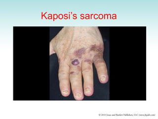
– Kaposi’s sarcoma: human herpesvirus 8 (HHV-8)
– Condylomas: papilloma virus; predisposes to cervical
carcinoma
– Chronic viral hepatitis: hepatitis B and C virus
– Nasopharyngeal carcinoma: Epstein-Barr virus also
causes infectious mononucleosis
Etiologic Factors inNeoplastic
Disease (2 of 2)
• Gene and chromosomal abnormalities
• Three large groups of genes play an important role
in regulating cell functions
• Mutations in these genes are associated with
tumor formation
– Proto-oncogenes
– Tumor-suppressor genes
– DNA repair genes
41.
Proto-oncogenes
• Normal “growthgenes” in the human
chromosomes that promote some aspects of cell
growth, differentiation, or mitotic activity
• Becomes an oncogene if mutation occurs or
genes are translocated to another chromosome
• Oncogene: abnormally functioning gene that
stimulates cell growth excessively, leading to
unrestricted cell proliferation
42.
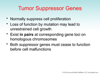
Tumor Suppressor Genes
•Normally suppress cell proliferation
• Loss of function by mutation may lead to
unrestrained cell growth
• Exist in pairs at corresponding gene loci on
homologous chromosomes
• Both suppressor genes must cease to function
before cell malfunctions
43.
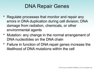
DNA Repair Genes
•Regulate processes that monitor and repair any
errors in DNA duplication during cell division; DNA
damage from radiation, chemicals, or other
environmental agents
• Mutation: any change in the normal arrangement of
DNA nucleotides on the DNA chain
• Failure in function of DNA repair genes increase the
likelihood of DNA mutations within the cell
44.
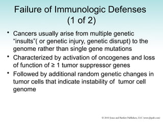
Failure of ImmunologicDefenses
(1 of 2)
• Cancers usually arise from multiple genetic
“insults”( or genetic injury, genetic disrupt) to the
genome rather than single gene mutations
• Characterized by activation of oncogenes and loss
of function of ≥ 1 tumor suppressor genes
• Followed by additional random genetic changes in
tumor cells that indicate instability of tumor cell
genome
45.
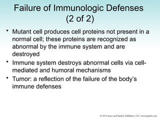
Failure of ImmunologicDefenses
(2 of 2)
• Mutant cell produces cell proteins not present in a
normal cell; these proteins are recognized as
abnormal by the immune system and are
destroyed
• Immune system destroys abnormal cells via cell-
mediated and humoral mechanisms
• Tumor: a reflection of the failure of the body’s
immune defenses
46.
Heredity and Tumors(1 of 2)
• Predisposition apparently results from
multifactorial inheritance pattern
• Individual at risk has inherited set of genes that
influence hormonal or enzyme-regulated
biochemical process in the body that can increase
susceptibility to a specific cancer
• Example: breast cancer
– 80% to 90%: no family history of the disease
– 10% linked to gene mutations
47.
Diagnosis of Tumors(1 of 2)
• Recognize early warning signs and symptoms
• Complete medical history and physical
examination
• Laboratory procedures
– Examination of rectum and colon(explain)
– Vaginal examination and Pap smear in women
– Examination of esophagus and stomach
– X-ray studies
– Abnormal smear: slides of abnormal cells shed from
surface of tumors
– Cytologic diagnosis: from smears, needle aspiration,
biopsy
– Frozen section: slides prepared and stained for rapid
histologic diagnosis
48.
Diagnosis of Tumors(2 of 2)
• Tumor associated antigen tests: some cancers secrete
substances that can be detected in the blood by lab
tests
• CEA (carcinoembrionic antigen): present in amounts
related to the size of tumor and its possible spread
– Produced by most malignant tumors of the GI tract, pancreas,
breast
• Alpha fetoprotein: normally produced by fetal tissues in
the placenta but not adult cells; elevated in primary
carcinoma of the liver
• Human chorionic gonadotropin: normally produced by
placenta; elevated in testicular carcinoma
• Acid-phosphatase: normally produced by prostate
epithelial cells, may be elevated in prostate cancer
Chemotherapy
• Eliminates cellsthat divide frequently
• Cancer cells + rapidly dividing normal cells
found in the:
– Mouth, skin, hair, bone marrow, digestive tract,
kidneys, bladder
– Lungs, nervous system, reproductive system
• Normal cells recover quickly, side effects
disappear gradually
• How soon the patient will feel better depends
on overall health, types of anticancer drugs
used
51.
Side Effects ofChemotherapy
(1 of 2)
• Anemia: extreme fatigue, weakness, tiredness,
paleness, dizziness experienced by more than
half of patients; reduces bone marrow’s ability
to make red blood cells
• Constipation: drugs, decrease in physical
activity, unbalanced diet
• Depression: physical and emotional stress
• Diarrhea: drugs affect cells that line intestines
• Fatigue
52.
Side Effects ofChemotherapy
(2 of 2)
• Hair loss (alopecia)
• Infection due to reduced ability of bone marrow
to produce white blood cells
• Loss of appetite (anorexia)
• Mouth, gum, and throat problems; sores
• Nausea and vomiting
• Sexual problems
– Males: affect sperm cells; temporary/permanent
infertility
– Women: irregular menstrual periods; vaginal
infections; menopause-like symptoms
53.
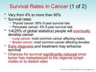
Survival Rates inCancer (1 of 2)
• Vary from 4% to more than 95%
• Survival rates:
– Thyroid cancer: 95% 5-year survival rate
– Pancreatic cancer: 4% 5-year survival rate
• 1/4(25% of global statistics) people will eventually
develop cancer
– Lung cancer: most common cancer affecting males
– Breast cancer: most common cancer affecting females
• Early diagnosis and treatment may enhance
survival
• Chances for survival significantly reduced once
tumor has metastasized to the regional lymph
nodes or to distant sites
54.
Survival Rates inCancer (2 of 2)
• 5-year survival does not indicate cure; some types
recur, can even be fatal
• Tumor may have already spread by time of
diagnosis and initial treatment, but metastatic
deposits held in check by immune defense
mechanisms
• Recurrence: failure of body’s defenses, reactivation
of tumor; some malignant tumors recur and can be
fatal many years after initial treatment
• Breast cancer and malignant melanoma prone to
late recurrences
• Breast cancer: 65%: 5-year survival rate and 50%
10-year survival rate