In this study of 277 post-menopausal women, the researchers found:
1) Higher bone mineral density was associated with increased severity of disc space narrowing at both the lumbar spine and femoral sites.
2) There was no association found between bone mineral density and severity of osteophytes.
3) Biochemical markers of bone resorption (CTX levels) decreased with increased severity of disc space narrowing, but were not associated with osteophyte severity. This suggests disc space narrowing may have a protective effect against bone loss through decreased bone resorption.
Analytical Study of Clinicopathological Data of Saudi Patients with Osteoarth...Prof. Hesham N. Mustafa
SUMMARY: Knee osteoarthritis (OA) is a common disabling disease. Epidemiological studies have revealed various risk
factors for OA, including sex, aging, obesity, occupational illnesses, and chronic diseases. Here we evaluate the clinical, pathological,
and radiological findings of knee OA in a subset of Saudi patients who were subjected to total knee replacement (TKA). The study
population included 30 Saudi patients with knee OA who were operated by TKA (from June 2014 to December 2015) in the Department
of Orthopedics, Faculty of Medicine, King Abdulaziz University, Saudi Arabia. Patient’s clinical and radiological data were collected
from the hospital files. Pathological examination of the excised superior articular surface of tibia and femoral condyles were done.
Pearson Chi-squared analysis was used to test for differences between the variables in associated risk factors. There were more women
than men. Sixty per cent of patients were older than 60 years [mean age, 59.2 (females) and 61.7 (men) years-old]. All patients exceeded
obesity class 1, with females being more obese than males. Pathological examination of the superior articular surface of tibia and femoral
condyles showed high score lesions, which was more apparent in females than in males. Radiological findings showed that most lesions
were high grade. The findings of this study will help to understand the pathogenesis of OA and improve treatment decision making
relevant to TKA in knee OA in Saudi Arabia and elsewhere.
KEY WORDS: Osteoarthritis; Knee; Arthroplasty.
Clinical and epidemiological profile of patients undergoing total hip arthro...David Sadigursky
Clinical and epidemiological profile of patients undergoing total hip arthroplasty.
Rheumatology and Orthopedic Medicine
Rheumatol Orthop Med, 2017 doi: 10.15761/ROM.1000120
Sanni Ali's presentation from Osteoporosis 2016: Antidiabetic medication use and the risk of fracture amongst type 2 diabetic patients: a nested case-control study
Find out more at: https://nos.org.uk/conference
A comparative study on the clinical and functional outcome of limb salvage su...NAAR Journal
The aim of this study was to analyze the survival, recurrence, complications as well as the quality of life (QOL) in tibial osteosarcoma (OSA) patients managed by limb salvage surgery (LSS), either by a prosthesis, resection or graft or by amputation. 106 tibial osteosarcoma patients were enrolled where 39 had custom-designed endoprosthetic arthroplasty (LSS1), 36 underwent resection and bone graft (LSS2) while only 31 underwent amputation. A Comparison was done based on post-operative survival rates, postoperative recurrence, and complications. The impact of the patient’s QOL was also evaluated.
The Incidence of Traumatic Posterior and Combined Labral Tears in Patients Un...Lennard Funk
Presentation at ISAKOS, 2019
There were 442 primary arthroscopic labral repair procedures performed over the three-year period. The total cohort had a mean age of 25.91±9.09 years (range, 14-67 years) and consisted of 89.6% males. There was no significant difference in mean age or gender between the isolated anterior, posterior or combined groups (p=0.383 and p=0.541, respectively).
• Of the 442 patients who underwent a shoulder labral repair, isolated anterior labral pathology occurred in 52.9% (n=234), with posterior and combined labral tears accounting for 16.3% (n=72) and 30.8%, respectively (n=136) (Table 3).
• Patients were stratified as either sporting or non-sporting; 74.9% of patients were categorised as sporting (n=331) and had a mean age of 24.91±5.69 years, which was significantly lower than the mean age of 35.40±11.94 years in the non-sporting population (p<0.001). In the non-sporting population 68.5% (n=76) of patients had isolated anterior labral tears with 12.6% (n=14) posterior and 18.9% (n=21) combined. In the sporting population isolated anterior labral tears accounted for 47.7% (n=158), posterior 17.5% (n=58) and combined labral tears 34.7% (n=115). The sporting population had a significantly greater proportion of posterior and combined labral tears with the non-sporting population a significantly greater proportion of anterior labral tears (p=0.013).
• Rugby players had the greatest incidence of shoulder instability within the sporting cohort accounting for 231 cases. Of the 231 cases, 47.2% were isolated anterior labral tears, 12.6% isolated posterior and 40.3% combined lesions.
Posterior and combined shoulder labral tears are more prevalent than previously reported in the civilian population. The rates are higher in young, sporting populations and especially in contact sports such as rugby.
Frequency of Osteoporotic Fractures, Parameters of Bone Mineral Density and T...CrimsonPublishersOPROJ
Frequency of Osteoporotic Fractures, Parameters of Bone Mineral Density and Trabecular Bone Score in Postmenopausal Women by Grygorieva N* in Orthopedic Research Online Journal
Osteoarthritis is the most common disease of women after menopause. There are many factors to develop the disease. Hormones play important role to in this context. The objective of the present study is to determine whether the levels of thyroid and sex hormones are associated with osteoarthritis (OA) in postmenopausal women. Forty three patients suffering from OA and twenty control subjects were included in this study. Thyroid and sex hormones were measured in the serum by enzyme linked immunosorbent assay technique. In OA patients serum estrogen levels were low as compared to control subjects(p<0.001), but these patients did not show any significant change in thyroid hormones and progesterone hormone levels when compared with control subjects. The findings suggest that estrogen deficiency after menopause may contribute to develop OA in postmenopausal women.
Dr Steve Cummings presentation from Osteoporosis 2016: Patients receiving bisphosphonates should not take holidays from treatment.
Find out more at: https://nos.org.uk/conference
Crimson Publishers-Abdominal Pain Caused by Bilateral Acetabular Fractures Se...CrimsonPublishersOPROJ
Abdominal Pain Caused by Bilateral Acetabular Fractures Secondary to an Epileptic Seizure Case Report and Review of the Literature by EJP Jansen in Orthopedic Research Online Journal
—Kyphosis and lordosis changes might be related to back extensor weakness and osteoporosis. The purpose of this study was to find out the correlations between thoracic kyphosis, lumbar lordosis with back extensor strength (BES) and bone mineral density (BMD). Methods: Thoracic kyphosis, lumbar lordosis, maximal isometric strength of the back extensors and BMD of the lumbar vertebral were evaluated in 47 elderly (50-75 years old)women. BMD of the lumbar vertebral was measured using Dual-Energy X-Ray Absorptiometry (DEXA) and kyphosis and lordosis degree were assayed using a flexible ruler. The maximal isometric strength of the back extensors was measured using an isometric manual muscle tester (MMT). Data were analyzed using ANOVA and independent t-test at p≤0.05 level of acceptance. Results: A significant reverse correlation was shown between BES and kyphosis (p=0.044, r=-0.30). No significant correlation were found between BES and lordosis degree, nor between lumbar vertebral BMD and, both, kyphosis and lordosis degrees. However, there was a significant difference in BES between three groups with various degree of kyphosis (p≤ 0.05). Conclusion: It can be concluded that the severity of thoracic kyphosis may be influenced by BES. So, stronger back extensor can prevent thoracic kyphosis despite decreased BMD.
Abstract— Cerebral palsy (CP) is the most common physical disability of childhood. Children with CP frequently grow slowly and are more prone to fractures. So this study was aimed to explore relationship of bone mineral density (BMD) with cerebral palsy by case-control study. This study was conducted at Department of Physical Medicine and Rehabilitation of Sawai Man Singh Medical College, Jaipur. Hip bone and spine bone was used to assess BMD. Bone mineral density was measured by DEXA in both groups i.e. study group and control group after ensuring the comparability of both groups. Difference in means of BMD in both the groups was inferred by unpaired student's’ test of significance. It was found in this study that bone mineral density of hip well as spine was significantly lowered in cerebral palsy cases.
CLINICAL EVALUATION OF THE EFFECT OF OMEGA-3 FATTY ACIDS ON OSTEOPOROTIC FEMA...Mohamed A. Galal
Mohamed A. Galal ; Mushira A. Dahaba, ; Basma M. Zaki
and Hanaa M. Elshenawy. CLINICAL EVALUATION OF THE EFFECT OF OMEGA-3 FATTY ACIDS ON OSTEOPOROTIC FEMALES HAVING CHRONIC PERIODONTITIS. Cairo Dental Journal (30)Number (1), 1:10January, 2014.
A Retrospective Study to Investigate Association among Age, BMI and BMD in th...IOSR Journals
Bone strength (and, hence, fracture risk) is dependent on many qualities of bone, of which bone mineral density (BMD) is the most commonly measured. Association between advancing age and lower body mass index (BMI) is an important risk factor in the occurrence of low BMD. This study was aimed at evaluation of the association among age, BMI and status of BMD among 159 age matched postmenopausal women who underwent Dual-Energy X-ray Absorptimetry (DEXA) scan. The study population was divided into three groups on the basis of body mass index (BMI) as normal weight, obese and severely obese. The mean bone mineral density (BMD) of obese and severely obese postmenopausal women was found to be significantly higher (P value < 0.001) as compared to the mean BMD of normal weight women. Significant negative correlation was found between the age and BMI except in severely obese group (P value < 0.05). Age and BMD in all the three groups correlated negatively (P value < 0.01) in all the three groups. BMD and BMI in the normal weight group significantly correlated negatively (P value < 0.05) while a very weak positive but insignificant correlation existed between the same in the obese and severely obese postmenopausal women. The study revealed that with advancing age BMD is lowered and that higher BMI might have a positive influence (although not significant as observed in the present study) on the BMD. Other factors like exposure to sunlight, calcium intake, diet etc should also be investigated which could not be probed in the present study as it was a retrospective analysis.
Significance of Trace Element Quantities in Osteomyelitis and Osteosarcoma_Cr...CrimsonpublishersCancer
To clarify the role of trace elements (TE) in the etiology and the pathogenesis of osteomyelitis (OM) and osteosarcoma (OS), a nondestructive neutron activation analysis were performed. The Ag, Co, Cr, Fe, Hg, Rb, Sb, Se, and Zn contents were measured in three groups of samples: normal bone samples from 27 persons with intact bone, and also in samples, obtained from open biopsies or after operation of 10 patients with OM and 27 patients with OS. The difference in the results between TE contents in the three groups was evaluated by the parametric Student’s t-test and non-parametric Wilcoxon-Mann-Whitney U-test. In the OM tissue the mean contents of Co, Cr, Fe, Se, and Zn are respectively 1.8, 1.7, 1.8, 1.7, and 1.5 times higher than those in normal bone tissues In the OS tissue the mean mass fractions of Co, Cr, Fe, Sb, Se, and Zn are respectively 4.6, 2.0, 4.8 2.4, 11.0, and 2.4 times higher while the mean mass fraction of Rb is more than 40% lower than in normal bone tissues. In the OS tissue the mean mass fractions of Co, Fe, Se, and Zn are significantly higher (2.6, 2.6, 6.2, and 1.6 times, respectively) and the mean mass fraction of Rb is more than 2 times lower than in inflamed bone. In addition, many inter-correlations between TE contents found in the control group were no longer evident in the inflamed and tumor transformed bone. Thus, considerable changes in TE content and their relationships were found in OM and OS and possible causes and effects of these alterations are discussed.
EVALUATION OF THE EFFICIENCY OF BISPHOSPHONATES IN THE TREATMENT OF OSTEOPORO...indexPub
Osteoporosis (OP) is a widespread metabolic disease of the skeleton, leading to decreased bone strength and increased risk of fractures. OP is a disease of varying nature that affects all age groups, but is most common in older people. For a long time, doctors did not have serious tools to treat this insidious disease and mainly dealt with its consequences - fractures.
Explore natural remedies for syphilis treatment in Singapore. Discover alternative therapies, herbal remedies, and lifestyle changes that may complement conventional treatments. Learn about holistic approaches to managing syphilis symptoms and supporting overall health.
Factory Supply Best Quality Pmk Oil CAS 28578–16–7 PMK Powder in Stockrebeccabio
Factory Supply Best Quality Pmk Oil CAS 28578–16–7 PMK Powder in Stock
Telegram: bmksupplier
signal: +85264872720
threema: TUD4A6YC
You can contact me on Telegram or Threema
Communicate promptly and reply
Free of customs clearance, Double Clearance 100% pass delivery to USA, Canada, Spain, Germany, Netherland, Poland, Italy, Sweden, UK, Czech Republic, Australia, Mexico, Russia, Ukraine, Kazakhstan.Door to door service
Hot Selling Organic intermediates
These lecture slides, by Dr Sidra Arshad, offer a quick overview of physiological basis of a normal electrocardiogram.
Learning objectives:
1. Define an electrocardiogram (ECG) and electrocardiography
2. Describe how dipoles generated by the heart produce the waveforms of the ECG
3. Describe the components of a normal electrocardiogram of a typical bipolar leads (limb II)
4. Differentiate between intervals and segments
5. Enlist some common indications for obtaining an ECG
Study Resources:
1. Chapter 11, Guyton and Hall Textbook of Medical Physiology, 14th edition
2. Chapter 9, Human Physiology - From Cells to Systems, Lauralee Sherwood, 9th edition
3. Chapter 29, Ganong’s Review of Medical Physiology, 26th edition
4. Electrocardiogram, StatPearls - https://www.ncbi.nlm.nih.gov/books/NBK549803/
5. ECG in Medical Practice by ABM Abdullah, 4th edition
6. ECG Basics, http://www.nataliescasebook.com/tag/e-c-g-basics
ARTIFICIAL INTELLIGENCE IN HEALTHCARE.pdfAnujkumaranit
Artificial intelligence (AI) refers to the simulation of human intelligence processes by machines, especially computer systems. It encompasses tasks such as learning, reasoning, problem-solving, perception, and language understanding. AI technologies are revolutionizing various fields, from healthcare to finance, by enabling machines to perform tasks that typically require human intelligence.
Anti ulcer drugs and their Advance pharmacology ||
Anti-ulcer drugs are medications used to prevent and treat ulcers in the stomach and upper part of the small intestine (duodenal ulcers). These ulcers are often caused by an imbalance between stomach acid and the mucosal lining, which protects the stomach lining.
||Scope: Overview of various classes of anti-ulcer drugs, their mechanisms of action, indications, side effects, and clinical considerations.
Ethanol (CH3CH2OH), or beverage alcohol, is a two-carbon alcohol
that is rapidly distributed in the body and brain. Ethanol alters many
neurochemical systems and has rewarding and addictive properties. It
is the oldest recreational drug and likely contributes to more morbidity,
mortality, and public health costs than all illicit drugs combined. The
5th edition of the Diagnostic and Statistical Manual of Mental Disorders
(DSM-5) integrates alcohol abuse and alcohol dependence into a single
disorder called alcohol use disorder (AUD), with mild, moderate,
and severe subclassifications (American Psychiatric Association, 2013).
In the DSM-5, all types of substance abuse and dependence have been
combined into a single substance use disorder (SUD) on a continuum
from mild to severe. A diagnosis of AUD requires that at least two of
the 11 DSM-5 behaviors be present within a 12-month period (mild
AUD: 2–3 criteria; moderate AUD: 4–5 criteria; severe AUD: 6–11 criteria).
The four main behavioral effects of AUD are impaired control over
drinking, negative social consequences, risky use, and altered physiological
effects (tolerance, withdrawal). This chapter presents an overview
of the prevalence and harmful consequences of AUD in the U.S.,
the systemic nature of the disease, neurocircuitry and stages of AUD,
comorbidities, fetal alcohol spectrum disorders, genetic risk factors, and
pharmacotherapies for AUD.
Prix Galien International 2024 Forum ProgramLevi Shapiro
June 20, 2024, Prix Galien International and Jerusalem Ethics Forum in ROME. Detailed agenda including panels:
- ADVANCES IN CARDIOLOGY: A NEW PARADIGM IS COMING
- WOMEN’S HEALTH: FERTILITY PRESERVATION
- WHAT’S NEW IN THE TREATMENT OF INFECTIOUS,
ONCOLOGICAL AND INFLAMMATORY SKIN DISEASES?
- ARTIFICIAL INTELLIGENCE AND ETHICS
- GENE THERAPY
- BEYOND BORDERS: GLOBAL INITIATIVES FOR DEMOCRATIZING LIFE SCIENCE TECHNOLOGIES AND PROMOTING ACCESS TO HEALTHCARE
- ETHICAL CHALLENGES IN LIFE SCIENCES
- Prix Galien International Awards Ceremony
TEST BANK for Operations Management, 14th Edition by William J. Stevenson, Ve...kevinkariuki227
TEST BANK for Operations Management, 14th Edition by William J. Stevenson, Verified Chapters 1 - 19, Complete Newest Version.pdf
TEST BANK for Operations Management, 14th Edition by William J. Stevenson, Verified Chapters 1 - 19, Complete Newest Version.pdf
Couples presenting to the infertility clinic- Do they really have infertility...Sujoy Dasgupta
Dr Sujoy Dasgupta presented the study on "Couples presenting to the infertility clinic- Do they really have infertility? – The unexplored stories of non-consummation" in the 13th Congress of the Asia Pacific Initiative on Reproduction (ASPIRE 2024) at Manila on 24 May, 2024.
HOT NEW PRODUCT! BIG SALES FAST SHIPPING NOW FROM CHINA!! EU KU DB BK substit...GL Anaacs
Contact us if you are interested:
Email / Skype : kefaya1771@gmail.com
Threema: PXHY5PDH
New BATCH Ku !!! MUCH IN DEMAND FAST SALE EVERY BATCH HAPPY GOOD EFFECT BIG BATCH !
Contact me on Threema or skype to start big business!!
Hot-sale products:
NEW HOT EUTYLONE WHITE CRYSTAL!!
5cl-adba precursor (semi finished )
5cl-adba raw materials
ADBB precursor (semi finished )
ADBB raw materials
APVP powder
5fadb/4f-adb
Jwh018 / Jwh210
Eutylone crystal
Protonitazene (hydrochloride) CAS: 119276-01-6
Flubrotizolam CAS: 57801-95-3
Metonitazene CAS: 14680-51-4
Payment terms: Western Union,MoneyGram,Bitcoin or USDT.
Deliver Time: Usually 7-15days
Shipping method: FedEx, TNT, DHL,UPS etc.Our deliveries are 100% safe, fast, reliable and discreet.
Samples will be sent for your evaluation!If you are interested in, please contact me, let's talk details.
We specializes in exporting high quality Research chemical, medical intermediate, Pharmaceutical chemicals and so on. Products are exported to USA, Canada, France, Korea, Japan,Russia, Southeast Asia and other countries.
Tom Selleck Health: A Comprehensive Look at the Iconic Actor’s Wellness Journeygreendigital
Tom Selleck, an enduring figure in Hollywood. has captivated audiences for decades with his rugged charm, iconic moustache. and memorable roles in television and film. From his breakout role as Thomas Magnum in Magnum P.I. to his current portrayal of Frank Reagan in Blue Bloods. Selleck's career has spanned over 50 years. But beyond his professional achievements. fans have often been curious about Tom Selleck Health. especially as he has aged in the public eye.
Follow us on: Pinterest
Introduction
Many have been interested in Tom Selleck health. not only because of his enduring presence on screen but also because of the challenges. and lifestyle choices he has faced and made over the years. This article delves into the various aspects of Tom Selleck health. exploring his fitness regimen, diet, mental health. and the challenges he has encountered as he ages. We'll look at how he maintains his well-being. the health issues he has faced, and his approach to ageing .
Early Life and Career
Childhood and Athletic Beginnings
Tom Selleck was born on January 29, 1945, in Detroit, Michigan, and grew up in Sherman Oaks, California. From an early age, he was involved in sports, particularly basketball. which played a significant role in his physical development. His athletic pursuits continued into college. where he attended the University of Southern California (USC) on a basketball scholarship. This early involvement in sports laid a strong foundation for his physical health and disciplined lifestyle.
Transition to Acting
Selleck's transition from an athlete to an actor came with its physical demands. His first significant role in "Magnum P.I." required him to perform various stunts and maintain a fit appearance. This role, which he played from 1980 to 1988. necessitated a rigorous fitness routine to meet the show's demands. setting the stage for his long-term commitment to health and wellness.
Fitness Regimen
Workout Routine
Tom Selleck health and fitness regimen has evolved. adapting to his changing roles and age. During his "Magnum, P.I." days. Selleck's workouts were intense and focused on building and maintaining muscle mass. His routine included weightlifting, cardiovascular exercises. and specific training for the stunts he performed on the show.
Selleck adjusted his fitness routine as he aged to suit his body's needs. Today, his workouts focus on maintaining flexibility, strength, and cardiovascular health. He incorporates low-impact exercises such as swimming, walking, and light weightlifting. This balanced approach helps him stay fit without putting undue strain on his joints and muscles.
Importance of Flexibility and Mobility
In recent years, Selleck has emphasized the importance of flexibility and mobility in his fitness regimen. Understanding the natural decline in muscle mass and joint flexibility with age. he includes stretching and yoga in his routine. These practices help prevent injuries, improve posture, and maintain mobilit
MANAGEMENT OF ATRIOVENTRICULAR CONDUCTION BLOCK.pdfJim Jacob Roy
Cardiac conduction defects can occur due to various causes.
Atrioventricular conduction blocks ( AV blocks ) are classified into 3 types.
This document describes the acute management of AV block.
Pulmonary Thromboembolism - etilogy, types, medical- Surgical and nursing man...VarunMahajani
Disruption of blood supply to lung alveoli due to blockage of one or more pulmonary blood vessels is called as Pulmonary thromboembolism. In this presentation we will discuss its causes, types and its management in depth.
2. conditions is still controversial [1,2] even after years of
research since the first results indicate an apparent
inverse relationship [3,4]. Indeed, many studies have
shown an association between high bone mineral density
at the spine and hip and OA of the hips, knees or hands
[5-8]. However, there are conflicting findings in the few
published studies on the association between bone mass
and degenerative disease in the spine, the latter being
characterized by disc space narrowing (DSN) and the
presence of vertebral osteophytes. Most [9-13], though
not all [14-16] studies that examined the association
between osteophytes and bone mass at the spine and
distant sites including the hip, suggest that they are
linked to an increased bone mass. Results are also dis-
cordant about the association between DSN and BMD
at distant sites. In a population of patients with OA of
the hip, isolated DSN without osteophytes was not asso-
ciated with high bone mass [17]. In contrast, in a gen-
eral population, those with isolated DSN have a higher
BMD in the spine (but not in the hip) than those with-
out [9].
In order to understand the underlying mechanism of
the interaction between bone mass and OA, noninvasive
biochemical assays for markers of bone resorption
(which the CTX-I is the most specific and sensitive
one), and bone formation have been developed and
enabled estimation of bone turnover. Few studies of bio-
chemical markers have been reported in subjects with
spinal OA, results have also been conflictual [18-20].
We undertook this study to determine the association
between radiographic features of lumbar disc degenera-
tion, namely osteophytes and DSN, and BMD at differ-
ent measured sites, as well as to investigate the
underlying mechanism at the tissue level through assess-
ment of biochemical markers of bone metabolism.
Methods
Subjects
The study involved 277 consecutive ambulatory post-
menopausal women living in urban centre of Morocco
and sent to our outpatient Bone Densitometry Center.
Recruitment was based on voluntary enrolment. Writ-
ten informed consent was obtained from all subjects
and the study was approved by the Ethical Committee
of El Ayachi University Hospital of Rabat-Sale. We
excluded from the study all patients with a history of:
(1) taking drugs known to influence bone metabolism
in the past two years, such as vitamin D, calcium, cor-
ticosteroids, bisphosphonates, sodium fluoride, raloxi-
fene, strontium ranelate, teriparatide and hormone
replacement therapy; (2) musclo skeletal, thyroid, para-
thyroid, adrenal, hepatic, or renal disease; (3) malig-
nancy; and (4) hysterectomy.
Data collection and measurements
Each patient completed a questionnaire on sociodemo-
graphic parameters and osteoporosis risk factors such as
female sex, age higher than 60 years, family history of
osteoporosis, early menopause, low BMI, smoking,
sedentary lifestyle, long term (≥3 months) corticosteroid
use and excessive alcohol consumption. Weight and
height were measured without clothes or shoes at the
time of bone densitometry measurements. The body
mass index (BMI) was calculated as body weight divided
by height squared (Kg/m2
).
Bone mineral density (BMD) measurements
Lumbar spine, trochanter, femoral neck and total hip
BMD were measured by dual-energy X-ray absorptiome-
try with a Lunar prodigy densitometer. Only vertebras
with scoliosis have been excluded from BMD test. Daily
quality control was carried out by measurement of a
Lunar phantom. At the time of the study, phantom
measurements showed stable results. The in vivo preci-
sion error for dual-energy X ray absorptiometry,
expressed as coefficient of variation, was 0.9% at the
lumbar spine and 1% at the femoral neck. Both T and Z
scores were obtained. In the T-score calculations, the
manufacturer’s ranges for European reference popula-
tion were used because of the absence of a Moroccan
data base.
Assessment of lumbar spine degeneration
Lumbar spine radiographs were taken according to a
standard protocol with the film centred at L2 (Figure 1).
The radiographs were subsequently evaluated by a single
observer for the presence of the individual radiographic
features of disc degeneration. Each vertebral level from
L1/2 to L4/5 was assessed for the presence and severity
of osteophytes and DSN, using a semiquantitative score
(grade 0, none; grade 1, mild; grade 2, moderate; grade 3,
severe) [21]. We defined, for each radiographic feature,
two summary statistics: ‘’MAX’’, which was the grade of
the most severely affected vertebral level per subject
(from L1/2 to L4/5) and which could range from 0 to 3,
and ‘’SUM’’, the sum of the four vertebral specific grades
per subject which thus could range from 0 to 12.
Biochemical measurements
Morning fasting blood was collected from every subject for
the measurement of the following parameters: osteocalcin
and C-terminal cross-linking telopeptide of type I (CTX).
Both parameters were measured by immunochemolumi-
nometric assay (Elecsys, Roche diagnostics, Mannheim,
Germany). Intra- and interassay variances were 5% and
7%, and the normal range were 15-46 ng/ml for osteocal-
cin, and 0.3-0.6 ng/ml for CTX.
Ichchou et al. BMC Women’s Health 2010, 10:25
http://www.biomedcentral.com/1472-6874/10/25
Page 2 of 7
3. Statistical analysis
Statistical analysis was performed with SPSS for Win-
dows 13.0 (SPSS Inc., Chicago, IL, USA). Population
descriptions are expressed as mean ± standard deviation
(SD) for continuous variables and as percentage distri-
butions for discrete variables.
Linear regression was used to determine the associa-
tion between each of the two radiographic features
(using both MAX and SUM) and BMD (dependent vari-
able) at spine and femoral sites. Adjustments were made
for age and BMI. We examined MAX as a continuous
variable to test for any trend of increasing bone mass
with increasing grade of radiographic feature. We exam-
ined SUM as a continuous variable to test for any trend
of increasing bone mass with increasing grade of feature
with the results expressed as b coefficients and 95%
confidence intervals.
Partial correlation after adjusting for age and BMI was
performed to determine the strength of the association
between each of the radiographic features (using both
MAX and SUM) and CTX and Osteocalcin. p values
lower than 0.05 were considered as statistically
significant.
Results
Clinical characteristics
Characteristics of participants enrolled in this cross sec-
tional study are shown in table 1. Mean age and BMI
were 58.7 ± 7.7 years and 29.08 ± 4.35 kg/m2
respec-
tively. 123 women (44.4%) had overweight (BMI > 25)
and 101 (36.5%) were obese. 88.44% of the 277 included
in the study had spine osteoarthritis and 31.2% were
osteoporotic. 43.5% had osteopenia, 46.6% of them had
at least a prevalent vertebral fracture and 12.3% had a
history of low trauma peripheral fractures. The preva-
lence of osteophytes and lumbar DSN was 87.5% and
47.2% respectively. Most of the patients (78.8%) had a
grade 1 of osteophytes. For DSN, 37.8%, 7.2% and 2.2%
of the patients had a grade 1, 2 and 3 respectively.
Mean level of CTX I and osteocalcin was 0.49 ± 0.25
and 24.56 ± 13.56 respectively.
Maximum grade of radiographic feature (MAX) and BMD
✓ Lumbar spine BMD
The association between BMD at the lumbar spine and
the maximum grade of each radiographic feature per
subject (MAX) is shown in table 2. After adjusting for
age and BMI, lumbar spine BMD increased with
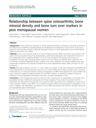
Figure 1 Osteophytosis and DSN demonstrated on a lateral
lumbar spine X-ray.
Table 1 Subject characteristics
Variable All subjects (N = 277)
Mean ± SD
Age (years) 58.7 ± 7.7
Age of menopause (years) 47.30 ± 5.28
Weight (kg) 71.38 ± 11.60
Height (m) 1,56 ± 0,06
BMI (Kg/m2
) 29.08 ± 4.35
Lumbar spine BMD (g/cm2
) 0.966 ± 0.159
Femoral neck BMD (g/cm2
) 0.848 ± 0.130
Femoral trochanter BMD (g/cm2
) 0.693 ± 0.120
Total femoral BMD (g/cm2
) 0.892 ± 0.134
CTX I 0.49 ± 0.25
Osteocalcin 24.56 ± 13.65
n %
Prevalence of osteoporosis 84 31,2
Prevalence of spine osteoarthritis 245 88.44
BMD: bone mineral density
BMI: body mass index
DSN: disc space narrowing
CTX: C-terminal cross-linking telopeptide of type I collagen
Ichchou et al. BMC Women’s Health 2010, 10:25
http://www.biomedcentral.com/1472-6874/10/25
Page 3 of 7
4. increasing grade of disc space narrowing. For example,
the mean age-adjusted lumbar spine BMD rises from
0.95 g/cm2
for patients without DSN to 1.118 g/cm2
for
patients with grade 3 of DSN (p = 0.001). This trend of
increasing BMD with increasing grade of DSN persisted
after adjusting for BMI. However, there was no associa-
tion between lumbar spine BMD and osteophytes.
✓ Femoral BMD
The association between the MAX of each radiographic
features and BMD at the neck, tochanter, and total
femoral is shown in table 2. After adjusting for age,
patients with grade 2 of DSN had an increased BMD at
all measured sites than other patients. For example, the
mean age-adjusted femoral neck BMD was 0.845 g/cm2
for patients without DSN and 0.870 g/cm2
for patients
with grade 2 of DSN (p = 0.003). This trend of increas-
ing BMD in patients with grade 2 of DSN persisted after
adjusting for BMI. In contrast, there was no association
between femoral BMD and osteophytes.
Summary score for radiographic features (SUM) and BMD
The influence of the radiographic features as assessed
using the total score (SUM) across all the four interverteb-
ral levels, on BMD at all measured sites, is shown in table
3. Results are expressed as b coefficients which may be
interpreted as the absolute change in BMD (g/cm2
) per
unit change in score. BMD at all measured sites increased
with SUM DSN. For example, after age adjustment, lum-
bar BMD increased by a value of 0.02 g/cm2
for each unit
change in the total DSN score. The observed associations
remain unchanged after adjustment for age and BMI.
However, there was no association between BMD at all
measured sites and SUM osteophytes.
Relationship between osteoarthritis and bone turn over
markers
A significant decrease in CTX-I levels associated with
lumbar spine disc degeneration was observed (table 4).
Indeed, after adjustment for age and BMI there was a
significant negative correlation between CTX and MAX
DSN (r adjusted = -0.192, p = 0. 026). The level of CTX
was also negatively associated with SUM DSN (r
adjusted = -0.209, p = 0.019). However, no significant
correlation was found between CTX and MAX or SUM
osteophytes and between osteocalcin; and both SUM
and MAX DSN or osteophytes.
Discussion
Our data show that in post menopausal women increas-
ing severity of disc space narrowing, but not osteo-
phytes, is related to increasing bone mineral density at
all measured sites. The severity of disc space narrowing
Table 2 Maximum grade (MAX) of individual radiographic features and BMD at different measured sites in 277
postmenopausal women
Lumbar spine BMD Trochanter BMD Femoral neck BMD Femoral total BMD
n (%) Mean
(SD)
Multivariate
analysis b (95%
CI)
Mean
(SD)
Multivariate
analysis b (95%
CI)
Mean
(SD)
Multivariate
analysis b (95%
CI)
Mean
(SD)
Multivariate
analysis b (95%
CI)
Osteophytes
Grade 0 34 (12.5) 0.983
(0.187)
1 0.710
(0.125)
1 0.884
(0.125)
1 0.925
(0.149)
1
Grade 1 219 (78.8) 0.963
(0.153)
-0.01 (-0.05 to 0.05) 0.693
(0.121)
-0.01 (-0.04 to 0.03) 0.845
(0.134)
-0.02 (-0.07 to 0.02) 0.889
(0.132)
-0.01 (-0.01 to 0.02)
Grade 2 11 (4.0) 0.937
(0.217)
0.02 (-0.07 to 0.13) 0.643
(0.103)
-0.01 (-0.08 to 0.06) 0.822
(0.884)
-0.01 (-0.08 to 0.08) 0.849
(0.110)
0.01 (-0.07 to 0.08)
Grade 3 13 (4.7) 0.989
(0.140)
-0.01 (-0.08 to 0.06) 0.693
(0.120)
-0.01 (-0.08 to 0.06) 0.831
(0.992)
-0.03 (-0.11 to 0.04) 0.891
(0.128)
-0.01 (-0.08 to 0.06)
DSN
Grade 0 146 (52.8) 0.953
(0.156)
1 0.686
(0.121)
1 0.845
(0.144)
1 0.896
(0.137)
1
Grade 1 105 (37.8) 0.974
(0.255)
0.02 (-0.01 to 0.06) 0.674
(0.114)
0.01 (-0.01 to 0.04) 0.849
(0.110)
0.01 (-0.02 to 0.04) 0.885
(0.130)
-0.01 (-0.03 to 0.02)
Grade 2 20 (7.2) 0.966
(0.232)
0.07 (-0.01 to 0.14)* 0.689
(0.152)
0.05 (-0.01 to 0.10)* 0.870
(0.137)
0.08 (0.02 to 0.14)* 0.893
(0.146)
0.06 (0.01 to 0.12)*
Grade 3 6 (2.2) 1.118
(0.861)
0.19 (0.07 to 0.30)* 0.683
(0.120)
0.01 (-0.06 to 0.10) 0.843
(0.931)
0.02 (-0.07 to 0.12) 0.909
(0.107)
0.05 (-0.04 to 0.14)
*p < 0.05
Adjustment for age and BMI in the multivariate analysis
BMD: bone mineral density
Ichchou et al. BMC Women’s Health 2010, 10:25
http://www.biomedcentral.com/1472-6874/10/25
Page 4 of 7
5. was also associated to a decrease in bone resorption,
without any effect on bone formation.
Marked differences in the prevalence of spinal degen-
eration features occur in association with older age,
female sex, post menopausal women and obesity. In our
study, the prevalence of spine osteoarthritis was high
(88.44%). 78.7% and 47.7% of patients had at least one
osteophyte or at least one DSN respectively. This can be
explained by age (Mean (SD) age was 58.7 ± 7.7), sex,
overweight (44.4% had BMI > 25) and obesity (36.5% of
the patients). Mean (SD) BMI was 29.08 ± 4.35.
On the other hand, several studies observe an inverse
relationship between OP and spine OA, the latter being
considered as possibly delaying the development of OP
[10,18,19,21]. Our data showed that increasing BMD in
spine OA is more related to DSN than to osteophytes.
Indeed, after adjusting for age and BMI, no association
was found between BMD at all measured sites and the
severity of osteophytes. This can be explained by the
fact that the majority of the patients (78.8%) had only a
grade 1 of osteophytes which corresponds to a mild
involvement of this radiographic feature. The influence
of osteophytes on BMD has been the focus of various
studies, which showed, in contrast to our findings
[6,10,12,16-18], that spinal BMD was greater in verteb-
rae with osteophytes. Several other studies have exam-
ined the association between osteophytes and bone mass
at distant sites including the hip and most [6,9-13,18],
though not all [14-16] suggest that they are also linked
to an increased bone mass. However, it is important to
note that spine disc degeneration can hinder the inter-
pretation of spine BMD: osteophytes cannot be distin-
guished from vertebral bone mineral using BMD area
measurements and may even in some cases overestimate
the measurements of bone mass in the affected areas.
Therefore, it has been suggested that spine BMD is not
a relevant surrogate marker for the assessment of osteo-
porosis in the spine in patients with osteoarthritis [9,12].
Relatively little however is known about the associa-
tion between bone mass and DSN and there are also
conflicting findings in the few published studies on the
subject [12-14,16,17]. Thus, in a population of patients
with OA of the hip, isolated DSN without osteophytes
was not associated with high bone mass [17]. In con-
trast, Pye et al showed an association between DSN and
increasing BMD at the spine but not at the hip [9]. Our
data show that increasing severity of DSN is associated
with increasing BMD at all measured sites and support,
through the DSN results, the hypothesis that degenera-
tive disc disease is inversely linked with osteoporosis.
The mechanism is unknown though several are possi-
ble; including confounding by environmental or constitu-
tional factors, hormonal, metabolic, and genetic factors
[22,23]. Our results remained unchanged after adjust-
ment for age and BMI, suggesting that they do not play a
major role in explaining the observed associations.
Few studies of biochemical markers have been
reported in subjects with spinal OA, with discordant
results [18-20]. Peel et al [18] and El Miedany et al [19]
have shown that spinal OA is associated with a general-
ized increase in BMD and decreased levels of serum and
urinary biochemical markers of bone formation and
bone resorption in patients with spine disc degeneration.
It has been suggested that the protective effect of spinal
OA against OP may be mediated through decreased rate
of bone turnover. Garnero et al [20] in a large cohort of
untreated postmenopausal women participating in the
OFELY prospective study have shown recently that lum-
bar spine DSN, but not osteophytes, is strongly asso-
ciated with increased CTX-II degradation, independently
of age and BMI. Our results agree partially with what
has been reported by Peel et al. and El Miedany et al
[18,19] who found in their studies a decrease in bone
Table 3 Total scores (SUM) of individual radiographic features and BMD at different measured sites, after adjusting
for age and BMI, in 277 postmenopausal women
Lumb spine BMD
b (95% CI)
Trochanter BMD
b (95% CI)
Femoral neck BMD
b (95% CI)
Total femoral BMD
b (95% CI)
Osteophyte -0.01 (-0.01 to 0.01) -0.01 (-0.01 to 0.01) -0.01 (-0.01 to 0.01) -0.01 (-0.01 to 0.01)
DSN 0.02 (0.01 to 0.03)* 0.01 (0.01 to 0.02)* 0.01 (0.01 to 0.02)* 0.01 (0.01 to 0.02)*
*p < 0.05
BMD: bone mineral density
DSN: disc space narrowing
CI: confidence interval
Table 4 Partial Correlation showing a significant negative
correlation between CTX and DSN in 277
postmenopausal women
CTX r adjusted Osteocalcin r adjusted
MAX osteophyte -0.097 -0.032
SUM osteophyte -0.137 -0.135
MAX DSN -0.192* -0.036
SUM DSN -0.209* -0.075
*p < 0.05
Adjustement for age and BMI
CTX: C-terminal cross-linking telopeptide of type I collagen
DSN: disc space narrowing
Ichchou et al. BMC Women’s Health 2010, 10:25
http://www.biomedcentral.com/1472-6874/10/25
Page 5 of 7
6. resorption markers in women with spinal OA. However,
we did not find any effect of spine disc degeneration on
bone formation. Moreover, no association was found
between the severity of osteophytes and either increas-
ing BMD or CTX levels.
Our study is limited by its cross-sectional design and
the use of a semiquantitative score to classify the radio-
graphic features of disc degeneration. As with any subjec-
tive evaluation, this is subject to errors of interpretation
which may result in misclassification. However, this
defect applies to all studies in this field because the same
grading systems are used universally. Moreover, only
9.4% of our subjects had disc space narrowing that was
associated with a significantly increased bone mineral
density and further studies on a larger population are
necessary to confirm our findings. Finally, and as men-
tioned above, spine BMD is not a relevant surrogate mar-
ker for the assessment of osteoporosis in the spine in
patients with osteoarthritis. Indeed, measurements of
BMD taken by DXA are the most accurate procedure for
the diagnosis of osteoporosis nowadays. However, these
measurements are two-dimensional and when made with
an anterior-posterior projection, the most used incidence,
this procedure has the disadvantage of measuring the
density of all the mineral components encountered in the
X-ray pathway, including osteophytes, bone sclerosis,
disk space narrowing, spondylolisthesis, vertebral frac-
tures, and vascular and extra-vertebral calcifications.
Moreover, some studies has actually shown that obesity
and these alterations can influence bone mineral density
results. Although this defect applies to all studies in this
field because DXA is used universally, it would be inter-
esting to study OA in other places.
Conclusion
This study showed that in post menopausal women the
severity of disc narrowing, but not osteophytes, was
associated with a generalized increase in BMD and a
decreased rate of bone resorption. These results are
consistent with the hypothesis that osteoarthritis,
through disc space narrowing, has a protective effect
against bone loss, mediated by a lower rate of bone
resorption. However, spine BMD is not a relevant surro-
gate marker for the assessment of osteoporosis in the
spine in patients with osteoarthritis and debate as to the
relationship between OA and OP is still open because of
the contradictory data in the literature.
Acknowledgements
This work was supported by grants from the University Mohammed V,
Souissi, Rabat-Morocco.
The University Hospital Center of Rabat-Morocco supported the bone
mineral density measures.
Author details
1
Laboratory of Information and Research on Bone Diseases (LIRPOS).
Department of Rheumatology, El Ayachi hospital, University Hospital of
Rabat-Sale, Morocco. 2
Laboratory of Biostatistical, Clinical and
Epidemiological Research (LBRCE). Faculty of Medicine and Pharmacy, Rabat,
Morocco.
Authors’ contributions
LI participated in study design and drafted the manuscript. FA conceived
the original idea for the study, supervised its design, performed the
statistical analysis and gave critical comments on the draft manuscript. SR
enrolled patients, participated in data acquisition and critical revision of the
manuscript. LB enrolled patients, participated in data acquisition and critical
revision of the manuscript. IH enrolled patients, participated in data
acquisition and critical revision of the manuscript. FZA enrolled patients,
participated in data acquisition and critical revision of the manuscript. HK
enrolled patients, participated in data acquisition and critical revision of the
manuscript. LE enrolled patients, participated in data acquisition and critical
revision of the manuscript. RA conceived the study and performed the
statistical analysis. NHH participated in the study design, coordinated the
study and gave critical comments on the draft manuscript. All authors read
and approved the final manuscript.
Competing interests
The authors declare that they have no competing interests.
Received: 6 February 2010 Accepted: 8 August 2010
Published: 8 August 2010
References
1. Dequeker J, Aerssens J, Luyten FP: Osteoarthritis and osteoporosis: clinical
and research evidence of inverse relationship. Aging Clin Exp Res 2004,
15:426-39.
2. Sambrook P, Naganathan V: What is the relationship between
osteoarthritis and osteoporosis? Baillieres Clin Rheumatol 1997, 11:695-710.
3. Foss MVL, Byers PD: Bone density, osteoarthritis of the hip, and fracture
of the upper end of the femur. Ann Rheum Dis 1972, 31:259-64.
4. Roh YS, Dequeker J, Mulier JC: Cortical bone remodeling and bone mass
in primary osteoarthritis of the hip. Invest Radiol 1973, 8:251-4.
5. Gevers G, Dequeker J, Geusens P: Physical and histomorphological
characteristics of iliac crest bone differ according to the grade of
osteoarthritis at the hand. Bone 1989, 10:173-7.
6. Reid IR, Evans MC, Ames R, Wattie DJ: The influence of osteophytes and
aortic calcification on spinal mineral density in postmenopausal women.
J Clin Endocrinol Metab 1991, 72:1372-6.
7. Äström J, Beertema J: Reduced risk of hip fractures in the mothers of
patients with osteoarthritis of the hip. J Bone Joint Surg Br 1992, 74:270-5.
8. Hannan MT, Anderson JJ, Zhang Y: Bone mineral density and knee
osteoarthritis in elderly men and women. The Framingham Study.
Arthritis Rheum 1993, 36:1671-6.
9. Pye SR, Reid DM, Adams JE, Silman AJ, O’Neill TW: Radiographic features
of lumbar disc degeneration and bone mineral density in men and
women. Ann Rheum Dis 2006, 65:234-8.
10. Masud T, Langley S, Wiltshire P, Doyle DV, Spector TD: Effect of spinal
osteophytosis on bone mineral density measurements in vertebral
osteoporosis. BMJ 1993, 307:172-3.
11. Hart DJ, Mootoosamy I, Doyle DV, Spector TD: The relationship between
osteoarthritis and osteoporosis in the general population: the Chingford
Study. Ann Rheum Dis 1994, 53:158-62.
12. Jones G, Nguyen T, Sambrook PN, Kelly PJ, Eisman JA: A longitudinal study
of the effect of spinal degenerative disease on bone density in the
elderly. J Rheumatol 1995, 22:932-6.
13. Miyakoshi N, Itoi E, Murai H, Wakabayashi I, Ito H, Minato T: Inverse relation
between osteoporosis and spondylosis in postmenopausal women as
evaluated by bone mineral density and semiquantitative scoring of
spinal degeneration. Spine 2003, 28:492-5.
14. Liu G, Peacock M, Eilam O, Dorulla G, Braunstein E, Johnston CC: Effect of
osteoarthritis in the lumbar spine and hip on bone mineral density and
diagnosis of osteoporosis in elderly men and women. Osteoporos Int
1997, 7:564-9.
Ichchou et al. BMC Women’s Health 2010, 10:25
http://www.biomedcentral.com/1472-6874/10/25
Page 6 of 7
7. 15. Dalle Carbonare L, Giannini S, Sartori L, Nobile M, Ciuffreda M, Silva-Netto F,
Arlot ME, Crepaldi G: Lumbar osteoarthritis, bone mineral density, and
quantitative ultrasound. Aging (Milan) 2000, 12:360-5.
16. Muraki S, Yamamoto S, Ishibashi H, Horiuchi T, Hosoi T, Orimo H,
Nakamura K: Impact of degenerative spinal diseases on bone mineral
density of the lumbar spine in elderly women. Osteoporos Int 2004,
15:724-8.
17. Nevitt MC, Lane NE, Scott JC, Hochberg MC, Pressman AR, Genant HK,
Cummings SR: Radiographic osteoarthritis of the hip and bone mineral
density. The Study of Osteoporotic Fractures Research Group. Arthritis
Rheum 1995, 38:907-16.
18. Peel NF, Barrington NA, Blumsohn A, Colwell A, Hannon R, Eastell R: Bone
mineral density and bone turnover in spinal osteoarthrosis. Ann Rheum
Dis 1995, 54:867-71.
19. El Miedany YM, Mehanna AN, El Baddini MA: Altered bone mineral
metabolism in patients with osteoarthritis. Joint Bone Spine 2000,
67:900-7.
20. Garnero P, Sornay-Rendu E, Arlot M, Christiansen C, Delmas PD: Association
between Spine Disc Degeneration and Type II Collagen Degradation in
Postmenopausal Women, The OFELY Study. Arthritis Rheum 2004,
50:3137-44.
21. Verstraeten A, Ermen HV, Haghebaert G, Nijs J, Geusens P, Dequeker J:
Osteoarthritis retards the development of osteoporosis: observation of
the coexistence of osteoarthritis and osteoporosis. Clin Orthop 1991,
264:169-77.
22. Dequeker J, Mohan S, Finkelman RD, Aerssens J, Baylink DJ: Generalised
osteoarthritis associated with increased insulin-like growth factors types
I and II and transforming growth factor b in cortical bone from the iliac
crest. Arthritis Rheum 1993, 36:1702-8.
23. Holderbaum D, Haqqi TM, Moskowitz RW: Genetics and osteoarthritis:
exposing the iceberg. Arthritis Rheum 1999, 42:397-405.
Pre-publication history
The pre-publication history for this paper can be accessed here:
http://www.biomedcentral.com/1472-6874/10/25/prepub
doi:10.1186/1472-6874-10-25
Cite this article as: Ichchou et al.: Relationship between spine
osteoarthritis, bone mineral density and bone turn over markers in post
menopausal women. BMC Women’s Health 2010 10:25.
Submit your next manuscript to BioMed Central
and take full advantage of:
• Convenient online submission
• Thorough peer review
• No space constraints or color figure charges
• Immediate publication on acceptance
• Inclusion in PubMed, CAS, Scopus and Google Scholar
• Research which is freely available for redistribution
Submit your manuscript at
www.biomedcentral.com/submit
Ichchou et al. BMC Women’s Health 2010, 10:25
http://www.biomedcentral.com/1472-6874/10/25
Page 7 of 7
![conditions is still controversial [1,2] even after years of
research since the first results indicate an apparent
inverse relationship [3,4]. Indeed, many studies have
shown an association between high bone mineral density
at the spine and hip and OA of the hips, knees or hands
[5-8]. However, there are conflicting findings in the few
published studies on the association between bone mass
and degenerative disease in the spine, the latter being
characterized by disc space narrowing (DSN) and the
presence of vertebral osteophytes. Most [9-13], though
not all [14-16] studies that examined the association
between osteophytes and bone mass at the spine and
distant sites including the hip, suggest that they are
linked to an increased bone mass. Results are also dis-
cordant about the association between DSN and BMD
at distant sites. In a population of patients with OA of
the hip, isolated DSN without osteophytes was not asso-
ciated with high bone mass [17]. In contrast, in a gen-
eral population, those with isolated DSN have a higher
BMD in the spine (but not in the hip) than those with-
out [9].
In order to understand the underlying mechanism of
the interaction between bone mass and OA, noninvasive
biochemical assays for markers of bone resorption
(which the CTX-I is the most specific and sensitive
one), and bone formation have been developed and
enabled estimation of bone turnover. Few studies of bio-
chemical markers have been reported in subjects with
spinal OA, results have also been conflictual [18-20].
We undertook this study to determine the association
between radiographic features of lumbar disc degenera-
tion, namely osteophytes and DSN, and BMD at differ-
ent measured sites, as well as to investigate the
underlying mechanism at the tissue level through assess-
ment of biochemical markers of bone metabolism.
Methods
Subjects
The study involved 277 consecutive ambulatory post-
menopausal women living in urban centre of Morocco
and sent to our outpatient Bone Densitometry Center.
Recruitment was based on voluntary enrolment. Writ-
ten informed consent was obtained from all subjects
and the study was approved by the Ethical Committee
of El Ayachi University Hospital of Rabat-Sale. We
excluded from the study all patients with a history of:
(1) taking drugs known to influence bone metabolism
in the past two years, such as vitamin D, calcium, cor-
ticosteroids, bisphosphonates, sodium fluoride, raloxi-
fene, strontium ranelate, teriparatide and hormone
replacement therapy; (2) musclo skeletal, thyroid, para-
thyroid, adrenal, hepatic, or renal disease; (3) malig-
nancy; and (4) hysterectomy.
Data collection and measurements
Each patient completed a questionnaire on sociodemo-
graphic parameters and osteoporosis risk factors such as
female sex, age higher than 60 years, family history of
osteoporosis, early menopause, low BMI, smoking,
sedentary lifestyle, long term (≥3 months) corticosteroid
use and excessive alcohol consumption. Weight and
height were measured without clothes or shoes at the
time of bone densitometry measurements. The body
mass index (BMI) was calculated as body weight divided
by height squared (Kg/m2
).
Bone mineral density (BMD) measurements
Lumbar spine, trochanter, femoral neck and total hip
BMD were measured by dual-energy X-ray absorptiome-
try with a Lunar prodigy densitometer. Only vertebras
with scoliosis have been excluded from BMD test. Daily
quality control was carried out by measurement of a
Lunar phantom. At the time of the study, phantom
measurements showed stable results. The in vivo preci-
sion error for dual-energy X ray absorptiometry,
expressed as coefficient of variation, was 0.9% at the
lumbar spine and 1% at the femoral neck. Both T and Z
scores were obtained. In the T-score calculations, the
manufacturer’s ranges for European reference popula-
tion were used because of the absence of a Moroccan
data base.
Assessment of lumbar spine degeneration
Lumbar spine radiographs were taken according to a
standard protocol with the film centred at L2 (Figure 1).
The radiographs were subsequently evaluated by a single
observer for the presence of the individual radiographic
features of disc degeneration. Each vertebral level from
L1/2 to L4/5 was assessed for the presence and severity
of osteophytes and DSN, using a semiquantitative score
(grade 0, none; grade 1, mild; grade 2, moderate; grade 3,
severe) [21]. We defined, for each radiographic feature,
two summary statistics: ‘’MAX’’, which was the grade of
the most severely affected vertebral level per subject
(from L1/2 to L4/5) and which could range from 0 to 3,
and ‘’SUM’’, the sum of the four vertebral specific grades
per subject which thus could range from 0 to 12.
Biochemical measurements
Morning fasting blood was collected from every subject for
the measurement of the following parameters: osteocalcin
and C-terminal cross-linking telopeptide of type I (CTX).
Both parameters were measured by immunochemolumi-
nometric assay (Elecsys, Roche diagnostics, Mannheim,
Germany). Intra- and interassay variances were 5% and
7%, and the normal range were 15-46 ng/ml for osteocal-
cin, and 0.3-0.6 ng/ml for CTX.
Ichchou et al. BMC Women’s Health 2010, 10:25
http://www.biomedcentral.com/1472-6874/10/25
Page 2 of 7](data:image/gif;base64,R0lGODlhAQABAIAAAAAAAP///yH5BAEAAAAALAAAAAABAAEAAAIBRAA7)