SlideShare a Scribd company logo
1 of 9
Download to read offline
Effect of Rho and ADP-ribosylation Factor GTPases on
Phospholipase D Activity in Intact Human
Adenocarcinoma A549 Cells*
(Received for publication, February 12, 1999)
Elisabetta Meacci‡§¶, Valeria Vasta§, Jonathan P. Moormanʈ, David A. Bobakʈ, Paola Bruni§,
Joel Moss‡**, and Martha Vaughan‡
From the ‡Pulmonary-Critical Care Medicine Branch, NHLBI, National Institutes of Health, Bethesda, Maryland 20892,
the §Dipartimento di Scienze Biochimiche, Universita` di Firenze, Viale G. B. Morgagni 50, 50134 Firenze, Italy, and the
ʈDepartment of Medicine, University of Virginia School of Medicine, Charlottesville, Virginia 22908
Phospholipase D (PLD) has been implicated as a cru-
cial signaling enzyme in secretory pathways. Two 20-
kDa guanine nucleotide-binding proteins, Rho and ADP-
ribosylation factor (ARF), are involved in the regulation
of secretion and can activate PLD in vitro. We investi-
gated in intact (human adenocarcinoma A549 cells) the
role of RhoA and ARF in activation of PLD by phorbol
12-myristate 13-acetate, bradykinin, and/or sphingosine
1-phosphate. To express recombinant Clostridium botu-
linum C3 exoenzyme (using double subgenomic recom-
binant Sindbis virus C3), an ADP-ribosyltransferase
that inactivates Rho, or dominant-negative Rho contain-
ing asparagine at position 19 (using double subgenomic
recombinant Sindbis virus Rho19N), cells were infected
with Sindbis virus, a novel vector that allows rapid, high
level expression of heterologous proteins. Expression of
C3 toxin or Rho19N increased basal and decreased phor-
bol 12-myristate 13-acetate-stimulated PLD activity.
Bradykinin or sphingosine 1-phosphate increased PLD
activity with additive effects that were abolished in cells
expressing C3 exoenzyme or Rho19N. In cells expressing
C3, modification of Rho appeared to be incomplete, sug-
gesting the existence of pools that differed in their ac-
cessibility to the enzyme. Similar results were obtained
with cells scrape-loaded in the presence of C3; however,
results with virus infection were more reproducible. To
assess the role of ARF, cells were incubated with brefel-
din A (BFA), a fungal metabolite that disrupts Golgi
structure and inhibits enzymes that catalyze ARF acti-
vation by accelerating guanine nucleotide exchange.
BFA disrupted Golgi structure, but did not affect basal
or agonist-stimulated PLD activity, i.e. it did not alter a
rate-limiting step in PLD activation. It also had no effect
on Rho-stimulated PLD activity, indicating that RhoA
action did not involve a BFA-sensitive pathway. A novel
PLD activation mechanism, not sensitive to BFA and
involving RhoA, was identified in human airway epithe-
lial cells by use of a viral infection technique that pre-
serves cell responsiveness.
Phospholipase D (PLD),1
an important effector in receptor-
mediated signal transduction pathways, catalyzes the hydrol-
ysis of the most abundant membrane phospholipid, phosphati-
dylcholine, to generate choline and phosphatidic acid. In intact
cells, choline is rapidly phosphorylated to phosphocholine,
which plays a role in cell proliferation (1). Phosphatidic acid
also serves as a second messenger in the regulation of secre-
tion, DNA synthesis, and cell proliferation (2). PLD activity
appears to be regulated, and can be activated by both phos-
phatidylinositol 4,5-bisphosphate (3) and phosphatidylinositol
1,4,5-trisphosphate (4) in vitro. Phosphatidylinositol 4,5-bis-
phosphate was essential for PLD activation in intact cells (5).
Consistent with a regulatory role for protein kinase C (PKC),
phorbol esters markedly activated PLD (6, 7), and PKC inhib-
itors abolished agonist-induced PLD activity (8) in some cells.
Both native and recombinant PLD were activated by ϳ20-
kDa guanine nucleotide-binding proteins including ADP-ribo-
sylation factors (ARFs) (3), which are critical for vesicular
trafficking (9), and Rho, a GTPase that regulates several cel-
lular functions including cytoskeletal organization (10). RhoA
was required for guanosine 5Ј-O-(3-thio)triphosphate stimula-
tion of PLD activity in neutrophil membranes (11). In rat
fibroblasts, Clostridium botulinum C3 exoenzyme, which ADP-
ribosylates and inactivates RhoA (12), inhibited activation of
PLD by lysophosphatidic acid (13). Similarly, toxin B from
Clostridium difficile, which inactivates Rho family GTPases by
monoglucosylation (14), abolished carbachol activation of PLD
in HEK cells (15) and blocked PLD activation initiated via the
IgE receptor in rat basophilic leukemia cells (16). More re-
cently, it was demonstrated that activation of PLD via ␣2-
adrenergic receptors in broken PC12 cell preparations required
RhoA and PKC activation (17).
ARF restored guanosine 5Ј-O-(3-thio)triphosphate-depend-
ent activation of PLD in cells depleted of cytosolic components
(11, 18) and activated PLD associated with partially purified
Golgi membranes (19). ARF proteins have also been implicated
in the activation of PLD by insulin (20). Brefeldin A (BFA), a
fungal metabolite that causes disruption of Golgi structure, can
block ARF activation by inhibiting the guanine nucleotide-
exchange protein that accelerates replacement of ARF-bound
GDP by GTP (21–23). This causes dissociation of ARF and
␤COP from Golgi membranes (in vivo and in vitro) before
* This work was supported in part by National Institutes of Health
Grant R01 GM 54572 (to D. A. B.). The costs of publication of this
article were defrayed in part by the payment of page charges. This
article must therefore be hereby marked “advertisement” in accordance
with 18 U.S.C. Section 1734 solely to indicate this fact.
¶ Current address: Dipartimento di Scienze Biochimiche, Universita`
degli Studi di Firenze, Viale G.B. Morgagni 50, 50134 Firenze, Italy.
** To whom correspondence should be addressed: Pulmonary-Critical
Care Medicine Branch, Rm. 5N-307, Bldg. 10, 10 Center Dr., MSC 1434,
National Institutes of Health, Bethesda, MD 20892-1434. Tel.: 301-496-
4554; Fax: 301-402-1610.
1
The abbreviations used are: PLD, phospholipase D; PMA, phorbol
12-myristate 13-acetate; BK, bradykinin; SPP, sphingosine 1-phos-
phate; PtdEtOH, phosphatidylethanol; ARF, ADP-ribosylation factor;
dsSIN, double subgenomic recombinant Sindbis virus; CAT, chloram-
phenicol acetyltransferase; MOI, multiplicity of infection; PKC, protein
kinase C; PBS, phosphate-buffered saline; IL, interleukin; BFA,
brefeldin A.
THE JOURNAL OF BIOLOGICAL CHEMISTRY Vol. 274, No. 26, Issue of June 25, pp. 18605–18612, 1999
Printed in U.S.A.
This paper is available on line at http://www.jbc.org 18605
byguestonOctober21,2016http://www.jbc.org/Downloadedfrom
redistribution of Golgi components to the endoplasmic reticu-
lum (24). BFA inhibited carbachol stimulation of PLD activity
in HEK cells expressing muscarinic M3 receptors (25), but not
PMA stimulation of PLD in HL-60 cells (26). Although BFA
treatment has been used frequently to identify effects in cells
that are ARF-dependent, the absence of BFA inhibition does
not exclude a role for ARF, which can also be activated by
BFA-insensitive guanine nucleotide-exchange proteins (27). In
addition, BFA has actions that are apparently independent of
ARF, such as stimulation of ADP-ribosylation (28), which fur-
ther complicates the interpretation of effects observed in intact
cells.
Although there is evidence that the same PLD might be
activated by ARF, Rho, and/or PKC (19), some forms of PLD are
clearly not regulated by PKC or guanine nucleotide-binding
proteins. Several studies do support a role for ARF and Rho in
PLD activation (29). For example, ARF and Rho acted syner-
gistically in the activation of brain membrane PLD (7). Rho,
ARF, and PKC were implicated in PLD activation in HL-60 (30)
and brain cell membranes (7); clearly, the effects in disrupted
and intact cells may be very different. To evaluate the role of
Rho in intact cells, a novel Sindbis virus vector was used to
induce expression of C3 and Rho proteins in A549 cells (human
adenocarcinoma cells). From the observations reported here, it
appears that a rate-limiting step in PLD stimulation by PMA,
BK, and SPP in A549 cells involves RhoA but not a BFA-
sensitive ARF pathway.
EXPERIMENTAL PROCEDURES
Materials
Palmitic acid (9,10–3
H) (30–60 Ci/mmol), used to label cellular phos-
pholipids, was purchased from NEN Life Science Products, standard
lipids were from Avanti Polar Lipids, solvents were from Fluka, and
silica gel 60 plates for thin layer chromatography were from Merck.
PMA, BK, SPP, BFA, and other chemicals were from Sigma. Mouse
monoclonal antibodies against a synthetic peptide with RhoA sequence
were from Santa Cruz Biotechnology, and monoclonal antibodies
against Golgi 58-kDa protein were from Sigma. Rabbit antiserum
raised against bacterially synthesized C3 was a generous gift of Dr. M.
Popoff (Institut Pasteur, Paris, France). Secondary antibodies (goat
anti-mouse and anti-rabbit immunoglobulin G1 conjugated with fluo-
rescein and rhodamine-B, respectively) were from BIOSOURCE. Hu-
man lung carcinoma A549 cells were from the American Type Culture
Collection (ATCC, Manassas, VA). All materials for cell culture were
from Biofluid.
Methods
Cell Culture—A549 cells were grown in Dulbecco’s modified Eagle’s
medium supplemented with 10% heat-inactivated fetal bovine serum,
penicillin (5 units/ml), streptomycin (5 ␮g/ml), and 2 mM glutamine.
Transphosphatidylation Assay of PLD in Intact Cells—PLD activity
was assayed by measuring [3
H]phosphatidylethanol ([3
H]PtdEtOH)
produced via PLD-catalyzed transphosphatidylation in cells previously
labeled with [3
H]palmitic acid (31). Cells were grown in medium con-
taining bovine serum albumin, 1 mg/ml, for 24 h with [3
H]palmitic acid
(10 ␮Ci/ml) added for the last 18 h. The medium was then aspirated and
after washing, cells were incubated in fresh medium for 2 h. To initiate
experiments, 1% ethanol was added 5 min before agonists. Incubations,
at 37 °C, were terminated by washing monolayers twice with ice-cold
PBS and immediately adding 1 ml of ice-cold methanol. Cells were
collected by scraping and dishes were washed with an additional 1 ml of
ice-cold methanol. Lipids were then extracted essentially as described
by Bligh and Dyer (32) except that water was replaced with 1 M NaCl.
The lower phase was dried under a stream of nitrogen, and the residue
was dissolved in a small volume of chloroform/methanol (2:1). [3
H]
PtdEtOH was separated from other phospholipids by TLC on silica gel
60 plates with the organic phase of the solvent mixture, chloroform/
methanol/acetic acid (90:10:10, v/v/v), and unlabeled PtdEtOH as a
FIG. 1. Effect of infection with re-
combinant Sindbis virus vector en-
coding C. botulinum C3 exoenzyme
on morphology of A549 cells. A549
cells were incubated at 37 °C for 1 h with
recombinant Sindbis virus (MOI ϭ 20)
encoding C. botulinum C3 exoenzyme (ds-
SIN:C3) before photography (A). Virus
was removed after 1 h of exposure, and
2 h later, cell morphology appeared nor-
mal (B). A Nikon optical microscope was
used for photography.
Regulation of PLD by Rho18606
byguestonOctober21,2016http://www.jbc.org/Downloadedfrom
marker. The appropriate lipid spots, detected by exposure to iodine
vapor, were marked and scraped into scintillation vials, followed by 0.5
ml of methanol and liquid scintillation fluid before radioassay.
Sindbis Vectors Expressing C3 and Mutant Rho—Shuttle phagemids
and dsSIN vectors were constructed as described by Hahn et al. (33) and
Huang (34). Generation of dsSIN:C3, dsSIN:Rho19N, and dsSIN:CAT,
which encode C3 exoenzyme, the negative dominant mutant of RhoA
(Rho19N), and chloramphenicol acetyltransferase (dsSIN:CAT), respec-
tively, has been described (35, 36). Viral stocks, 109
plaque-forming
units/ml, were divided into portions sufficient for a single experiment
and stored at Ϫ80 °C.
dsSIN Infections—Cells, plated at a density of 5–7 ϫ 105
cells/plate
and labeled when indicated with [3
H]palmitic acid, were washed with
PBS and diluted in PBS containing 1 mM CaCl2 and 2 mM MgCl2. After
incubation at 37 °C for 1 h with the indicated amount of dsSIN recom-
binant, PBS was removed, appropriate culture medium was added, and
infected cells were incubated for 2 h before addition of agonists and
processing for PLD assay, Western analysis, or ADP-ribosylation
experiments.
Scrape Loading—A549 cells, grown to confluence on 100-mm Petri
dishes, were washed twice with PBS and 2 ml of scraping buffer (114
mM KCl, 1 mM NaCl, 5.5 mM MgCl2, 10 mM Tris-HCl). C3 transferase (5
␮g/ml) or vehicle was added in 0.5 ml of scraping buffer and the cells
were gently scraped, suspended in Dulbecco’s modified Eagle’s medium/
10% fetal bovine serum, and split among six-well dishes. After 6–7 h of
incubation, we estimated ϳ70% viable cells. Cells were labeled with
[3
H]glycerol (5 ␮Ci/ml) in serum-free medium overnight and used for
PLD assay as described above.
Western Blotting—To evaluate protein expression from recombinant
Sindbis virus, cultured cells were harvested at specified times after
infection and lysed by sonification twice for 15 s on ice in Buffer A
containing 50 mM Tris, pH 7.5, 150 mM NaCl, 1 mM EGTA, and protease
inhibitors (1 mM 4-(2-aminoethyl)benzenesulfonyl fluoride, 0.3 ␮M apro-
tinin, and leupeptin and pepstatin, each 10 ␮g/ml). Total cellular pro-
tein was quantified, and samples (20–50 ␮g) were subjected to SDS-
polyacrylamide gel electrophoresis in 16% gels. Proteins were
transferred to nitrocellulose membranes, which were incubated over-
night in 20 mM Tris-HCl (pH 7.6), 137 mM NaCl, 0.1% Tween-20 (TTBS
(0.1% Tween 20 in Tris-buffered (pH 7.6) saline)) and 0.2% I-block
(Tropix, Bedford, MA). Hybridization with mouse monoclonal antibod-
ies against RhoA or rabbit antiserum raised against bacterially synthe-
sized C3 (2 h room temperature) was followed by washing with TTBS
and incubation with peroxidase-conjugated goat anti-mouse or anti-
rabbit IgG. Proteins were detected by enhanced chemiluminescence
(ECL, Amersham Pharmacia Biotech).
ADP-ribosylation Assay—After dsSIN infection, cells were har-
vested, washed, and lysed in Buffer A (106
cells/ml). Assays (total
volume, 50 ␮l) containing 20 mM Tris-HCl (pH 7.5), 5 mM MgCl2, 1 mM
dithiothreitol, 5 ␮M NAD, 0.5 ␮Ci of [32
P]NAD, 2 mM ADP-ribose, and
lysate sample plus C3 exoenzyme or recombinant RhoA as a His6 (6ϫ
histidine, Calbiochem) fusion protein synthesized in Escherichia coli, as
indicated, were incubated at 37 °C for 1 h. Proteins were precipitated
with 20% trichloroacetic acid and separated by SDS-polyacrylamide gel
electrophoresis; dried gels were exposed to autoradiography film.
Indirect Immunofluorescence—For indirect immunofluorescence ex-
periments, A549 cells were grown on sterilized glass coverslips placed
in six-well dishes with conditions as described for PLD assays. After
incubation with BFA (5 ␮g/ml for 30 min) or infection with recombinant
Sindbis virus (3 h earlier), cells were washed three times with Dulbec-
co’s PBS and fixed with 2% paraformaldehyde in Dulbecco’s PBS for 30
min at room temperature followed by further incubation sequentially
for 2 min each with 10, 25, 50, 75, and 100% ethanol before permeabi-
lization by a 30-min incubation with 0.05% saponin and 1 mg/ml bovine
serum albumin in PBS without Ca2ϩ
and Mg2ϩ
. Cells were then incu-
bated overnight with monoclonal antibodies against Golgi 58-K (a 58-
kDa protein used as Golgi marker) or RhoA, or polyclonal antibodies
against C3, washed four times, and incubated for 1 h with goat anti-
mouse fluorescein- or rhodamine B-conjugated IgG. After washing in
PBS containing 0.05% saponin and 1% bovine serum albumin, the
coverslips were mounted on Mowoil (Hoescht), examined with a Leica
confocal microscope equipped with appropriate filters, and scanned
with the SCANware program.
Interleukin-6 (IL-6) Secretion—For IL-6 determination, confluent
cells, treated or not with 5 ␮g/ml BFA for 30 min, were incubated for 2 h
in 1 ml of Dulbecco’s modified Eagle’s medium without or with 100 nM
PMA. IL-6 in the medium was then quantified with an immunoassay kit
(R & D Systems, Inc., Minneapolis, MN).
RESULTS
Expression of Recombinant Proteins in Cells—Infection of
A549 cells with dsSIN:C3 led to cell rounding, consistent with
reported effects of microinjected C3 (37). Two hours after re-
moval of virus, cell morphology had returned to normal (Fig. 1).
Intracellular C3 exoenzyme was detected as early as 2 h after
infection with an MOI of 3 (Fig. 2A). Because expression was
maximal with an MOI of 20, this condition was used for all
other experiments. No reaction of anti-C3 antibodies with pro-
teins in dsSIN:CAT-infected A549 cells was found, consistent
with the specificity of these antibodies. The expression of C3 in
A549 cells was accompanied by a loss of immunodetectable
ϳ20-kDa RhoA (Fig. 2B), probably because, as had been sug-
gested for rat1 fibroblasts (13), ADP-ribosylation of RhoA in-
creases its susceptibility to degradation. A significant level of
immunoreactive Rho19N protein (a dominant negative mutant
that contains asparagine at position 19), was detected by anti-
RhoA antibodies 2 h after viral infection (MOI of 20). A faint
FIG. 2. Expression of recombinant C3 exoenzyme, Rho19N,
and CAT in A549 cells. A, effect of MOI on C3 exoenzyme expression.
A549 cells were incubated for 1 h with Sindbis virus encoding CAT or
indicated amounts (MOI) of Sindbis virus encoding C. botulinum C3
exoenzyme (dsSIN:C3). Fresh virus-free medium was added to infected
cells 2 h before they were harvested for Western analysis. Proteins (20
␮g) were separated by SDS-polyacrylamide gel electrophoresis in 16%
gel. Rabbit antiserum raised against bacterially expressed C3 was used
to detect C3 exoenzyme. B, effect of expression of dsSIN:C3 on endog-
enous RhoA. Experiments were performed as described above except
that 50 ␮g of lysate proteins were used for Western analysis, and RhoA
was detected using mouse monoclonal antibody raised against a syn-
thetic peptide corresponding to amino acids 120–150 of human RhoA.
C, expression of Rho19N. Proteins (20 ␮g) from cells infected with
dsSIN:Rho19N were processed as described above, and RhoA was de-
tected with the mouse monoclonal anti-RhoA antibody.
TABLE I
Effect of Rho proteins on basal and C3-induced PLD activity
Serum-starved cells were incubated overnight with [3
H]palmitic acid
(10 ␮Ci/ml) and infected with dsSIN:CAT or dsSIN:C3 as described
under “Experimental Procedures” or scraped without or with C3 trans-
ferase (5 ␮g/ml) and labeled with [3
H]glycerol (5 ␮Ci/ml) in serum-free
medium. Cells were processed for PLD assay as noted under “Experi-
mental Procedures.” Data are means Ϯ S.E. of values from three ex-
periments with duplicate assays.
Treatment of cells PLD activity
Infected Scraped No C3 Plus C3
% [3
H]PtdEtOH/phosphatidylcholine
No No 0.20 Ϯ 0.04
dsSIN:CAT No 0.22 Ϯ 0.01
dsSIN:Rho19N No 0.40 Ϯ 0.03a
dsSIN:C3 No 0.43 Ϯ 0.08a
No Yes 0.31 Ϯ 0.03 0.47 Ϯ 0.05a
a
Expression of C3 or Rho19N or C3 treatment significantly increased
PLD activity (P Ͻ 0.05) by Student’s t test.
Regulation of PLD by Rho 18607
byguestonOctober21,2016http://www.jbc.org/Downloadedfrom
band was observed in dsSIN:CAT-infected cells, indicating a
low level of endogenous RhoA in A549 cells (Fig. 2C).
Effects of Rho Proteins on Basal PLD Activity—The involve-
ment of Rho in PLD activation was demonstrated by infecting
A549 cells with recombinant Sindbis virus vectors to express
mutant Rho protein or C. botulinum C3 exoenzyme, an ADP-
ribosyltransferase that inactivates Rho. Expression of either
C3 exoenzyme or the dominant negative Rho19N in A549 cells
by infection with dsSIN:C3 or dsSIN:Rho19N, respectively,
resulted in a doubling of basal PLD activity (Table I). The
increase was similar in A549 cells scrape-loaded in the pres-
ence of C3 transferase. The effect of C3 after scrape loading was
less than that after Sindbis infection (ϳ50% stimulation versus
ϳ100%). Both experiments, however, suggest that Rho proteins
play a role in the regulation of PLD activity in human epithelial
cells and may have a negative effect on the enzyme activity,
restraining or minimizing the level of PLD in unstimulated
cells, perhaps by inhibiting a specific PLD or interfering with
its activation.
Effects of PMA, Bradykinin, and Sphingosine 1-Phosphate on
PLD Activity—PMA activation of PLD was appreciable at 15
min and reached a maximum after 30 min of stimulation (data
not shown). With 1 ␮M PMA, PLD activity of A549 cells was
increased 42 Ϯ 4.1-fold after 30 min (Fig. 3A). The effect of 50
nM PMA was equal to that of 1 ␮M and 1 nM PMA: it increased
activity 100% (data not shown). BK and SPP, which act
through G protein (heterotrimeric guanine nucleotide-binding
protein)-coupled receptors, also stimulated PLD activity (ap-
proximately 2-fold); maximal effects were achieved with 1 ␮M
and 10 ␮M, respectively; these effects were additive (Fig. 3B).
Effects of Rho Proteins on Agonist-stimulated PLD Activi-
ty—As shown in Fig. 3A, expression of either dsSIN:C3 or
dsSIN:Rho19N reduced but did not abolish PMA stimulation of
PLD activity, indicating that Rho proteins are involved in a
PKC-dependent pathway. In cells infected with dsSIN:C3, BK
or SPP failed to increase PLD activity (Fig. 3B). BK plus SPP,
however, induced an ϳ80% increase in PLD activity, suggest-
ing that the combination of agonists was able to overcome, in
part, the inhibitory effects of C3, perhaps acting through Rho
that had not been ADP-ribosylated by the exoenzyme. In
scrape-loaded cells, the effect of C3 in reducing PLD stimula-
tion by agonists was less than it was in infected cells, albeit
statistically significant; in cells expressing C3, PMA stimula-
tion was reduced 25 Ϯ 2% versus 38 Ϯ 4% (n ϭ 3), and BK plus
SPP stimulation was reduced 20 Ϯ 1.5% versus 40 Ϯ 9% (n ϭ
3). These data are consistent with the conclusion that for dem-
onstration of C3 effects, viral infection was more effective than
scrape loading.
Incomplete Modification of Endogenous RhoA by C3 Trans-
ferase—To explore the possibility that the incomplete reduction
of PLD stimulation by C3 was due to differences in the acces-
sibility of intracellular pools of Rho, we quantified ADP-ribo-
sylation of RhoA in cell lysates. First to prove that C3 trans-
ferase expressed in dsSIN:C3 infected cells was enzymatically
active, 100 ng of exogenous RhoA (His6-Rho) and [14
C]NAD
were added to cell lysates (Fig. 4). A clear band of 20 kDa was
detected, indicating that the C3 transferase was able to cata-
lyze ADP-ribosylation of RhoA. Then to verify whether the
intracellular pools of Rho were completely modified by the
expressed C3 transferase, we performed experiments using
exogenous C3 exoenzyme. ADP-ribosylation of endogenous Rho
was not detected when C3 exoenzyme was added at final con-
centration of 0.025 ␮g/ml to lysates of cells infected with dsSIN:
CAT or dsSIN:C3. After addition of higher concentration of C3
(0.5 ␮g/ml) (Fig. 5), a single radiolabeled band, corresponding
in migration to endogenous Rho, was detected. The band at
ϳ20 kDa was more intense in lane 4 than in lane 2, consistent
with the larger amount of the overexpressed dominant nega-
tive Rho19N. The presence of a band in lane 6, although per-
haps somewhat weaker than that in lane 2, suggested that
even when the expression of C3 seemed maximal, modification
of the endogenous Rho was not complete. Similarly, some en-
dogenous Rho was inaccessible to the C3 in dsSIN:C3-infected
cells that were treated with agonists (Fig. 6). Those data could
explain the incomplete inhibition of PMA stimulation of PLD
and the significant, albeit attenuated, effects of BK and SPP on
FIG. 3. Effect of Rho protein on stimulated PLD activity. Cells were infected with Sindbis virus encoding CAT, C. botulinum C3 exoenzyme,
or the dominant-negative mutant Rho19N. Virus was removed after 1 h, and 2 h later, cells were incubated with vehicle (0.1% Me2SO) or 1 ␮M PMA
for 30 min (A) or with 1 ␮M BK, 10 ␮M SPP, or 1 ␮M BK plus 10 ␮M SPP for 10 min (B) before assay of PLD activity. PLD activity is expressed relative
to that of nonstimulated, noninfected cells. [3
H]PtdEtOH formed as a percentage of total [3
H]phosphatidylcholine was 0.20 Ϯ 0.04 and 0.22 Ϯ 0.01%
in uninfected cells and in dsSIN:CAT infected cells, respectively; viral infection did not significantly affect basal PLD activity or the stimulation
by PMA. Data are means Ϯ S.E. of values from at least four experiments with duplicate assays. There was no significant difference between
noninfected and dsSIN-infected cells in amounts of [3
H]palmitic acid in total phospholipids (1.8 Ϯ 0.20 ϫ 106
and 2.1 Ϯ 0.26 ϫ 106
dpm,
respectively). Effects of C3 and Rho19N expression on PMA-stimulated PLD activity and C3 expression on receptor-stimulated PLD activity were
significant (p Ͻ 0.05, Student’s t test).
Regulation of PLD by Rho18608
byguestonOctober21,2016http://www.jbc.org/Downloadedfrom
PLD activity in cells expressing C3 exoenzyme. The extent of
ADP-ribosylation of endogenous Rho in dsSIN:C3-infected cells
varied from one experiment to another, but it was always
statistically significant when data were quantified by densi-
tometry (data not shown). Because of the presence of unmodi-
fied RhoA, indirect immunofluorescence was used to localize C3
exoenzyme and RhoA in A549 cells. In dsSIN:C3-infected cells,
C3 transferase was concentrated in the Golgi compartment and
widely distributed in the cytosol (Fig. 7, panel 4). Endogenous
RhoA in unstimulated cells appeared predominantly in Golgi
and in endoplasmic reticulum (Fig. 7, panels 5 and 6). Thus,
indirect immunofluorescence indicated colocalization of C3 and
endogenous RhoA in Golgi and endoplasmic reticulum
compartments.
Effects of BFA on Basal, PMA-stimulated, and Receptor-
stimulated PLD Activity—The effects of BFA on Golgi structure
and PLD activity in A549 cells were evaluated. Incubation for
1 h with BFA (5 ␮g/ml) induced a drastic redistribution of the
Golgi 58-kDa immunoreactivity (Fig. 8, panels 2 and 3). Only a
slight effect on Golgi structure was detected in PMA-treated
cells (panel 4). In similar experiments, BFA significantly de-
creased PMA-stimulated secretion of IL-6 (control, 435 Ϯ 45
pg/ml; 1 ␮M PMA, 1414 Ϯ 65 pg/ml; PMA plus BFA, 431 Ϯ 10
pg/ml). Both these findings indicated that A549 cells were fully
sensitive to the well known effects of BFA on Golgi structure
and function. As shown in Table II, incubation of A549 cells
with BFA (5 ␮g/ml) for 30 min or 3 h (data not shown) did not
significantly affect basal PLD activity or the stimulation by
PMA, BK, SPP, or BK plus SPP. Nor was a significant effect of
BFA observed with submaximal concentrations of these ago-
nists (5 nM BK or 30 nM SPP). Synergistic effects of Rho and
ARF proteins on PLD activity have been reported (7, 29). Using
C3 exoenzyme, we investigated whether the Rho-dependent
PLD activity was modified by BFA treatment. As shown in
Table II, incubation with BFA (5 ␮g/ml, 30 min) had no signif-
icant effect on C3-induced PLD activity (Table II). No signifi-
cant effect of BFA was also seen in cells scraped-loaded in the
presence of C3 and stimulated with PMA (3.4 Ϯ 0.38 versus
3.8 Ϯ 0.31, n ϭ 3) or BK plus SPP (1.4 Ϯ 0.25 versus 1.8 Ϯ 0.12,
n ϭ 3).
DISCUSSION
To investigate the roles of Rho and ARF in the regulation of
PLD in human airway epithelial cells, cultured A549 cells were
treated with PMA, a direct activator of PKC, and with agonists
that act through G protein-coupled receptors, i.e. bradykinin
(BK), a proinflammatory peptide shown to play a critical role in
the development of airway hyper-responsiveness, and SPP,
which has been implicated in several biological responses (38).
Involvement of 20-kDa GTP-binding proteins in PMA- or re-
ceptor-mediated activation of PLD has been reported (19), al-
though it seems clear that the pathways utilized can differ with
different agonists and cell types.
C. botulinum exoenzyme C3 ADP-ribosylates Asn-41 in Rho,
which is within the putative effector domain of the molecule
(12). Because this covalent modification specifically inactivates
Rho, C3 can be a useful probe of Rho function and has been
used in A549 cells to investigate the role of the Rho GTPase in
bradykinin-stimulated nuclear factor-␬B activation and IL-1b
gene expression (39). Unfortunately, C3 does not enter intact
cells, or does so only poorly, making it inconvenient to use in
living cells to probe Rho function. Electroporation, permeabili-
zation, prolonged incubation (days), and scrape loading proce-
dures have been used to effect intracellular delivery of C3 (13,
15, 37). To avoid these manipulations and preserve fully re-
sponsive cells, in this study, A549 cells were infected with
recombinant Sindbis virus encoding C3 exoenzyme (dsSIN:C3)
or the dominant negative Rho19N (dsSIN:Rho19N). Infection of
cells with these vectors resulted in a high level of expression of
recombinant proteins with minimal cytotoxicity (33). The cells,
FIG. 4. Effect of expression of C3 exoenzyme on ADP-ribosyla-
tion of RhoA. Samples of lysate from cells infected with dsSIN:CAT or
dsSIN:C3 were incubated in ADP-ribosylation assays as described un-
der “Experimental Procedures” for 1 h at 37 °C without or with 0.025 ␮g
of C3 exoenzyme or 100 ng of recombinant RhoA (His6-RhoA) as indi-
cated, before precipitation and separation of proteins (15 ␮g) by SDS-
polyacrylamide gel electrophoresis. The dried gel was exposed over-
night for autoradiography.
FIG. 5. ADP-ribosylation of endogenous Rho in A549 cells. Sam-
ples (20 ␮g) of lysates from cells infected with dsSIN:CAT, dsSIN:
Rho19N, or dsSIN:C3 were incubated with [32
P]NAD with or without
C3 exoenzyme (0.5 ␮g) and processed as described in Fig. 4.
FIG. 6. Effect of agonists on ADP-ribosylation of RhoA in in-
fected A549 cells. Lysates (20 ␮g) of A549 cells infected with dsSIN:
CAT or dsSIN:C3, some of which had been stimulated with 1 ␮M PMA,
BK, or 10 ␮M SPP, were incubated with [32
P]NAD with or without C3
exoenzyme (0.5 ␮g) and/or His6-RhoA (100 ng) for 1 h at 37 °C. Samples
were processed as described in Fig. 4.
Regulation of PLD by Rho 18609
byguestonOctober21,2016http://www.jbc.org/Downloadedfrom
2 h after removal of virus, demonstrated apparently normal
Golgi localization of p58. Viral infection did not alter cell me-
tabolism nonspecifically, at least in so far as was manifest by
effects on incorporation of radiolabeled palmitic acid into phos-
pholipids or on phosphatidylethanol formation (basal or ago-
nist-stimulated). Moreover, lysates from infected cells were
still able to ADP-ribosylate recombinant His6-Rho added in
vitro. When Sindbis infection was used in studies of basic cell
functions, such as posttranslational processing (40, 41) or pro-
grammed cell death (36), no cytotoxicity was reported. We
observed that cell viability was, in fact, higher than with scrape
loading (Ͼ90% versus 60–70%). A disadvantage of the Sindbis
expression system, however, is that some cell types cannot be
used for long term experiments (longer than 8 h) because virus
replication inhibits host protein synthesis. To check for effects
on cell metabolism, protein synthesis was examined by incu-
bating cells with [3
H]methionine for 3 h after removal of the
virus; an approximately 10% inhibition of protein labeling was
observed (data not shown). In A549 cells, expression of C3
exoenzyme was rapid and efficient as confirmed by Western
analysis and changes in cell morphology during virus
incubation.
RhoA is present in A549 cells at very low levels, but was
detectable in homogenates after radiolabeling in vitro with
[32
P]NAD and exogenous C3 exoenzyme. C3 expression was
accompanied by a decrease in the amount of immunoreactive
RhoA, as previously demonstrated in fibroblasts treated with
C3 exoenzyme (13), probably because C3-catalyzed ADP-ribo-
sylation accelerated its degradation. Loss of RhoA was ob-
served under conditions in which not all of the RhoA had been
modified by C3. As shown here, the loss of RhoA protein in cells
expressing C3 was associated with PLD activation. Because a
similar increase in activity was observed in cells expressing the
dominant negative Rho19N, it was presumably not due to a
nonspecific action of C3 on other types of Rho proteins. More-
over, the C3-induced increase in PLD activity was not due to an
effect of the viral infection because a significant increase was
also observed in cells scrape-loaded with C3. Together, these
findings indicate that the regulation of PLD in A549 cells
involves specifically RhoA and is the first evidence of negative
regulation of PLD by RhoA in intact cells.
Inactivation of RhoA-dependent pathways with C3 or a dom-
inant negative RhoA (Rho19N) enhanced basal PLD. The only
prior reports of effects of inactivation of Rho on PLD activity in
FIG. 7. Immunolocalization of C3
exoenzyme (A) and endogenous Rho
(B). Cells infected with dsSIN:C3 virus
were fixed and double-labeled with mouse
monoclonal antibodies against Golgi 58-
kDa protein (panel 2) and rabbit anti-
serum against C3 (panel 4), which were
detected using anti-mouse fluorescein-
and anti-rabbit rhodamine-B-conjugated
IgG1, respectively. The two images are
superimposed in panel 3. Monoclonal an-
ti-RhoA and fluorescein-conjugated im-
munoglobulin IgG1 were used to detect
endogenous RhoA in unstimulated A549
cells (panel 5). Nuclei (panel 6) were
stained with 4Ј,6-diamidino-2-phenylin-
dole (blue). The coverslips were mounted
on Mowoil (Hoescht) and examined with a
Leica confocal microscope.
Regulation of PLD by Rho18610
byguestonOctober21,2016http://www.jbc.org/Downloadedfrom
the absence of added effectors involved PC12 and HEK cells.
Incubation of isolated membranes or permeabilized cells with
the exoenzyme decreased or did not affect the basal activity,
respectively (7, 17). Under conditions where basal PLD was
increased, activation of PLD by PMA was reduced about 50%
by infection of cells with dsSIN:C3 or dsSIN:Rho19N; PLD was
not affected by infection with dsSIN:CAT, a recombinant virus
encoding chloramphenicol acetyltransferase, indicating that
the cells were fully responsive after infection and consistent
with a specific role for RhoA. Partial reduction of PMA-stimu-
lated PLD by C3 has also been reported in rat1 fibroblasts (13).
We found that even when expression was optimal and all cells
expressed C3 exoenzyme, a significant fraction of Rho protein
was not ADP-ribosylated.
Although activation of PLD by ARF has been widely inves-
tigated in cell-free systems (3, 18, 19, 42), evidence for its
occurrence in cells is limited (18, 20, 43). In many cells, proc-
essing and secretion of proteins are blocked by brief incubation
with BFA, a fatty acid metabolite widely used to inhibit ARF
action (44, 45). Natural resistance to BFA has been reported in
PtK-1 and MDCK cell lines (46–48), and mutant CHO cells
resistant to BFA have been isolated and characterized (49). The
A549 cells used in the studies reported here were fully sensitive
to effects of BFA on Golgi structure and function. Immunoflu-
orescence microscopy using antibodies raised against the 58-
kDa protein showed redistribution of this Golgi marker in cells
treated for 1 h with BFA, 5 ␮g/ml. A significant decrease in IL-6
secretion was also observed. Similar treatment caused no sig-
nificant alteration of PLD activity in A549 cells incubated with
or without agonists, suggesting that BFA-sensitive guanine
nucleotide-exchange proteins for ARF that are involved in the
maintenance of Golgi structure and function do not directly
influence or are not rate-limiting in the regulation of PLD
activity. Similarly, Guillemain and Exton (26) recently re-
ported that treatment of differentiated HL-60 cells with BFA
for up to 6 h markedly altered the distribution of the trans-
Golgi marker enzyme galactosyltransferase activity but did not
affect PLD activation by formyl Met-Leu-Phe, ATP, or PMA.
Because PLD activity was assayed in intact cells, inhibition of
an ARF-dependent component by BFA treatment might be too
small a fraction of the total to be detected. BFA also failed to
affect PLD activity in cells expressing C3. Several explanations
FIG. 8. Effect of BFA on immunolo-
calization of Golgi 58-kDa protein.
A549 cells grown on glass coverslips and
processed in parallel with cells used for
PLD assay were incubated without (pan-
els 1 and 4) or with 5 ␮g/ml BFA (panels 2
and 3), followed by incubation with 1 ␮M
PMA for 30 min (panels 3 and 4), before
fixation (2% paraformaldehyde) and per-
meabilization (0.05% saponin). Cells were
then incubated overnight with mono-
clonal antibodies against Golgi 58-kDa
protein, washed in PBS containing 0.05%
saponin and bovine serum albumin (1 mg/
ml), and incubated for 1 h with goat anti-
mouse rhodamine-B-conjugated IgG.
TABLE II
Effects of brefeldin A on stimulation of PLD in A549 cells by C3
transferase, PMA, BK, and SPP, in A549 cells
Serum-starved cells were labeled with [3
H]glycerol (5 ␮Ci/ml) in
serum-free medium. BFA (5 ␮g/ml) or vehicle (methanol 0.1%) was
added for 30 min before stimulation with agonists for 10 min and in the
presence of 2% ethanol. For C3-treatment experiments, cells were
scraped with C3 transferase (5 ␮g/ml) and processed for PLD assay as
described under “Experimental Procedures.” Data are means Ϯ S.E. of
values from at least three separate experiments with duplicate assays.
Effects of BFA on basal and stimulated PLD activity were not signifi-
cant (p Ͼ 0.1) by Student’s t test.
Activators
Phospholipase D activity
No BFA Plus BFA
% [3
H]PtdEtOH
None 0.28 Ϯ 0.04 0.44 Ϯ 0.09
C3 (5 ␮g/ml) 0.47 Ϯ 0.05a
0.65 Ϯ 0.08a
PMA, 1 ␮M 2.80 Ϯ 0.17a
2.40 Ϯ 0.19a
BK, 5 nM 1.03 Ϯ 0.20a
0.82 Ϯ 0.10a
BK, 1 ␮M 1.25 Ϯ 0.13a
1.01 Ϯ 0.10a
SPP, 30 nM 0.65 Ϯ 0.04a
0.61 Ϯ 0.05a
SPP, 1 ␮M 1.20 Ϯ 0.20a
1.55 Ϯ 0.15a
BK plus SPP 1.90 Ϯ 0.40a
2.11 Ϯ 0.38a
a
Significant effects on respective control (p Ͻ 0.05).
Regulation of PLD by Rho 18611
byguestonOctober21,2016http://www.jbc.org/Downloadedfrom
for the negative findings are possible, e.g. the effect of BFA on
ARF activation may be incomplete so that sufficient active ARF
remains to permit full PLD activation, but not secretion. It
seems equally possible that activation of the ARF required for
PLD activation may be catalyzed by a BFA-insensitive guanine
nucleotide-exchange protein, such as cytohesin-1. Alterna-
tively, PLD stimulation by active ARF in intact cells may be
prevented by an inhibitor such as arfaptin (50). In any case, our
observations are consistent with the notion that BFA-sensitive
activation of ARF is not directly involved in the rapid stimula-
tion of PLD induced by PMA, BK, or SPP, and also with the
existence of a BFA-insensitive pathway that involves a PLD
isoform not required for IL-6 secretion.
Because inactivation of RhoA was accompanied by an in-
crease of PLD activity, there appears to be a Rho-dependent
inhibitory component(s) in A549 cells. This was not affected by
BFA treatment, indicating that RhoA did not require BFA-
sensitive ARF activation to regulate PLD activity. Both BFA
treatment and Rho inactivation modify the cytoskeleton. Ef-
fects of C3 exoenzyme and Rho19N are not, however, simply
due to a general disruption of signal transduction as a conse-
quence of altered cytoskeletal organization or function, but to a
more direct involvement of the Rho GTPase, which is consist-
ent, also with the apparent contribution to PLD activation of a
pool of Rho that is not completely accessible to C3 toxin. The
data reported here do not exclude roles for ARF proteins in the
regulation of PLD, but they indicate that a rate-limiting step in
PLD stimulation by PMA, BK, and SPP and in basal PLD
regulation does not involve BFA-sensitive pathways. Seem-
ingly different roles for RhoA in the regulation of basal and
agonist-enhanced PLD activity in human A549 cells were dem-
onstrated. Together, the data support the existence of a novel
mechanism of PLD activation that involves RhoA, is not sen-
sitive to BFA, and may be localized in a specific subcellular
compartment.
REFERENCES
1. Cuadrado, A., Carnero, A., Dolfi, F., Jimenez, B., and Lacal, L. C. (1993)
Oncogene 8, 2959–2968
2. Fukami, K., and Takenawa, T. (1992) J. Biol. Chem. 267, 10988–10994
3. Brown, H. A., Gutowski, S., Moomaw, C. R., Slaughter, C., and Sternweis, P. C.
(1993) Cell 75, 1137–1144
4. Hammond, S. M., Jenco, J. M., Nakashima, S., Cadwallader, K., Gu, Q., Cook,
S., Nozawa, Y., Prestwich, G. D., Frohman, M. A., and Morris, A. J. (1997)
J. Biol. Chem. 272, 3860–3868
5. Pertile, P., Liscovitch, M., Chalifa, V., and Cantley, L. C. (1995) J. Biol. Chem.
270, 5130–5135
6. Lopez, I., Burns, D. J., and Lambeth, J. D. (1995) J. Biol. Chem. 270,
19465–19472
7. Singer, W. D., Brown, H. A., Jiang, X., and Sternweis, P. C. (1996) J. Biol.
Chem. 271, 4504–4510
8. Yeo, E.-J., Kazlauskas, A., and Exton, J. H. (1994) J. Biol. Chem. 269,
27823–27826
9. Moss, J., and Vaughan, M. (1995) J. Biol. Chem. 270, 12327–12330
10. Tapon, N., and Hall, A. (1997) Curr. Opin. Cell Biol. 9, 86–92
11. Bowman, E. P., Uhlinger, D. J., and Lambeth, J. D. (1993) J. Biol. Chem. 268,
21509–21512
12. Sekine, A., Fujiwara, M., and Narumiya, S. (1989) J. Biol. Chem. 264,
8602–8605
13. Malcolm, K. C., Elliott, C. M., and Exton, J. H. (1996) J. Biol. Chem. 271,
13135–13139
14. Just, I., Selzer, J., Wilm, M., Von Eichel-Streiber, C., Mann, M., and Aktories,
K. (1995) Nature 375, 500–503
15. Schmidt, M., Rumenapp, U., Bienek, C., Keller, J., Von Eichel-Streiber, C., and
Jakobs, K. H., (1996) J. Biol. Chem. 271, 2422–2426
16. Ojio, K., Banno, Y., Nakashima, S., Kato, N., Watanabe, K., Lyerly, D. M., and
Nozawa, Y. (1996) Biochem. Biophys. Res. Commun. 224, 591–596
17. Jinsi-Parimoo, A., and Deth, R. C. (1997) J. Biol. Chem. 272, 14556–14561
18. Cockcroft, S., Thomas, G. M. H., Fensome, A., Geny, B., Cunningham, E., Gout,
I., Hiles, I., Totty, N. F., Truong, O., and Hsuan, J. J. (1994) Science 263,
523–526
19. Exton, J. H. (1997) J. Biol. Chem. 272, 15579–15582
20. Karnam, P., Standaert, M. L., Galloway, L., and Farese, R. V. (1997) J. Biol.
Chem. 272, 6136–6140
21. Donaldson, J. G., Finazzi, D., and Klausner, R. D. (1992) Nature 360, 350–352
22. Helms, J. B., and Rothman, J. A. (1992) Nature 360, 352–354
23. Morinaga, N., Tsai, S.-C., Moss, J., and Vaughan, M. (1996) Proc. Natl. Acad.
Sci. U. S. A. 92, 12856–12860
24. Donaldson, J. G., Cassel, D., Kahn, R. A., and Klausner, R. D. (1992) Proc.
Natl. Acad. Sci. U. S. A. 89, 6409–6412
25. Rumenapp, U., Geiszt, M., Wahn, F., Schmidt, M., and Jakobs, K. H. (1995)
Eur. J. Biochem. 234, 240–244
26. Guillemain, I., and Exton, J. H. (1997) Eur. J. Biochem. 249, 812–819
27. Meacci, E., Tsai, S.-C., Adamik, R., Moss, J., and Vaughan, M. (1997) Proc.
Natl. Acad. Sci. U. S. A. 94, 1745–1748
28. De Matteis, M. A., Di Girolamo, M., Colanzi, A., Pallas, M., Di Tuillo, G.,
McDonald, L. J., Moss, J., Santini, G., Bannykh, S., Corda, D., and Luini, A.
(1994) Proc. Natl. Acad. Sci. U. S. A. 91, 1114–1118
29. Fensome, A., Whatmore, J., Morgan, C., Jones, D., and Cockcroft, S. (1998)
J. Biol. Chem. 273, 13157–13164
30. Ohguchi, K., Banno, Y., Nakashima, S., and Nozawa, Y. (1996) J. Biol. Chem.
271, 4366–4372
31. Morris, A. J., Frohman, M. A., and Engebrecht, J. (1997) Anal. Biochem. 252,
1–9
32. Bligh, E. G., and Dyer, W. J. (1959) Can. J. Biochem. Physiol. 37, 911–917
33. Hahn, C. S., Hahn, Y. S., Braciale, T. J., and Rice, C. M. (1992) Proc. Natl.
Acad. Sci. U. S. A. 89, 2679–2683
34. Huang, H. V. (1996) Curr. Opin. Biotechnol. 7, 531–535
35. Moorman, J. P., Bobak, D. A., and Hahn, C. S. (1996) J. Immunol. 156,
4146–4153
36. Bobak, D., Moorman, J., Guanzon, A., Gutmer, L., and Hahn, C. (1997) Onco-
gene 15, 2179–2189
37. Paterson, H. F., Self, A. J., Garrett, M. D., Just, I., Aktories, K., and Hall, A.
(1990) J. Cell Biol. 111, 1001–1007
38. Van Brocklyn, J. R., Lee, M. J., Menzeleev, R., Olivera, A., Edsall, L., Cuvillier,
O., Thomas, D. M., Coopman, P. J. P., Thangada, S., Liu, C. H., Hla, T., and
Spiegel, S. (1998) J. Cell Biol. 142, 229–240
39. Pan, Z. K., Ye, R. D., Christiansen, S. C., Jagels, M. A., Bokoch, G. M., and
Zuraw, B. L. (1998) J. Immunol. 160, 3038–3045
40. Li, G., Barbieri, M. A., and Stahl, P. D. (1995) Arch. Biochem. Biophys. 316,
529–534
41. Piper, R. C., Tai, C., Kulesza, P., Pang, S., Warnock, D., Baenziger, J., Slot, J.
W., Geuze, H. J., Puri, C., and James, D. E. (1993) J. Cell Biol. 121,
1221–1232
42. Massenburg, D., Han, J.-S., Liyanage, W. A., Patton, W., Moss, J., and
Vaughan, M. (1994) Proc. Natl. Acad. Sci. U. S. A. 91, 11718–11722
43. Luo, J.-Q., Liu, X., Frankel, P., Rotunda, T., Ramos, M., Flom, J., Jiang, H.,
Feig, L. A., Morris, A. J., Kahn, R. A., and Foster, D. A. (1998) Proc. Natl.
Acad. Sci. U. S. A. 95, 3632–3637
44. Misumi, Y., Misumi, Y., Miki, K., Takatsuki, G., Tamura, G., and Ikehara, Y.
(1986) J. Biol. Chem. 261, 11398–11403
45. Bershadsky, A. D., and Futerman, A. H. (1994) Proc. Natl. Acad. Sci. U. S. A.
91, 5686–5689
46. Ktistakis, N. T., Roth, M. G., and Bloom, G. S. (1991) J. Cell Biol. 113,
1009–1023
47. Hunziker, W., Whitney, J. A., and Mellman, I. (1991) Cell 67, 617–627
48. Low, S. H., Tang, B. L., Wong, S. H., and Hong, W. (1992) J. Cell Biol. 118,
51–62
49. Yan, J.-P., Colon, M. E., Beebe, L. A., and Melanc¸on, P. (1994) J. Cell Biol. 126,
65–75
50. Tsai, S.-C., Adamik, R., Hong, J.-X., Moss, J., Vaughan, M., Ranoh, H., and
Exton, J. H. (1998) J. Biol. Chem. 273, 20697–20701
Regulation of PLD by Rho18612
byguestonOctober21,2016http://www.jbc.org/Downloadedfrom
Joel Moss and Martha Vaughan
Elisabetta Meacci, Valeria Vasta, Jonathan P. Moorman, David A. Bobak, Paola Bruni,
Intact Human Adenocarcinoma A549 Cells
Effect of Rho and ADP-ribosylation Factor GTPases on Phospholipase D Activity in
doi: 10.1074/jbc.274.26.18605
1999, 274:18605-18612.J. Biol. Chem.
http://www.jbc.org/content/274/26/18605Access the most updated version of this article at
Alerts:
When a correction for this article is posted•
When this article is cited•
to choose from all of JBC's e-mail alertsClick here
http://www.jbc.org/content/274/26/18605.full.html#ref-list-1
This article cites 50 references, 33 of which can be accessed free at
byguestonOctober21,2016http://www.jbc.org/Downloadedfrom

More Related Content

What's hot

Mouse zar1 like (xm 359149) colocalizes with m-rna processing components and ...
Mouse zar1 like (xm 359149) colocalizes with m-rna processing components and ...Mouse zar1 like (xm 359149) colocalizes with m-rna processing components and ...
Mouse zar1 like (xm 359149) colocalizes with m-rna processing components and ...wujunbo1015
 
Technology used for High Level Expression and Purification of Recombinant Pro...
Technology used for High Level Expression and Purification of Recombinant Pro...Technology used for High Level Expression and Purification of Recombinant Pro...
Technology used for High Level Expression and Purification of Recombinant Pro...SookYee1234
 
2014 - cIAP1 regulates TNF-mediated cdc42 activation and filopodia formation
2014 - cIAP1 regulates TNF-mediated cdc42 activation and filopodia formation2014 - cIAP1 regulates TNF-mediated cdc42 activation and filopodia formation
2014 - cIAP1 regulates TNF-mediated cdc42 activation and filopodia formationSimon Gemble
 
Regulation of atp7 a gene expression by the grx1 as an inducer in menkes d...
Regulation of atp7 a gene expression by the    grx1 as an inducer in menkes d...Regulation of atp7 a gene expression by the    grx1 as an inducer in menkes d...
Regulation of atp7 a gene expression by the grx1 as an inducer in menkes d...Pranamee Sarma
 
Glucocorticoids modulate micro rna expression and processing during lymphocyt...
Glucocorticoids modulate micro rna expression and processing during lymphocyt...Glucocorticoids modulate micro rna expression and processing during lymphocyt...
Glucocorticoids modulate micro rna expression and processing during lymphocyt...wangdong2336
 
NAGLU_IGF2 in vitro studies
NAGLU_IGF2 in vitro studiesNAGLU_IGF2 in vitro studies
NAGLU_IGF2 in vitro studiesShih-hsin Kan
 
Postdoc_research_Day2010
Postdoc_research_Day2010Postdoc_research_Day2010
Postdoc_research_Day2010Jan Peters
 
Schindler and Schekman 2009 PNAS
Schindler and Schekman 2009 PNASSchindler and Schekman 2009 PNAS
Schindler and Schekman 2009 PNASAdam Schindler
 
Ddtdpcr array poster
Ddtdpcr array posterDdtdpcr array poster
Ddtdpcr array posterElsa von Licy
 
A reading report for <A Secreted Slit2 Fragment Regulates Adipose Tissue Ther...
A reading report for <A Secreted Slit2 Fragment Regulates Adipose Tissue Ther...A reading report for <A Secreted Slit2 Fragment Regulates Adipose Tissue Ther...
A reading report for <A Secreted Slit2 Fragment Regulates Adipose Tissue Ther...星云 王
 
Molecular Cloning of the Structural Gene for Exopolygalacturonate
Molecular Cloning of the Structural Gene for ExopolygalacturonateMolecular Cloning of the Structural Gene for Exopolygalacturonate
Molecular Cloning of the Structural Gene for ExopolygalacturonateAlan Brooks
 
ShRNA-specific regulation of FMNL2 expression in P19 cells
ShRNA-specific regulation of FMNL2 expression in P19 cellsShRNA-specific regulation of FMNL2 expression in P19 cells
ShRNA-specific regulation of FMNL2 expression in P19 cellsYousefLayyous
 
First aid for usmle step 1 with uworld and nbme notes sample
First aid for usmle step 1 with uworld and nbme notes sampleFirst aid for usmle step 1 with uworld and nbme notes sample
First aid for usmle step 1 with uworld and nbme notes sampleusmlematerialsnet
 
Wang b et al 2007
Wang b et al 2007Wang b et al 2007
Wang b et al 2007asrpm
 
Protein and peptide affinity tags
Protein and peptide affinity tagsProtein and peptide affinity tags
Protein and peptide affinity tagsAhmad Ali khan
 

What's hot (20)

Cristina Clement L08 presentation YI APS 2015 Orlando Florida
Cristina Clement L08 presentation YI APS 2015 Orlando FloridaCristina Clement L08 presentation YI APS 2015 Orlando Florida
Cristina Clement L08 presentation YI APS 2015 Orlando Florida
 
Mouse zar1 like (xm 359149) colocalizes with m-rna processing components and ...
Mouse zar1 like (xm 359149) colocalizes with m-rna processing components and ...Mouse zar1 like (xm 359149) colocalizes with m-rna processing components and ...
Mouse zar1 like (xm 359149) colocalizes with m-rna processing components and ...
 
Technology used for High Level Expression and Purification of Recombinant Pro...
Technology used for High Level Expression and Purification of Recombinant Pro...Technology used for High Level Expression and Purification of Recombinant Pro...
Technology used for High Level Expression and Purification of Recombinant Pro...
 
2014 - cIAP1 regulates TNF-mediated cdc42 activation and filopodia formation
2014 - cIAP1 regulates TNF-mediated cdc42 activation and filopodia formation2014 - cIAP1 regulates TNF-mediated cdc42 activation and filopodia formation
2014 - cIAP1 regulates TNF-mediated cdc42 activation and filopodia formation
 
Regulation of atp7 a gene expression by the grx1 as an inducer in menkes d...
Regulation of atp7 a gene expression by the    grx1 as an inducer in menkes d...Regulation of atp7 a gene expression by the    grx1 as an inducer in menkes d...
Regulation of atp7 a gene expression by the grx1 as an inducer in menkes d...
 
Glyco special hart
Glyco special hartGlyco special hart
Glyco special hart
 
Glucocorticoids modulate micro rna expression and processing during lymphocyt...
Glucocorticoids modulate micro rna expression and processing during lymphocyt...Glucocorticoids modulate micro rna expression and processing during lymphocyt...
Glucocorticoids modulate micro rna expression and processing during lymphocyt...
 
NAGLU_IGF2 in vitro studies
NAGLU_IGF2 in vitro studiesNAGLU_IGF2 in vitro studies
NAGLU_IGF2 in vitro studies
 
Postdoc_research_Day2010
Postdoc_research_Day2010Postdoc_research_Day2010
Postdoc_research_Day2010
 
Schindler and Schekman 2009 PNAS
Schindler and Schekman 2009 PNASSchindler and Schekman 2009 PNAS
Schindler and Schekman 2009 PNAS
 
Ddtdpcr array poster
Ddtdpcr array posterDdtdpcr array poster
Ddtdpcr array poster
 
A reading report for <A Secreted Slit2 Fragment Regulates Adipose Tissue Ther...
A reading report for <A Secreted Slit2 Fragment Regulates Adipose Tissue Ther...A reading report for <A Secreted Slit2 Fragment Regulates Adipose Tissue Ther...
A reading report for <A Secreted Slit2 Fragment Regulates Adipose Tissue Ther...
 
Molecular Cloning of the Structural Gene for Exopolygalacturonate
Molecular Cloning of the Structural Gene for ExopolygalacturonateMolecular Cloning of the Structural Gene for Exopolygalacturonate
Molecular Cloning of the Structural Gene for Exopolygalacturonate
 
ShRNA-specific regulation of FMNL2 expression in P19 cells
ShRNA-specific regulation of FMNL2 expression in P19 cellsShRNA-specific regulation of FMNL2 expression in P19 cells
ShRNA-specific regulation of FMNL2 expression in P19 cells
 
First aid for usmle step 1 with uworld and nbme notes sample
First aid for usmle step 1 with uworld and nbme notes sampleFirst aid for usmle step 1 with uworld and nbme notes sample
First aid for usmle step 1 with uworld and nbme notes sample
 
Stable infected HEK293 OATP Cells for Transporter Analysis
Stable infected HEK293 OATP Cells for Transporter AnalysisStable infected HEK293 OATP Cells for Transporter Analysis
Stable infected HEK293 OATP Cells for Transporter Analysis
 
Recombinant protein
Recombinant protein Recombinant protein
Recombinant protein
 
Final GSK John A L Short Report
Final GSK John A L Short ReportFinal GSK John A L Short Report
Final GSK John A L Short Report
 
Wang b et al 2007
Wang b et al 2007Wang b et al 2007
Wang b et al 2007
 
Protein and peptide affinity tags
Protein and peptide affinity tagsProtein and peptide affinity tags
Protein and peptide affinity tags
 

Similar to Effect of Rho and ADP-ribosylation Factor GTPases on Phospholipase D in Lung Cells

Identification of Potent Phosphodiesterase Inhibitors that Demonstrate Cyclic...
Identification of Potent Phosphodiesterase Inhibitors that Demonstrate Cyclic...Identification of Potent Phosphodiesterase Inhibitors that Demonstrate Cyclic...
Identification of Potent Phosphodiesterase Inhibitors that Demonstrate Cyclic...Trang Luc
 
Environmental Factor - July 2014_ Intramural papers of the month
Environmental Factor - July 2014_ Intramural papers of the monthEnvironmental Factor - July 2014_ Intramural papers of the month
Environmental Factor - July 2014_ Intramural papers of the monthXunhai 郑训海
 
UNIT 3 7 QUESTION.pdf
UNIT 3  7 QUESTION.pdfUNIT 3  7 QUESTION.pdf
UNIT 3 7 QUESTION.pdfKAREEMABDUL14
 
Ras presentation complete
Ras presentation completeRas presentation complete
Ras presentation completeIrsalanasif
 
Disrupted development and altered hormone signaling in male Padi2:Padi4 doubl...
Disrupted development and altered hormone signaling in male Padi2:Padi4 doubl...Disrupted development and altered hormone signaling in male Padi2:Padi4 doubl...
Disrupted development and altered hormone signaling in male Padi2:Padi4 doubl...Cornell University
 
oncogenes and tumour supressor genes
oncogenes and tumour supressor genesoncogenes and tumour supressor genes
oncogenes and tumour supressor genesNivedha Vedhina
 
Poster Filippa Pettersson ICDT 2010
Poster Filippa Pettersson ICDT 2010Poster Filippa Pettersson ICDT 2010
Poster Filippa Pettersson ICDT 2010FilippaP
 
E-cadherin-based complexes regulate cell behaviour
E-cadherin-based complexes regulate cell behaviourE-cadherin-based complexes regulate cell behaviour
E-cadherin-based complexes regulate cell behaviouroncoportal.net
 
Aditya - Cell Cycle Article Presentation.pptx
Aditya - Cell Cycle Article Presentation.pptxAditya - Cell Cycle Article Presentation.pptx
Aditya - Cell Cycle Article Presentation.pptxArPratiwiHasanuddin1
 
Patel_et_al-2016-Journal_of_Cellular_and_Molecular_Medicine
Patel_et_al-2016-Journal_of_Cellular_and_Molecular_MedicinePatel_et_al-2016-Journal_of_Cellular_and_Molecular_Medicine
Patel_et_al-2016-Journal_of_Cellular_and_Molecular_MedicineGaurang Trivedi
 

Similar to Effect of Rho and ADP-ribosylation Factor GTPases on Phospholipase D in Lung Cells (20)

1-s2.0-S1357272513001234-main
1-s2.0-S1357272513001234-main1-s2.0-S1357272513001234-main
1-s2.0-S1357272513001234-main
 
pap paper pdf
pap paper pdfpap paper pdf
pap paper pdf
 
Identification of Potent Phosphodiesterase Inhibitors that Demonstrate Cyclic...
Identification of Potent Phosphodiesterase Inhibitors that Demonstrate Cyclic...Identification of Potent Phosphodiesterase Inhibitors that Demonstrate Cyclic...
Identification of Potent Phosphodiesterase Inhibitors that Demonstrate Cyclic...
 
Environmental Factor - July 2014_ Intramural papers of the month
Environmental Factor - July 2014_ Intramural papers of the monthEnvironmental Factor - July 2014_ Intramural papers of the month
Environmental Factor - July 2014_ Intramural papers of the month
 
Omara-Opyene et al 2004
Omara-Opyene et al 2004Omara-Opyene et al 2004
Omara-Opyene et al 2004
 
Xie X et al 2014
Xie X et al 2014Xie X et al 2014
Xie X et al 2014
 
publication 3
publication 3publication 3
publication 3
 
UNIT 3 7 QUESTION.pdf
UNIT 3  7 QUESTION.pdfUNIT 3  7 QUESTION.pdf
UNIT 3 7 QUESTION.pdf
 
Ras presentation complete
Ras presentation completeRas presentation complete
Ras presentation complete
 
Hui_MCB2004
Hui_MCB2004Hui_MCB2004
Hui_MCB2004
 
G12,13
G12,13G12,13
G12,13
 
Disrupted development and altered hormone signaling in male Padi2:Padi4 doubl...
Disrupted development and altered hormone signaling in male Padi2:Padi4 doubl...Disrupted development and altered hormone signaling in male Padi2:Padi4 doubl...
Disrupted development and altered hormone signaling in male Padi2:Padi4 doubl...
 
1506.full
1506.full1506.full
1506.full
 
oncogenes and tumour supressor genes
oncogenes and tumour supressor genesoncogenes and tumour supressor genes
oncogenes and tumour supressor genes
 
StAR
StARStAR
StAR
 
J. Biol. Chem.-2015-Maganti-9812-22
J. Biol. Chem.-2015-Maganti-9812-22J. Biol. Chem.-2015-Maganti-9812-22
J. Biol. Chem.-2015-Maganti-9812-22
 
Poster Filippa Pettersson ICDT 2010
Poster Filippa Pettersson ICDT 2010Poster Filippa Pettersson ICDT 2010
Poster Filippa Pettersson ICDT 2010
 
E-cadherin-based complexes regulate cell behaviour
E-cadherin-based complexes regulate cell behaviourE-cadherin-based complexes regulate cell behaviour
E-cadherin-based complexes regulate cell behaviour
 
Aditya - Cell Cycle Article Presentation.pptx
Aditya - Cell Cycle Article Presentation.pptxAditya - Cell Cycle Article Presentation.pptx
Aditya - Cell Cycle Article Presentation.pptx
 
Patel_et_al-2016-Journal_of_Cellular_and_Molecular_Medicine
Patel_et_al-2016-Journal_of_Cellular_and_Molecular_MedicinePatel_et_al-2016-Journal_of_Cellular_and_Molecular_Medicine
Patel_et_al-2016-Journal_of_Cellular_and_Molecular_Medicine
 

Effect of Rho and ADP-ribosylation Factor GTPases on Phospholipase D in Lung Cells

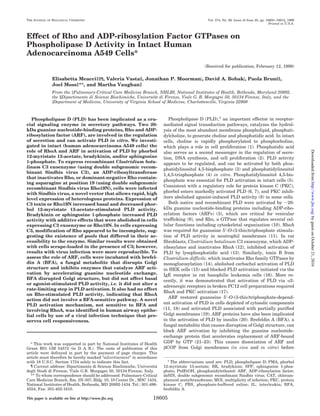
  • 1. Effect of Rho and ADP-ribosylation Factor GTPases on Phospholipase D Activity in Intact Human Adenocarcinoma A549 Cells* (Received for publication, February 12, 1999) Elisabetta Meacci‡§¶, Valeria Vasta§, Jonathan P. Moormanʈ, David A. Bobakʈ, Paola Bruni§, Joel Moss‡**, and Martha Vaughan‡ From the ‡Pulmonary-Critical Care Medicine Branch, NHLBI, National Institutes of Health, Bethesda, Maryland 20892, the §Dipartimento di Scienze Biochimiche, Universita` di Firenze, Viale G. B. Morgagni 50, 50134 Firenze, Italy, and the ʈDepartment of Medicine, University of Virginia School of Medicine, Charlottesville, Virginia 22908 Phospholipase D (PLD) has been implicated as a cru- cial signaling enzyme in secretory pathways. Two 20- kDa guanine nucleotide-binding proteins, Rho and ADP- ribosylation factor (ARF), are involved in the regulation of secretion and can activate PLD in vitro. We investi- gated in intact (human adenocarcinoma A549 cells) the role of RhoA and ARF in activation of PLD by phorbol 12-myristate 13-acetate, bradykinin, and/or sphingosine 1-phosphate. To express recombinant Clostridium botu- linum C3 exoenzyme (using double subgenomic recom- binant Sindbis virus C3), an ADP-ribosyltransferase that inactivates Rho, or dominant-negative Rho contain- ing asparagine at position 19 (using double subgenomic recombinant Sindbis virus Rho19N), cells were infected with Sindbis virus, a novel vector that allows rapid, high level expression of heterologous proteins. Expression of C3 toxin or Rho19N increased basal and decreased phor- bol 12-myristate 13-acetate-stimulated PLD activity. Bradykinin or sphingosine 1-phosphate increased PLD activity with additive effects that were abolished in cells expressing C3 exoenzyme or Rho19N. In cells expressing C3, modification of Rho appeared to be incomplete, sug- gesting the existence of pools that differed in their ac- cessibility to the enzyme. Similar results were obtained with cells scrape-loaded in the presence of C3; however, results with virus infection were more reproducible. To assess the role of ARF, cells were incubated with brefel- din A (BFA), a fungal metabolite that disrupts Golgi structure and inhibits enzymes that catalyze ARF acti- vation by accelerating guanine nucleotide exchange. BFA disrupted Golgi structure, but did not affect basal or agonist-stimulated PLD activity, i.e. it did not alter a rate-limiting step in PLD activation. It also had no effect on Rho-stimulated PLD activity, indicating that RhoA action did not involve a BFA-sensitive pathway. A novel PLD activation mechanism, not sensitive to BFA and involving RhoA, was identified in human airway epithe- lial cells by use of a viral infection technique that pre- serves cell responsiveness. Phospholipase D (PLD),1 an important effector in receptor- mediated signal transduction pathways, catalyzes the hydrol- ysis of the most abundant membrane phospholipid, phosphati- dylcholine, to generate choline and phosphatidic acid. In intact cells, choline is rapidly phosphorylated to phosphocholine, which plays a role in cell proliferation (1). Phosphatidic acid also serves as a second messenger in the regulation of secre- tion, DNA synthesis, and cell proliferation (2). PLD activity appears to be regulated, and can be activated by both phos- phatidylinositol 4,5-bisphosphate (3) and phosphatidylinositol 1,4,5-trisphosphate (4) in vitro. Phosphatidylinositol 4,5-bis- phosphate was essential for PLD activation in intact cells (5). Consistent with a regulatory role for protein kinase C (PKC), phorbol esters markedly activated PLD (6, 7), and PKC inhib- itors abolished agonist-induced PLD activity (8) in some cells. Both native and recombinant PLD were activated by ϳ20- kDa guanine nucleotide-binding proteins including ADP-ribo- sylation factors (ARFs) (3), which are critical for vesicular trafficking (9), and Rho, a GTPase that regulates several cel- lular functions including cytoskeletal organization (10). RhoA was required for guanosine 5Ј-O-(3-thio)triphosphate stimula- tion of PLD activity in neutrophil membranes (11). In rat fibroblasts, Clostridium botulinum C3 exoenzyme, which ADP- ribosylates and inactivates RhoA (12), inhibited activation of PLD by lysophosphatidic acid (13). Similarly, toxin B from Clostridium difficile, which inactivates Rho family GTPases by monoglucosylation (14), abolished carbachol activation of PLD in HEK cells (15) and blocked PLD activation initiated via the IgE receptor in rat basophilic leukemia cells (16). More re- cently, it was demonstrated that activation of PLD via ␣2- adrenergic receptors in broken PC12 cell preparations required RhoA and PKC activation (17). ARF restored guanosine 5Ј-O-(3-thio)triphosphate-depend- ent activation of PLD in cells depleted of cytosolic components (11, 18) and activated PLD associated with partially purified Golgi membranes (19). ARF proteins have also been implicated in the activation of PLD by insulin (20). Brefeldin A (BFA), a fungal metabolite that causes disruption of Golgi structure, can block ARF activation by inhibiting the guanine nucleotide- exchange protein that accelerates replacement of ARF-bound GDP by GTP (21–23). This causes dissociation of ARF and ␤COP from Golgi membranes (in vivo and in vitro) before * This work was supported in part by National Institutes of Health Grant R01 GM 54572 (to D. A. B.). The costs of publication of this article were defrayed in part by the payment of page charges. This article must therefore be hereby marked “advertisement” in accordance with 18 U.S.C. Section 1734 solely to indicate this fact. ¶ Current address: Dipartimento di Scienze Biochimiche, Universita` degli Studi di Firenze, Viale G.B. Morgagni 50, 50134 Firenze, Italy. ** To whom correspondence should be addressed: Pulmonary-Critical Care Medicine Branch, Rm. 5N-307, Bldg. 10, 10 Center Dr., MSC 1434, National Institutes of Health, Bethesda, MD 20892-1434. Tel.: 301-496- 4554; Fax: 301-402-1610. 1 The abbreviations used are: PLD, phospholipase D; PMA, phorbol 12-myristate 13-acetate; BK, bradykinin; SPP, sphingosine 1-phos- phate; PtdEtOH, phosphatidylethanol; ARF, ADP-ribosylation factor; dsSIN, double subgenomic recombinant Sindbis virus; CAT, chloram- phenicol acetyltransferase; MOI, multiplicity of infection; PKC, protein kinase C; PBS, phosphate-buffered saline; IL, interleukin; BFA, brefeldin A. THE JOURNAL OF BIOLOGICAL CHEMISTRY Vol. 274, No. 26, Issue of June 25, pp. 18605–18612, 1999 Printed in U.S.A. This paper is available on line at http://www.jbc.org 18605 byguestonOctober21,2016http://www.jbc.org/Downloadedfrom
  • 2. redistribution of Golgi components to the endoplasmic reticu- lum (24). BFA inhibited carbachol stimulation of PLD activity in HEK cells expressing muscarinic M3 receptors (25), but not PMA stimulation of PLD in HL-60 cells (26). Although BFA treatment has been used frequently to identify effects in cells that are ARF-dependent, the absence of BFA inhibition does not exclude a role for ARF, which can also be activated by BFA-insensitive guanine nucleotide-exchange proteins (27). In addition, BFA has actions that are apparently independent of ARF, such as stimulation of ADP-ribosylation (28), which fur- ther complicates the interpretation of effects observed in intact cells. Although there is evidence that the same PLD might be activated by ARF, Rho, and/or PKC (19), some forms of PLD are clearly not regulated by PKC or guanine nucleotide-binding proteins. Several studies do support a role for ARF and Rho in PLD activation (29). For example, ARF and Rho acted syner- gistically in the activation of brain membrane PLD (7). Rho, ARF, and PKC were implicated in PLD activation in HL-60 (30) and brain cell membranes (7); clearly, the effects in disrupted and intact cells may be very different. To evaluate the role of Rho in intact cells, a novel Sindbis virus vector was used to induce expression of C3 and Rho proteins in A549 cells (human adenocarcinoma cells). From the observations reported here, it appears that a rate-limiting step in PLD stimulation by PMA, BK, and SPP in A549 cells involves RhoA but not a BFA- sensitive ARF pathway. EXPERIMENTAL PROCEDURES Materials Palmitic acid (9,10–3 H) (30–60 Ci/mmol), used to label cellular phos- pholipids, was purchased from NEN Life Science Products, standard lipids were from Avanti Polar Lipids, solvents were from Fluka, and silica gel 60 plates for thin layer chromatography were from Merck. PMA, BK, SPP, BFA, and other chemicals were from Sigma. Mouse monoclonal antibodies against a synthetic peptide with RhoA sequence were from Santa Cruz Biotechnology, and monoclonal antibodies against Golgi 58-kDa protein were from Sigma. Rabbit antiserum raised against bacterially synthesized C3 was a generous gift of Dr. M. Popoff (Institut Pasteur, Paris, France). Secondary antibodies (goat anti-mouse and anti-rabbit immunoglobulin G1 conjugated with fluo- rescein and rhodamine-B, respectively) were from BIOSOURCE. Hu- man lung carcinoma A549 cells were from the American Type Culture Collection (ATCC, Manassas, VA). All materials for cell culture were from Biofluid. Methods Cell Culture—A549 cells were grown in Dulbecco’s modified Eagle’s medium supplemented with 10% heat-inactivated fetal bovine serum, penicillin (5 units/ml), streptomycin (5 ␮g/ml), and 2 mM glutamine. Transphosphatidylation Assay of PLD in Intact Cells—PLD activity was assayed by measuring [3 H]phosphatidylethanol ([3 H]PtdEtOH) produced via PLD-catalyzed transphosphatidylation in cells previously labeled with [3 H]palmitic acid (31). Cells were grown in medium con- taining bovine serum albumin, 1 mg/ml, for 24 h with [3 H]palmitic acid (10 ␮Ci/ml) added for the last 18 h. The medium was then aspirated and after washing, cells were incubated in fresh medium for 2 h. To initiate experiments, 1% ethanol was added 5 min before agonists. Incubations, at 37 °C, were terminated by washing monolayers twice with ice-cold PBS and immediately adding 1 ml of ice-cold methanol. Cells were collected by scraping and dishes were washed with an additional 1 ml of ice-cold methanol. Lipids were then extracted essentially as described by Bligh and Dyer (32) except that water was replaced with 1 M NaCl. The lower phase was dried under a stream of nitrogen, and the residue was dissolved in a small volume of chloroform/methanol (2:1). [3 H] PtdEtOH was separated from other phospholipids by TLC on silica gel 60 plates with the organic phase of the solvent mixture, chloroform/ methanol/acetic acid (90:10:10, v/v/v), and unlabeled PtdEtOH as a FIG. 1. Effect of infection with re- combinant Sindbis virus vector en- coding C. botulinum C3 exoenzyme on morphology of A549 cells. A549 cells were incubated at 37 °C for 1 h with recombinant Sindbis virus (MOI ϭ 20) encoding C. botulinum C3 exoenzyme (ds- SIN:C3) before photography (A). Virus was removed after 1 h of exposure, and 2 h later, cell morphology appeared nor- mal (B). A Nikon optical microscope was used for photography. Regulation of PLD by Rho18606 byguestonOctober21,2016http://www.jbc.org/Downloadedfrom
  • 3. marker. The appropriate lipid spots, detected by exposure to iodine vapor, were marked and scraped into scintillation vials, followed by 0.5 ml of methanol and liquid scintillation fluid before radioassay. Sindbis Vectors Expressing C3 and Mutant Rho—Shuttle phagemids and dsSIN vectors were constructed as described by Hahn et al. (33) and Huang (34). Generation of dsSIN:C3, dsSIN:Rho19N, and dsSIN:CAT, which encode C3 exoenzyme, the negative dominant mutant of RhoA (Rho19N), and chloramphenicol acetyltransferase (dsSIN:CAT), respec- tively, has been described (35, 36). Viral stocks, 109 plaque-forming units/ml, were divided into portions sufficient for a single experiment and stored at Ϫ80 °C. dsSIN Infections—Cells, plated at a density of 5–7 ϫ 105 cells/plate and labeled when indicated with [3 H]palmitic acid, were washed with PBS and diluted in PBS containing 1 mM CaCl2 and 2 mM MgCl2. After incubation at 37 °C for 1 h with the indicated amount of dsSIN recom- binant, PBS was removed, appropriate culture medium was added, and infected cells were incubated for 2 h before addition of agonists and processing for PLD assay, Western analysis, or ADP-ribosylation experiments. Scrape Loading—A549 cells, grown to confluence on 100-mm Petri dishes, were washed twice with PBS and 2 ml of scraping buffer (114 mM KCl, 1 mM NaCl, 5.5 mM MgCl2, 10 mM Tris-HCl). C3 transferase (5 ␮g/ml) or vehicle was added in 0.5 ml of scraping buffer and the cells were gently scraped, suspended in Dulbecco’s modified Eagle’s medium/ 10% fetal bovine serum, and split among six-well dishes. After 6–7 h of incubation, we estimated ϳ70% viable cells. Cells were labeled with [3 H]glycerol (5 ␮Ci/ml) in serum-free medium overnight and used for PLD assay as described above. Western Blotting—To evaluate protein expression from recombinant Sindbis virus, cultured cells were harvested at specified times after infection and lysed by sonification twice for 15 s on ice in Buffer A containing 50 mM Tris, pH 7.5, 150 mM NaCl, 1 mM EGTA, and protease inhibitors (1 mM 4-(2-aminoethyl)benzenesulfonyl fluoride, 0.3 ␮M apro- tinin, and leupeptin and pepstatin, each 10 ␮g/ml). Total cellular pro- tein was quantified, and samples (20–50 ␮g) were subjected to SDS- polyacrylamide gel electrophoresis in 16% gels. Proteins were transferred to nitrocellulose membranes, which were incubated over- night in 20 mM Tris-HCl (pH 7.6), 137 mM NaCl, 0.1% Tween-20 (TTBS (0.1% Tween 20 in Tris-buffered (pH 7.6) saline)) and 0.2% I-block (Tropix, Bedford, MA). Hybridization with mouse monoclonal antibod- ies against RhoA or rabbit antiserum raised against bacterially synthe- sized C3 (2 h room temperature) was followed by washing with TTBS and incubation with peroxidase-conjugated goat anti-mouse or anti- rabbit IgG. Proteins were detected by enhanced chemiluminescence (ECL, Amersham Pharmacia Biotech). ADP-ribosylation Assay—After dsSIN infection, cells were har- vested, washed, and lysed in Buffer A (106 cells/ml). Assays (total volume, 50 ␮l) containing 20 mM Tris-HCl (pH 7.5), 5 mM MgCl2, 1 mM dithiothreitol, 5 ␮M NAD, 0.5 ␮Ci of [32 P]NAD, 2 mM ADP-ribose, and lysate sample plus C3 exoenzyme or recombinant RhoA as a His6 (6ϫ histidine, Calbiochem) fusion protein synthesized in Escherichia coli, as indicated, were incubated at 37 °C for 1 h. Proteins were precipitated with 20% trichloroacetic acid and separated by SDS-polyacrylamide gel electrophoresis; dried gels were exposed to autoradiography film. Indirect Immunofluorescence—For indirect immunofluorescence ex- periments, A549 cells were grown on sterilized glass coverslips placed in six-well dishes with conditions as described for PLD assays. After incubation with BFA (5 ␮g/ml for 30 min) or infection with recombinant Sindbis virus (3 h earlier), cells were washed three times with Dulbec- co’s PBS and fixed with 2% paraformaldehyde in Dulbecco’s PBS for 30 min at room temperature followed by further incubation sequentially for 2 min each with 10, 25, 50, 75, and 100% ethanol before permeabi- lization by a 30-min incubation with 0.05% saponin and 1 mg/ml bovine serum albumin in PBS without Ca2ϩ and Mg2ϩ . Cells were then incu- bated overnight with monoclonal antibodies against Golgi 58-K (a 58- kDa protein used as Golgi marker) or RhoA, or polyclonal antibodies against C3, washed four times, and incubated for 1 h with goat anti- mouse fluorescein- or rhodamine B-conjugated IgG. After washing in PBS containing 0.05% saponin and 1% bovine serum albumin, the coverslips were mounted on Mowoil (Hoescht), examined with a Leica confocal microscope equipped with appropriate filters, and scanned with the SCANware program. Interleukin-6 (IL-6) Secretion—For IL-6 determination, confluent cells, treated or not with 5 ␮g/ml BFA for 30 min, were incubated for 2 h in 1 ml of Dulbecco’s modified Eagle’s medium without or with 100 nM PMA. IL-6 in the medium was then quantified with an immunoassay kit (R & D Systems, Inc., Minneapolis, MN). RESULTS Expression of Recombinant Proteins in Cells—Infection of A549 cells with dsSIN:C3 led to cell rounding, consistent with reported effects of microinjected C3 (37). Two hours after re- moval of virus, cell morphology had returned to normal (Fig. 1). Intracellular C3 exoenzyme was detected as early as 2 h after infection with an MOI of 3 (Fig. 2A). Because expression was maximal with an MOI of 20, this condition was used for all other experiments. No reaction of anti-C3 antibodies with pro- teins in dsSIN:CAT-infected A549 cells was found, consistent with the specificity of these antibodies. The expression of C3 in A549 cells was accompanied by a loss of immunodetectable ϳ20-kDa RhoA (Fig. 2B), probably because, as had been sug- gested for rat1 fibroblasts (13), ADP-ribosylation of RhoA in- creases its susceptibility to degradation. A significant level of immunoreactive Rho19N protein (a dominant negative mutant that contains asparagine at position 19), was detected by anti- RhoA antibodies 2 h after viral infection (MOI of 20). A faint FIG. 2. Expression of recombinant C3 exoenzyme, Rho19N, and CAT in A549 cells. A, effect of MOI on C3 exoenzyme expression. A549 cells were incubated for 1 h with Sindbis virus encoding CAT or indicated amounts (MOI) of Sindbis virus encoding C. botulinum C3 exoenzyme (dsSIN:C3). Fresh virus-free medium was added to infected cells 2 h before they were harvested for Western analysis. Proteins (20 ␮g) were separated by SDS-polyacrylamide gel electrophoresis in 16% gel. Rabbit antiserum raised against bacterially expressed C3 was used to detect C3 exoenzyme. B, effect of expression of dsSIN:C3 on endog- enous RhoA. Experiments were performed as described above except that 50 ␮g of lysate proteins were used for Western analysis, and RhoA was detected using mouse monoclonal antibody raised against a syn- thetic peptide corresponding to amino acids 120–150 of human RhoA. C, expression of Rho19N. Proteins (20 ␮g) from cells infected with dsSIN:Rho19N were processed as described above, and RhoA was de- tected with the mouse monoclonal anti-RhoA antibody. TABLE I Effect of Rho proteins on basal and C3-induced PLD activity Serum-starved cells were incubated overnight with [3 H]palmitic acid (10 ␮Ci/ml) and infected with dsSIN:CAT or dsSIN:C3 as described under “Experimental Procedures” or scraped without or with C3 trans- ferase (5 ␮g/ml) and labeled with [3 H]glycerol (5 ␮Ci/ml) in serum-free medium. Cells were processed for PLD assay as noted under “Experi- mental Procedures.” Data are means Ϯ S.E. of values from three ex- periments with duplicate assays. Treatment of cells PLD activity Infected Scraped No C3 Plus C3 % [3 H]PtdEtOH/phosphatidylcholine No No 0.20 Ϯ 0.04 dsSIN:CAT No 0.22 Ϯ 0.01 dsSIN:Rho19N No 0.40 Ϯ 0.03a dsSIN:C3 No 0.43 Ϯ 0.08a No Yes 0.31 Ϯ 0.03 0.47 Ϯ 0.05a a Expression of C3 or Rho19N or C3 treatment significantly increased PLD activity (P Ͻ 0.05) by Student’s t test. Regulation of PLD by Rho 18607 byguestonOctober21,2016http://www.jbc.org/Downloadedfrom
  • 4. band was observed in dsSIN:CAT-infected cells, indicating a low level of endogenous RhoA in A549 cells (Fig. 2C). Effects of Rho Proteins on Basal PLD Activity—The involve- ment of Rho in PLD activation was demonstrated by infecting A549 cells with recombinant Sindbis virus vectors to express mutant Rho protein or C. botulinum C3 exoenzyme, an ADP- ribosyltransferase that inactivates Rho. Expression of either C3 exoenzyme or the dominant negative Rho19N in A549 cells by infection with dsSIN:C3 or dsSIN:Rho19N, respectively, resulted in a doubling of basal PLD activity (Table I). The increase was similar in A549 cells scrape-loaded in the pres- ence of C3 transferase. The effect of C3 after scrape loading was less than that after Sindbis infection (ϳ50% stimulation versus ϳ100%). Both experiments, however, suggest that Rho proteins play a role in the regulation of PLD activity in human epithelial cells and may have a negative effect on the enzyme activity, restraining or minimizing the level of PLD in unstimulated cells, perhaps by inhibiting a specific PLD or interfering with its activation. Effects of PMA, Bradykinin, and Sphingosine 1-Phosphate on PLD Activity—PMA activation of PLD was appreciable at 15 min and reached a maximum after 30 min of stimulation (data not shown). With 1 ␮M PMA, PLD activity of A549 cells was increased 42 Ϯ 4.1-fold after 30 min (Fig. 3A). The effect of 50 nM PMA was equal to that of 1 ␮M and 1 nM PMA: it increased activity 100% (data not shown). BK and SPP, which act through G protein (heterotrimeric guanine nucleotide-binding protein)-coupled receptors, also stimulated PLD activity (ap- proximately 2-fold); maximal effects were achieved with 1 ␮M and 10 ␮M, respectively; these effects were additive (Fig. 3B). Effects of Rho Proteins on Agonist-stimulated PLD Activi- ty—As shown in Fig. 3A, expression of either dsSIN:C3 or dsSIN:Rho19N reduced but did not abolish PMA stimulation of PLD activity, indicating that Rho proteins are involved in a PKC-dependent pathway. In cells infected with dsSIN:C3, BK or SPP failed to increase PLD activity (Fig. 3B). BK plus SPP, however, induced an ϳ80% increase in PLD activity, suggest- ing that the combination of agonists was able to overcome, in part, the inhibitory effects of C3, perhaps acting through Rho that had not been ADP-ribosylated by the exoenzyme. In scrape-loaded cells, the effect of C3 in reducing PLD stimula- tion by agonists was less than it was in infected cells, albeit statistically significant; in cells expressing C3, PMA stimula- tion was reduced 25 Ϯ 2% versus 38 Ϯ 4% (n ϭ 3), and BK plus SPP stimulation was reduced 20 Ϯ 1.5% versus 40 Ϯ 9% (n ϭ 3). These data are consistent with the conclusion that for dem- onstration of C3 effects, viral infection was more effective than scrape loading. Incomplete Modification of Endogenous RhoA by C3 Trans- ferase—To explore the possibility that the incomplete reduction of PLD stimulation by C3 was due to differences in the acces- sibility of intracellular pools of Rho, we quantified ADP-ribo- sylation of RhoA in cell lysates. First to prove that C3 trans- ferase expressed in dsSIN:C3 infected cells was enzymatically active, 100 ng of exogenous RhoA (His6-Rho) and [14 C]NAD were added to cell lysates (Fig. 4). A clear band of 20 kDa was detected, indicating that the C3 transferase was able to cata- lyze ADP-ribosylation of RhoA. Then to verify whether the intracellular pools of Rho were completely modified by the expressed C3 transferase, we performed experiments using exogenous C3 exoenzyme. ADP-ribosylation of endogenous Rho was not detected when C3 exoenzyme was added at final con- centration of 0.025 ␮g/ml to lysates of cells infected with dsSIN: CAT or dsSIN:C3. After addition of higher concentration of C3 (0.5 ␮g/ml) (Fig. 5), a single radiolabeled band, corresponding in migration to endogenous Rho, was detected. The band at ϳ20 kDa was more intense in lane 4 than in lane 2, consistent with the larger amount of the overexpressed dominant nega- tive Rho19N. The presence of a band in lane 6, although per- haps somewhat weaker than that in lane 2, suggested that even when the expression of C3 seemed maximal, modification of the endogenous Rho was not complete. Similarly, some en- dogenous Rho was inaccessible to the C3 in dsSIN:C3-infected cells that were treated with agonists (Fig. 6). Those data could explain the incomplete inhibition of PMA stimulation of PLD and the significant, albeit attenuated, effects of BK and SPP on FIG. 3. Effect of Rho protein on stimulated PLD activity. Cells were infected with Sindbis virus encoding CAT, C. botulinum C3 exoenzyme, or the dominant-negative mutant Rho19N. Virus was removed after 1 h, and 2 h later, cells were incubated with vehicle (0.1% Me2SO) or 1 ␮M PMA for 30 min (A) or with 1 ␮M BK, 10 ␮M SPP, or 1 ␮M BK plus 10 ␮M SPP for 10 min (B) before assay of PLD activity. PLD activity is expressed relative to that of nonstimulated, noninfected cells. [3 H]PtdEtOH formed as a percentage of total [3 H]phosphatidylcholine was 0.20 Ϯ 0.04 and 0.22 Ϯ 0.01% in uninfected cells and in dsSIN:CAT infected cells, respectively; viral infection did not significantly affect basal PLD activity or the stimulation by PMA. Data are means Ϯ S.E. of values from at least four experiments with duplicate assays. There was no significant difference between noninfected and dsSIN-infected cells in amounts of [3 H]palmitic acid in total phospholipids (1.8 Ϯ 0.20 ϫ 106 and 2.1 Ϯ 0.26 ϫ 106 dpm, respectively). Effects of C3 and Rho19N expression on PMA-stimulated PLD activity and C3 expression on receptor-stimulated PLD activity were significant (p Ͻ 0.05, Student’s t test). Regulation of PLD by Rho18608 byguestonOctober21,2016http://www.jbc.org/Downloadedfrom
  • 5. PLD activity in cells expressing C3 exoenzyme. The extent of ADP-ribosylation of endogenous Rho in dsSIN:C3-infected cells varied from one experiment to another, but it was always statistically significant when data were quantified by densi- tometry (data not shown). Because of the presence of unmodi- fied RhoA, indirect immunofluorescence was used to localize C3 exoenzyme and RhoA in A549 cells. In dsSIN:C3-infected cells, C3 transferase was concentrated in the Golgi compartment and widely distributed in the cytosol (Fig. 7, panel 4). Endogenous RhoA in unstimulated cells appeared predominantly in Golgi and in endoplasmic reticulum (Fig. 7, panels 5 and 6). Thus, indirect immunofluorescence indicated colocalization of C3 and endogenous RhoA in Golgi and endoplasmic reticulum compartments. Effects of BFA on Basal, PMA-stimulated, and Receptor- stimulated PLD Activity—The effects of BFA on Golgi structure and PLD activity in A549 cells were evaluated. Incubation for 1 h with BFA (5 ␮g/ml) induced a drastic redistribution of the Golgi 58-kDa immunoreactivity (Fig. 8, panels 2 and 3). Only a slight effect on Golgi structure was detected in PMA-treated cells (panel 4). In similar experiments, BFA significantly de- creased PMA-stimulated secretion of IL-6 (control, 435 Ϯ 45 pg/ml; 1 ␮M PMA, 1414 Ϯ 65 pg/ml; PMA plus BFA, 431 Ϯ 10 pg/ml). Both these findings indicated that A549 cells were fully sensitive to the well known effects of BFA on Golgi structure and function. As shown in Table II, incubation of A549 cells with BFA (5 ␮g/ml) for 30 min or 3 h (data not shown) did not significantly affect basal PLD activity or the stimulation by PMA, BK, SPP, or BK plus SPP. Nor was a significant effect of BFA observed with submaximal concentrations of these ago- nists (5 nM BK or 30 nM SPP). Synergistic effects of Rho and ARF proteins on PLD activity have been reported (7, 29). Using C3 exoenzyme, we investigated whether the Rho-dependent PLD activity was modified by BFA treatment. As shown in Table II, incubation with BFA (5 ␮g/ml, 30 min) had no signif- icant effect on C3-induced PLD activity (Table II). No signifi- cant effect of BFA was also seen in cells scraped-loaded in the presence of C3 and stimulated with PMA (3.4 Ϯ 0.38 versus 3.8 Ϯ 0.31, n ϭ 3) or BK plus SPP (1.4 Ϯ 0.25 versus 1.8 Ϯ 0.12, n ϭ 3). DISCUSSION To investigate the roles of Rho and ARF in the regulation of PLD in human airway epithelial cells, cultured A549 cells were treated with PMA, a direct activator of PKC, and with agonists that act through G protein-coupled receptors, i.e. bradykinin (BK), a proinflammatory peptide shown to play a critical role in the development of airway hyper-responsiveness, and SPP, which has been implicated in several biological responses (38). Involvement of 20-kDa GTP-binding proteins in PMA- or re- ceptor-mediated activation of PLD has been reported (19), al- though it seems clear that the pathways utilized can differ with different agonists and cell types. C. botulinum exoenzyme C3 ADP-ribosylates Asn-41 in Rho, which is within the putative effector domain of the molecule (12). Because this covalent modification specifically inactivates Rho, C3 can be a useful probe of Rho function and has been used in A549 cells to investigate the role of the Rho GTPase in bradykinin-stimulated nuclear factor-␬B activation and IL-1b gene expression (39). Unfortunately, C3 does not enter intact cells, or does so only poorly, making it inconvenient to use in living cells to probe Rho function. Electroporation, permeabili- zation, prolonged incubation (days), and scrape loading proce- dures have been used to effect intracellular delivery of C3 (13, 15, 37). To avoid these manipulations and preserve fully re- sponsive cells, in this study, A549 cells were infected with recombinant Sindbis virus encoding C3 exoenzyme (dsSIN:C3) or the dominant negative Rho19N (dsSIN:Rho19N). Infection of cells with these vectors resulted in a high level of expression of recombinant proteins with minimal cytotoxicity (33). The cells, FIG. 4. Effect of expression of C3 exoenzyme on ADP-ribosyla- tion of RhoA. Samples of lysate from cells infected with dsSIN:CAT or dsSIN:C3 were incubated in ADP-ribosylation assays as described un- der “Experimental Procedures” for 1 h at 37 °C without or with 0.025 ␮g of C3 exoenzyme or 100 ng of recombinant RhoA (His6-RhoA) as indi- cated, before precipitation and separation of proteins (15 ␮g) by SDS- polyacrylamide gel electrophoresis. The dried gel was exposed over- night for autoradiography. FIG. 5. ADP-ribosylation of endogenous Rho in A549 cells. Sam- ples (20 ␮g) of lysates from cells infected with dsSIN:CAT, dsSIN: Rho19N, or dsSIN:C3 were incubated with [32 P]NAD with or without C3 exoenzyme (0.5 ␮g) and processed as described in Fig. 4. FIG. 6. Effect of agonists on ADP-ribosylation of RhoA in in- fected A549 cells. Lysates (20 ␮g) of A549 cells infected with dsSIN: CAT or dsSIN:C3, some of which had been stimulated with 1 ␮M PMA, BK, or 10 ␮M SPP, were incubated with [32 P]NAD with or without C3 exoenzyme (0.5 ␮g) and/or His6-RhoA (100 ng) for 1 h at 37 °C. Samples were processed as described in Fig. 4. Regulation of PLD by Rho 18609 byguestonOctober21,2016http://www.jbc.org/Downloadedfrom
  • 6. 2 h after removal of virus, demonstrated apparently normal Golgi localization of p58. Viral infection did not alter cell me- tabolism nonspecifically, at least in so far as was manifest by effects on incorporation of radiolabeled palmitic acid into phos- pholipids or on phosphatidylethanol formation (basal or ago- nist-stimulated). Moreover, lysates from infected cells were still able to ADP-ribosylate recombinant His6-Rho added in vitro. When Sindbis infection was used in studies of basic cell functions, such as posttranslational processing (40, 41) or pro- grammed cell death (36), no cytotoxicity was reported. We observed that cell viability was, in fact, higher than with scrape loading (Ͼ90% versus 60–70%). A disadvantage of the Sindbis expression system, however, is that some cell types cannot be used for long term experiments (longer than 8 h) because virus replication inhibits host protein synthesis. To check for effects on cell metabolism, protein synthesis was examined by incu- bating cells with [3 H]methionine for 3 h after removal of the virus; an approximately 10% inhibition of protein labeling was observed (data not shown). In A549 cells, expression of C3 exoenzyme was rapid and efficient as confirmed by Western analysis and changes in cell morphology during virus incubation. RhoA is present in A549 cells at very low levels, but was detectable in homogenates after radiolabeling in vitro with [32 P]NAD and exogenous C3 exoenzyme. C3 expression was accompanied by a decrease in the amount of immunoreactive RhoA, as previously demonstrated in fibroblasts treated with C3 exoenzyme (13), probably because C3-catalyzed ADP-ribo- sylation accelerated its degradation. Loss of RhoA was ob- served under conditions in which not all of the RhoA had been modified by C3. As shown here, the loss of RhoA protein in cells expressing C3 was associated with PLD activation. Because a similar increase in activity was observed in cells expressing the dominant negative Rho19N, it was presumably not due to a nonspecific action of C3 on other types of Rho proteins. More- over, the C3-induced increase in PLD activity was not due to an effect of the viral infection because a significant increase was also observed in cells scrape-loaded with C3. Together, these findings indicate that the regulation of PLD in A549 cells involves specifically RhoA and is the first evidence of negative regulation of PLD by RhoA in intact cells. Inactivation of RhoA-dependent pathways with C3 or a dom- inant negative RhoA (Rho19N) enhanced basal PLD. The only prior reports of effects of inactivation of Rho on PLD activity in FIG. 7. Immunolocalization of C3 exoenzyme (A) and endogenous Rho (B). Cells infected with dsSIN:C3 virus were fixed and double-labeled with mouse monoclonal antibodies against Golgi 58- kDa protein (panel 2) and rabbit anti- serum against C3 (panel 4), which were detected using anti-mouse fluorescein- and anti-rabbit rhodamine-B-conjugated IgG1, respectively. The two images are superimposed in panel 3. Monoclonal an- ti-RhoA and fluorescein-conjugated im- munoglobulin IgG1 were used to detect endogenous RhoA in unstimulated A549 cells (panel 5). Nuclei (panel 6) were stained with 4Ј,6-diamidino-2-phenylin- dole (blue). The coverslips were mounted on Mowoil (Hoescht) and examined with a Leica confocal microscope. Regulation of PLD by Rho18610 byguestonOctober21,2016http://www.jbc.org/Downloadedfrom
  • 7. the absence of added effectors involved PC12 and HEK cells. Incubation of isolated membranes or permeabilized cells with the exoenzyme decreased or did not affect the basal activity, respectively (7, 17). Under conditions where basal PLD was increased, activation of PLD by PMA was reduced about 50% by infection of cells with dsSIN:C3 or dsSIN:Rho19N; PLD was not affected by infection with dsSIN:CAT, a recombinant virus encoding chloramphenicol acetyltransferase, indicating that the cells were fully responsive after infection and consistent with a specific role for RhoA. Partial reduction of PMA-stimu- lated PLD by C3 has also been reported in rat1 fibroblasts (13). We found that even when expression was optimal and all cells expressed C3 exoenzyme, a significant fraction of Rho protein was not ADP-ribosylated. Although activation of PLD by ARF has been widely inves- tigated in cell-free systems (3, 18, 19, 42), evidence for its occurrence in cells is limited (18, 20, 43). In many cells, proc- essing and secretion of proteins are blocked by brief incubation with BFA, a fatty acid metabolite widely used to inhibit ARF action (44, 45). Natural resistance to BFA has been reported in PtK-1 and MDCK cell lines (46–48), and mutant CHO cells resistant to BFA have been isolated and characterized (49). The A549 cells used in the studies reported here were fully sensitive to effects of BFA on Golgi structure and function. Immunoflu- orescence microscopy using antibodies raised against the 58- kDa protein showed redistribution of this Golgi marker in cells treated for 1 h with BFA, 5 ␮g/ml. A significant decrease in IL-6 secretion was also observed. Similar treatment caused no sig- nificant alteration of PLD activity in A549 cells incubated with or without agonists, suggesting that BFA-sensitive guanine nucleotide-exchange proteins for ARF that are involved in the maintenance of Golgi structure and function do not directly influence or are not rate-limiting in the regulation of PLD activity. Similarly, Guillemain and Exton (26) recently re- ported that treatment of differentiated HL-60 cells with BFA for up to 6 h markedly altered the distribution of the trans- Golgi marker enzyme galactosyltransferase activity but did not affect PLD activation by formyl Met-Leu-Phe, ATP, or PMA. Because PLD activity was assayed in intact cells, inhibition of an ARF-dependent component by BFA treatment might be too small a fraction of the total to be detected. BFA also failed to affect PLD activity in cells expressing C3. Several explanations FIG. 8. Effect of BFA on immunolo- calization of Golgi 58-kDa protein. A549 cells grown on glass coverslips and processed in parallel with cells used for PLD assay were incubated without (pan- els 1 and 4) or with 5 ␮g/ml BFA (panels 2 and 3), followed by incubation with 1 ␮M PMA for 30 min (panels 3 and 4), before fixation (2% paraformaldehyde) and per- meabilization (0.05% saponin). Cells were then incubated overnight with mono- clonal antibodies against Golgi 58-kDa protein, washed in PBS containing 0.05% saponin and bovine serum albumin (1 mg/ ml), and incubated for 1 h with goat anti- mouse rhodamine-B-conjugated IgG. TABLE II Effects of brefeldin A on stimulation of PLD in A549 cells by C3 transferase, PMA, BK, and SPP, in A549 cells Serum-starved cells were labeled with [3 H]glycerol (5 ␮Ci/ml) in serum-free medium. BFA (5 ␮g/ml) or vehicle (methanol 0.1%) was added for 30 min before stimulation with agonists for 10 min and in the presence of 2% ethanol. For C3-treatment experiments, cells were scraped with C3 transferase (5 ␮g/ml) and processed for PLD assay as described under “Experimental Procedures.” Data are means Ϯ S.E. of values from at least three separate experiments with duplicate assays. Effects of BFA on basal and stimulated PLD activity were not signifi- cant (p Ͼ 0.1) by Student’s t test. Activators Phospholipase D activity No BFA Plus BFA % [3 H]PtdEtOH None 0.28 Ϯ 0.04 0.44 Ϯ 0.09 C3 (5 ␮g/ml) 0.47 Ϯ 0.05a 0.65 Ϯ 0.08a PMA, 1 ␮M 2.80 Ϯ 0.17a 2.40 Ϯ 0.19a BK, 5 nM 1.03 Ϯ 0.20a 0.82 Ϯ 0.10a BK, 1 ␮M 1.25 Ϯ 0.13a 1.01 Ϯ 0.10a SPP, 30 nM 0.65 Ϯ 0.04a 0.61 Ϯ 0.05a SPP, 1 ␮M 1.20 Ϯ 0.20a 1.55 Ϯ 0.15a BK plus SPP 1.90 Ϯ 0.40a 2.11 Ϯ 0.38a a Significant effects on respective control (p Ͻ 0.05). Regulation of PLD by Rho 18611 byguestonOctober21,2016http://www.jbc.org/Downloadedfrom
  • 8. for the negative findings are possible, e.g. the effect of BFA on ARF activation may be incomplete so that sufficient active ARF remains to permit full PLD activation, but not secretion. It seems equally possible that activation of the ARF required for PLD activation may be catalyzed by a BFA-insensitive guanine nucleotide-exchange protein, such as cytohesin-1. Alterna- tively, PLD stimulation by active ARF in intact cells may be prevented by an inhibitor such as arfaptin (50). In any case, our observations are consistent with the notion that BFA-sensitive activation of ARF is not directly involved in the rapid stimula- tion of PLD induced by PMA, BK, or SPP, and also with the existence of a BFA-insensitive pathway that involves a PLD isoform not required for IL-6 secretion. Because inactivation of RhoA was accompanied by an in- crease of PLD activity, there appears to be a Rho-dependent inhibitory component(s) in A549 cells. This was not affected by BFA treatment, indicating that RhoA did not require BFA- sensitive ARF activation to regulate PLD activity. Both BFA treatment and Rho inactivation modify the cytoskeleton. Ef- fects of C3 exoenzyme and Rho19N are not, however, simply due to a general disruption of signal transduction as a conse- quence of altered cytoskeletal organization or function, but to a more direct involvement of the Rho GTPase, which is consist- ent, also with the apparent contribution to PLD activation of a pool of Rho that is not completely accessible to C3 toxin. The data reported here do not exclude roles for ARF proteins in the regulation of PLD, but they indicate that a rate-limiting step in PLD stimulation by PMA, BK, and SPP and in basal PLD regulation does not involve BFA-sensitive pathways. Seem- ingly different roles for RhoA in the regulation of basal and agonist-enhanced PLD activity in human A549 cells were dem- onstrated. Together, the data support the existence of a novel mechanism of PLD activation that involves RhoA, is not sen- sitive to BFA, and may be localized in a specific subcellular compartment. REFERENCES 1. Cuadrado, A., Carnero, A., Dolfi, F., Jimenez, B., and Lacal, L. C. (1993) Oncogene 8, 2959–2968 2. Fukami, K., and Takenawa, T. (1992) J. Biol. Chem. 267, 10988–10994 3. Brown, H. A., Gutowski, S., Moomaw, C. R., Slaughter, C., and Sternweis, P. C. (1993) Cell 75, 1137–1144 4. Hammond, S. M., Jenco, J. M., Nakashima, S., Cadwallader, K., Gu, Q., Cook, S., Nozawa, Y., Prestwich, G. D., Frohman, M. A., and Morris, A. J. (1997) J. Biol. Chem. 272, 3860–3868 5. Pertile, P., Liscovitch, M., Chalifa, V., and Cantley, L. C. (1995) J. Biol. Chem. 270, 5130–5135 6. Lopez, I., Burns, D. J., and Lambeth, J. D. (1995) J. Biol. Chem. 270, 19465–19472 7. Singer, W. D., Brown, H. A., Jiang, X., and Sternweis, P. C. (1996) J. Biol. Chem. 271, 4504–4510 8. Yeo, E.-J., Kazlauskas, A., and Exton, J. H. (1994) J. Biol. Chem. 269, 27823–27826 9. Moss, J., and Vaughan, M. (1995) J. Biol. Chem. 270, 12327–12330 10. Tapon, N., and Hall, A. (1997) Curr. Opin. Cell Biol. 9, 86–92 11. Bowman, E. P., Uhlinger, D. J., and Lambeth, J. D. (1993) J. Biol. Chem. 268, 21509–21512 12. Sekine, A., Fujiwara, M., and Narumiya, S. (1989) J. Biol. Chem. 264, 8602–8605 13. Malcolm, K. C., Elliott, C. M., and Exton, J. H. (1996) J. Biol. Chem. 271, 13135–13139 14. Just, I., Selzer, J., Wilm, M., Von Eichel-Streiber, C., Mann, M., and Aktories, K. (1995) Nature 375, 500–503 15. Schmidt, M., Rumenapp, U., Bienek, C., Keller, J., Von Eichel-Streiber, C., and Jakobs, K. H., (1996) J. Biol. Chem. 271, 2422–2426 16. Ojio, K., Banno, Y., Nakashima, S., Kato, N., Watanabe, K., Lyerly, D. M., and Nozawa, Y. (1996) Biochem. Biophys. Res. Commun. 224, 591–596 17. Jinsi-Parimoo, A., and Deth, R. C. (1997) J. Biol. Chem. 272, 14556–14561 18. Cockcroft, S., Thomas, G. M. H., Fensome, A., Geny, B., Cunningham, E., Gout, I., Hiles, I., Totty, N. F., Truong, O., and Hsuan, J. J. (1994) Science 263, 523–526 19. Exton, J. H. (1997) J. Biol. Chem. 272, 15579–15582 20. Karnam, P., Standaert, M. L., Galloway, L., and Farese, R. V. (1997) J. Biol. Chem. 272, 6136–6140 21. Donaldson, J. G., Finazzi, D., and Klausner, R. D. (1992) Nature 360, 350–352 22. Helms, J. B., and Rothman, J. A. (1992) Nature 360, 352–354 23. Morinaga, N., Tsai, S.-C., Moss, J., and Vaughan, M. (1996) Proc. Natl. Acad. Sci. U. S. A. 92, 12856–12860 24. Donaldson, J. G., Cassel, D., Kahn, R. A., and Klausner, R. D. (1992) Proc. Natl. Acad. Sci. U. S. A. 89, 6409–6412 25. Rumenapp, U., Geiszt, M., Wahn, F., Schmidt, M., and Jakobs, K. H. (1995) Eur. J. Biochem. 234, 240–244 26. Guillemain, I., and Exton, J. H. (1997) Eur. J. Biochem. 249, 812–819 27. Meacci, E., Tsai, S.-C., Adamik, R., Moss, J., and Vaughan, M. (1997) Proc. Natl. Acad. Sci. U. S. A. 94, 1745–1748 28. De Matteis, M. A., Di Girolamo, M., Colanzi, A., Pallas, M., Di Tuillo, G., McDonald, L. J., Moss, J., Santini, G., Bannykh, S., Corda, D., and Luini, A. (1994) Proc. Natl. Acad. Sci. U. S. A. 91, 1114–1118 29. Fensome, A., Whatmore, J., Morgan, C., Jones, D., and Cockcroft, S. (1998) J. Biol. Chem. 273, 13157–13164 30. Ohguchi, K., Banno, Y., Nakashima, S., and Nozawa, Y. (1996) J. Biol. Chem. 271, 4366–4372 31. Morris, A. J., Frohman, M. A., and Engebrecht, J. (1997) Anal. Biochem. 252, 1–9 32. Bligh, E. G., and Dyer, W. J. (1959) Can. J. Biochem. Physiol. 37, 911–917 33. Hahn, C. S., Hahn, Y. S., Braciale, T. J., and Rice, C. M. (1992) Proc. Natl. Acad. Sci. U. S. A. 89, 2679–2683 34. Huang, H. V. (1996) Curr. Opin. Biotechnol. 7, 531–535 35. Moorman, J. P., Bobak, D. A., and Hahn, C. S. (1996) J. Immunol. 156, 4146–4153 36. Bobak, D., Moorman, J., Guanzon, A., Gutmer, L., and Hahn, C. (1997) Onco- gene 15, 2179–2189 37. Paterson, H. F., Self, A. J., Garrett, M. D., Just, I., Aktories, K., and Hall, A. (1990) J. Cell Biol. 111, 1001–1007 38. Van Brocklyn, J. R., Lee, M. J., Menzeleev, R., Olivera, A., Edsall, L., Cuvillier, O., Thomas, D. M., Coopman, P. J. P., Thangada, S., Liu, C. H., Hla, T., and Spiegel, S. (1998) J. Cell Biol. 142, 229–240 39. Pan, Z. K., Ye, R. D., Christiansen, S. C., Jagels, M. A., Bokoch, G. M., and Zuraw, B. L. (1998) J. Immunol. 160, 3038–3045 40. Li, G., Barbieri, M. A., and Stahl, P. D. (1995) Arch. Biochem. Biophys. 316, 529–534 41. Piper, R. C., Tai, C., Kulesza, P., Pang, S., Warnock, D., Baenziger, J., Slot, J. W., Geuze, H. J., Puri, C., and James, D. E. (1993) J. Cell Biol. 121, 1221–1232 42. Massenburg, D., Han, J.-S., Liyanage, W. A., Patton, W., Moss, J., and Vaughan, M. (1994) Proc. Natl. Acad. Sci. U. S. A. 91, 11718–11722 43. Luo, J.-Q., Liu, X., Frankel, P., Rotunda, T., Ramos, M., Flom, J., Jiang, H., Feig, L. A., Morris, A. J., Kahn, R. A., and Foster, D. A. (1998) Proc. Natl. Acad. Sci. U. S. A. 95, 3632–3637 44. Misumi, Y., Misumi, Y., Miki, K., Takatsuki, G., Tamura, G., and Ikehara, Y. (1986) J. Biol. Chem. 261, 11398–11403 45. Bershadsky, A. D., and Futerman, A. H. (1994) Proc. Natl. Acad. Sci. U. S. A. 91, 5686–5689 46. Ktistakis, N. T., Roth, M. G., and Bloom, G. S. (1991) J. Cell Biol. 113, 1009–1023 47. Hunziker, W., Whitney, J. A., and Mellman, I. (1991) Cell 67, 617–627 48. Low, S. H., Tang, B. L., Wong, S. H., and Hong, W. (1992) J. Cell Biol. 118, 51–62 49. Yan, J.-P., Colon, M. E., Beebe, L. A., and Melanc¸on, P. (1994) J. Cell Biol. 126, 65–75 50. Tsai, S.-C., Adamik, R., Hong, J.-X., Moss, J., Vaughan, M., Ranoh, H., and Exton, J. H. (1998) J. Biol. Chem. 273, 20697–20701 Regulation of PLD by Rho18612 byguestonOctober21,2016http://www.jbc.org/Downloadedfrom
  • 9. Joel Moss and Martha Vaughan Elisabetta Meacci, Valeria Vasta, Jonathan P. Moorman, David A. Bobak, Paola Bruni, Intact Human Adenocarcinoma A549 Cells Effect of Rho and ADP-ribosylation Factor GTPases on Phospholipase D Activity in doi: 10.1074/jbc.274.26.18605 1999, 274:18605-18612.J. Biol. Chem. http://www.jbc.org/content/274/26/18605Access the most updated version of this article at Alerts: When a correction for this article is posted• When this article is cited• to choose from all of JBC's e-mail alertsClick here http://www.jbc.org/content/274/26/18605.full.html#ref-list-1 This article cites 50 references, 33 of which can be accessed free at byguestonOctober21,2016http://www.jbc.org/Downloadedfrom