SlideShare a Scribd company logo
1 of 9
Download to read offline
Supplemental Figure 1
Figure S1 Characterization of podocyte markers by RT-PCR and Western blots in cultured podocytes
harvested from the urine of children with HIVAN. A. Primary podocytes (P-1), and two podocyte cell
lines (P-2 and P-3), generated from cells isolated from the urine of two children with HIVAN, were expanded
and tested for the expression of podocyte markers by RT-PCR. Lanes loaded with water (4), RNA derived
from human renal proximal tubular epithelial cells (RPTE), or a human kidney tissue sample (HK), served as
negative and positive controls respectively. The mRNA expression of WT-1, synaptopodin, nestin, nephrin,
and podocalyxin was detected in all podocyte clones (P1, P2, P3) and the HK, but not in RPTE cells. mRNA
for podocin and the endothelial cell marker Von Willebrand factor (vWF), were detected only in the HK.
GAPDH served as an internal control. Reaction for GAPDH without RT (-RT) was performed to rule out the
possibility of genomic DNA contamination. B. Western blots showing WT-1, synaptopodin and nestin
protein expression in the podocyte clones P-1, P-2, and P-3.
A
B
Supplemental Table 1 Pairs of primers used for the characterization of human podocytes in Figure S1.
Supplemental Figure 2
Figure S2 Sequence information corresponding to the Tat gene cloned from cultured renal epithelial
cells infected with HIV-1, and used to generate the adenoviral Tat-WT vector. The upper panel shows
the DNA sequence of the Tat gene cloned from cultured primary renal epithelial cells harvested from the
urine a child with HIVAN, and infected with primary HIV-1 isolates corresponding to the same child, as
previously described.29
In the lower panel, the protein sequence of the Tat-HIVAN gene is aligned with other
Tat genes, including pcDNA3.1-Tat(101), which was subcloned from pcDNA3.1/Tat101-Flag (PEV280, NIH
AIDS Research and Reference Reagent Program), and originally derived from the HIV-1 cDNA clone pCV1,
and the monocyte-tropic HIV-1 isolate ADA. We used the Clustal Omega multiple sequence alignment
program (http:// www.ebi.ac.uk/Tools /msa/clustalo/). The basic domain and RGD motif are indicated in
brackets. The Tat sequencing procedure was repeated three times, to rule out the possibility of a sequencing
error. Other Tat clones generated from the same cells, also showed a similar RGD motif deletion, suggesting
that the Tat-HIVAN sequence is unlikely derived from an inactive quasispecies.
Supplemental Figure 3
Figure S3 Translocation of Tat-HIVAN to the nucleus of cultured podocytes. Podocytes were
transfected with cDNA vectors corresponding to either the wild type Tat-HIVAN, described in Figure S2, or
Tat-HIVAN carrying a mutation in the basic domain (Tat-BDM1). Thirty six hours later, the cells were fixed
with 4% paraformaldehyde, and stained with an anti-Flag antibody followed by anti-mouse IgG Alexa Flour
488, to detect Tat-Flag. The intracellular distribution of Tat was assessed by confocal microscopy. The left
panels show the fluorescence microscopy studies, and the right panels show phase contrast microscopy. Tat-
WT was detected in the cytoplasm and the nucleus of cultured podocytes, while Tat-BDM1 remained
localized in the cytoplasm of these cells. These findings confirm that the wild type Tat-HIVAN can
translocate to the nucleus of cultured podocytes, and demonstrate the critical role of the Tat basic domain in
this process. The scale bar is 10 m.
Supplemental Figure 4
Figure S4 The wild type Tat-HIVAN, unlike the Tat-HIVAN carrying a mutation in the basic domain,
induces transactivation of the HIV-LTR in GHOST cells. GHOST cells were transiently transfected with
wild type Tat-HIVAN (Tat-WT) or Tat-HIVAN carrying mutations in the basic domain (Tat-BDM1 or Tat-
BDM2). Thirty six hours later, all cells were harvested for FACS analysis (upper panel) or Western blotting
(lower panel). As discussed in methods, in this reporter system, the expression of GFP is under the control of
the HIV-LTR and can be used as readout system to assess the transactivation activity of Tat.
Supplemental Figure 5
Figure S5 Induction of HIV-envelope (Env) gene expression in the kidney of young HIV-Tg26 mice
infected with Ad-Tat-WT. Newborn HIV-Tg26 mice (n =3 mice per group) were infected with adenoviral
vectors carrying wild type Tat-HIVAN (Ad-Tat-WT) or Tat-HIVAN carrying a mutation in the basic biding
domain (Ad-Tat-BDM1) as previously described.
36
Seven days later, all mice were sacrificed, and their
kidneys were harvested and processed for the RT-PCR studies. The upper panel shows representative RT-
PCR results corresponding to HIV-env mRNA expression in the kidney of young HIV-Tg26 mice infected
with Ad-Tat-WT or Ad-Tat-BDM1. The graph summarizes the results corresponding to three mice per group
(unpaired t test; ** p = 0.011).
Supplemental Figure 6
Figure 6S Adenoviral vectors carrying WT-Tat-HIVAN precipitated the development of HIVAN in
young HIV-Tg26 mice. Newborn heterozygous HIV-Tg26 mice were infected with adenoviral vectors carrying
A
B
A B C
D E F
C
either, Lac Z (rAd-LacZ), Tat-HIVAN with a mutation in the basic biding domain (rAd-Tat-BDM1), or wild
type Tat-HIVAN (rAd-Tat-WT) as previously described.36
Seven days later, all mice were sacrificed, and the
kidneys were processed for the RT-PCR studies. The upper panel shows representative RT-PCR results for
Tat mRNA in kidney samples collected from wild type (WT) and HIV-Tg26 mice infected with rAd-LacZ, rAd-
Tat-BDM1, or rAd-Tat-WT (n = 4 - 8 mice per group). The 301 bp Tat amplificaton product shown in panel
A, was sequenced to confirm the expression of Tat in the kidney. Panel B shows a representative Western
Blot of the immunoprecipitated Tat protein expressed in the kidney and liver of WT mice infected with rAD-
Tat-WT-Flag vector. Tat was detected with an anti-Flag antibody as described in the methods section. Panel
C shows representative renal sections stained with hematoxylin and eosin. All mice were infected at birth
with the corresponding adenoviral vectors and sacrificed at 35 days. At this time point, fifty per cent (4/8) of
the HIV-Tg26 mice infected with rAd-WT-Tat developed the complete renal histological HIVAN phenotype
(F). These findings represent a significant acceleration of the timing for developing HIVAN in our HIV-Tg26
mouse colony. In contrast, none (0/8*) of the HIV-Tg26 mice infected with rAd-LacZ (D) or rAd-Tat-BDM1
(E) developed the complete renal histological HIVAN phenotype at this time point. In addition, none (0/12**)
of the WT mice infected with rAd-LacZ , rAd-Tat-BDM1, or rAd-Tat-WT, showed significant renal
histological lesions at 35 days of life. (Chi-square test, * p < 0.02; ** p < 0.006). (A-F, original magnification
x 100)
Supplemental Figure 7
Figure7S Circulating FGF-2 induced albuminuria in wild type and HIV-Tg26 mice. Wild type and HIV-
Tg26 mice (4 weeks old), without pre-existing renal disease were infected with adenoviral viral vectors carrying
either a secreted form of human recombinant FGF-2 or the Lac-Z gene (n = 5 mice per group), as described in
the methods section.37
Urine samples were collected 28 days after the adenoviral infection. FGF-2 induced
significant albuminuria in both WT and HIV-Tg26 mice, although these changes were more significant in HIV-
Tg26 mice (* p< 0.01; ** p< 0.001, ANOVA).

More Related Content

What's hot

Rt2 profilerbrochure
Rt2 profilerbrochureRt2 profilerbrochure
Rt2 profilerbrochureElsa von Licy
 
Serum copper as a novel biomarker for resistance to thyroid hormone
Serum copper as a novel biomarker for resistance to thyroid hormoneSerum copper as a novel biomarker for resistance to thyroid hormone
Serum copper as a novel biomarker for resistance to thyroid hormonePrakamya Gupta
 
Ashia Drosophilia Poster
Ashia Drosophilia PosterAshia Drosophilia Poster
Ashia Drosophilia PosterAshia Williams
 
Role of Thromboxane A2 in the Induction of Apoptosis of Immature Thymocytes b...
Role of Thromboxane A2 in the Induction of Apoptosis of Immature Thymocytes b...Role of Thromboxane A2 in the Induction of Apoptosis of Immature Thymocytes b...
Role of Thromboxane A2 in the Induction of Apoptosis of Immature Thymocytes b...Federal University of Bahia
 
Activation of surrogate death receptor signalling triggers peroxynitrite depe...
Activation of surrogate death receptor signalling triggers peroxynitrite depe...Activation of surrogate death receptor signalling triggers peroxynitrite depe...
Activation of surrogate death receptor signalling triggers peroxynitrite depe...Saurabh Shekhar
 
同玩節海報事件
同玩節海報事件同玩節海報事件
同玩節海報事件lalacamp07
 
FGF-2 treatment of high passage human mesenchymal stem cells enhances chondro...
FGF-2 treatment of high passage human mesenchymal stem cells enhances chondro...FGF-2 treatment of high passage human mesenchymal stem cells enhances chondro...
FGF-2 treatment of high passage human mesenchymal stem cells enhances chondro...Boris Schmalz, MD
 
FGFBP1 pathways control after induction of a conditional transgene in a mouse...
FGFBP1 pathways control after induction of a conditional transgene in a mouse...FGFBP1 pathways control after induction of a conditional transgene in a mouse...
FGFBP1 pathways control after induction of a conditional transgene in a mouse...Anne Deslattes Mays
 
Work In Progress 03.17.11 Final Edit
Work In Progress 03.17.11 Final EditWork In Progress 03.17.11 Final Edit
Work In Progress 03.17.11 Final Editl_presser
 
Genetics, structure, and prevalence of FP967 T-DNA in flax
Genetics, structure, and prevalence of FP967 T-DNA in flaxGenetics, structure, and prevalence of FP967 T-DNA in flax
Genetics, structure, and prevalence of FP967 T-DNA in flaxJamille McLeod
 

What's hot (20)

pap paper pdf
pap paper pdfpap paper pdf
pap paper pdf
 
Rt2 profilerbrochure
Rt2 profilerbrochureRt2 profilerbrochure
Rt2 profilerbrochure
 
Serum copper as a novel biomarker for resistance to thyroid hormone
Serum copper as a novel biomarker for resistance to thyroid hormoneSerum copper as a novel biomarker for resistance to thyroid hormone
Serum copper as a novel biomarker for resistance to thyroid hormone
 
Gene expression on rat1 fibroblast cells after transformation by evi1
Gene expression on rat1 fibroblast cells after transformation by evi1Gene expression on rat1 fibroblast cells after transformation by evi1
Gene expression on rat1 fibroblast cells after transformation by evi1
 
Hippo pathoway
Hippo pathoway Hippo pathoway
Hippo pathoway
 
Simske Poster 2016-2
Simske Poster 2016-2Simske Poster 2016-2
Simske Poster 2016-2
 
Ashia Drosophilia Poster
Ashia Drosophilia PosterAshia Drosophilia Poster
Ashia Drosophilia Poster
 
cancerres
cancerrescancerres
cancerres
 
Xie X et al 2014
Xie X et al 2014Xie X et al 2014
Xie X et al 2014
 
Role of Thromboxane A2 in the Induction of Apoptosis of Immature Thymocytes b...
Role of Thromboxane A2 in the Induction of Apoptosis of Immature Thymocytes b...Role of Thromboxane A2 in the Induction of Apoptosis of Immature Thymocytes b...
Role of Thromboxane A2 in the Induction of Apoptosis of Immature Thymocytes b...
 
Activation of surrogate death receptor signalling triggers peroxynitrite depe...
Activation of surrogate death receptor signalling triggers peroxynitrite depe...Activation of surrogate death receptor signalling triggers peroxynitrite depe...
Activation of surrogate death receptor signalling triggers peroxynitrite depe...
 
4785
47854785
4785
 
12263-184470-1-PB
12263-184470-1-PB12263-184470-1-PB
12263-184470-1-PB
 
Fulltext
FulltextFulltext
Fulltext
 
Sfnge array poster
Sfnge array posterSfnge array poster
Sfnge array poster
 
同玩節海報事件
同玩節海報事件同玩節海報事件
同玩節海報事件
 
FGF-2 treatment of high passage human mesenchymal stem cells enhances chondro...
FGF-2 treatment of high passage human mesenchymal stem cells enhances chondro...FGF-2 treatment of high passage human mesenchymal stem cells enhances chondro...
FGF-2 treatment of high passage human mesenchymal stem cells enhances chondro...
 
FGFBP1 pathways control after induction of a conditional transgene in a mouse...
FGFBP1 pathways control after induction of a conditional transgene in a mouse...FGFBP1 pathways control after induction of a conditional transgene in a mouse...
FGFBP1 pathways control after induction of a conditional transgene in a mouse...
 
Work In Progress 03.17.11 Final Edit
Work In Progress 03.17.11 Final EditWork In Progress 03.17.11 Final Edit
Work In Progress 03.17.11 Final Edit
 
Genetics, structure, and prevalence of FP967 T-DNA in flax
Genetics, structure, and prevalence of FP967 T-DNA in flaxGenetics, structure, and prevalence of FP967 T-DNA in flax
Genetics, structure, and prevalence of FP967 T-DNA in flax
 

Viewers also liked

Docker Containers Deep Dive
Docker Containers Deep DiveDocker Containers Deep Dive
Docker Containers Deep DiveWill Kinard
 
Beauty bubbleonblabdropin (1)
Beauty bubbleonblabdropin (1)Beauty bubbleonblabdropin (1)
Beauty bubbleonblabdropin (1)William Slaise Jr
 
New.Novum.11.Lure.Lore.Research
New.Novum.11.Lure.Lore.ResearchNew.Novum.11.Lure.Lore.Research
New.Novum.11.Lure.Lore.ResearchHoward Moskowitz
 
DraytonHall Fall 2016 Interiors Collector's Ed
DraytonHall Fall 2016 Interiors Collector's EdDraytonHall Fall 2016 Interiors Collector's Ed
DraytonHall Fall 2016 Interiors Collector's EdNatalie Titcomb Wilson
 
5 одегова ашан_плюс слайд
5 одегова ашан_плюс слайд5 одегова ашан_плюс слайд
5 одегова ашан_плюс слайдEkaterina Abramova
 
Polls Are Becoming Increasingly Inaccurate. Why?
Polls Are Becoming Increasingly Inaccurate. Why?Polls Are Becoming Increasingly Inaccurate. Why?
Polls Are Becoming Increasingly Inaccurate. Why?Robert Pressel
 
1314惠州的環保設施及自然保護區兩天之旅
1314惠州的環保設施及自然保護區兩天之旅1314惠州的環保設施及自然保護區兩天之旅
1314惠州的環保設施及自然保護區兩天之旅baptistrainbow
 
Second multi-stakeholder partnerships for SDGs
Second multi-stakeholder partnerships for SDGsSecond multi-stakeholder partnerships for SDGs
Second multi-stakeholder partnerships for SDGsFelix Dodds
 

Viewers also liked (11)

Docker Containers Deep Dive
Docker Containers Deep DiveDocker Containers Deep Dive
Docker Containers Deep Dive
 
Beauty bubbleonblabdropin (1)
Beauty bubbleonblabdropin (1)Beauty bubbleonblabdropin (1)
Beauty bubbleonblabdropin (1)
 
New.Novum.11.Lure.Lore.Research
New.Novum.11.Lure.Lore.ResearchNew.Novum.11.Lure.Lore.Research
New.Novum.11.Lure.Lore.Research
 
Cv Denice
Cv DeniceCv Denice
Cv Denice
 
DraytonHall Fall 2016 Interiors Collector's Ed
DraytonHall Fall 2016 Interiors Collector's EdDraytonHall Fall 2016 Interiors Collector's Ed
DraytonHall Fall 2016 Interiors Collector's Ed
 
5 одегова ашан_плюс слайд
5 одегова ашан_плюс слайд5 одегова ашан_плюс слайд
5 одегова ашан_плюс слайд
 
Polls Are Becoming Increasingly Inaccurate. Why?
Polls Are Becoming Increasingly Inaccurate. Why?Polls Are Becoming Increasingly Inaccurate. Why?
Polls Are Becoming Increasingly Inaccurate. Why?
 
1314惠州的環保設施及自然保護區兩天之旅
1314惠州的環保設施及自然保護區兩天之旅1314惠州的環保設施及自然保護區兩天之旅
1314惠州的環保設施及自然保護區兩天之旅
 
Afghanistan
AfghanistanAfghanistan
Afghanistan
 
Second multi-stakeholder partnerships for SDGs
Second multi-stakeholder partnerships for SDGsSecond multi-stakeholder partnerships for SDGs
Second multi-stakeholder partnerships for SDGs
 
Not a Monster
Not a MonsterNot a Monster
Not a Monster
 

Similar to Xie's PaperASN2013070710SupplementaryData

Whole Transcriptome Analysis of Testicular Germ Cell Tumors
Whole Transcriptome Analysis of Testicular Germ Cell TumorsWhole Transcriptome Analysis of Testicular Germ Cell Tumors
Whole Transcriptome Analysis of Testicular Germ Cell TumorsThermo Fisher Scientific
 
c-Jun overexpression in CAR-T cells induces exhaustion resistance
c-Jun overexpression in CAR-T cells induces exhaustion resistancec-Jun overexpression in CAR-T cells induces exhaustion resistance
c-Jun overexpression in CAR-T cells induces exhaustion resistancelinyandeng1
 
Genome responses of trypanosome infected cattle
Genome responses of trypanosome infected cattleGenome responses of trypanosome infected cattle
Genome responses of trypanosome infected cattleLaurence Dawkins-Hall
 
Ablooglu, AJ (2010) Development
Ablooglu, AJ (2010) DevelopmentAblooglu, AJ (2010) Development
Ablooglu, AJ (2010) DevelopmentArarat Ablooglu
 
2004 paper primer ctx dissemination of ctx m-type -lactamases among clinical ...
2004 paper primer ctx dissemination of ctx m-type -lactamases among clinical ...2004 paper primer ctx dissemination of ctx m-type -lactamases among clinical ...
2004 paper primer ctx dissemination of ctx m-type -lactamases among clinical ...JairAlexanderTllez
 
2011 - Cellular inhibitor of apoptosis protein-1 (cIAP1) can regulate E2F1 tr...
2011 - Cellular inhibitor of apoptosis protein-1 (cIAP1) can regulate E2F1 tr...2011 - Cellular inhibitor of apoptosis protein-1 (cIAP1) can regulate E2F1 tr...
2011 - Cellular inhibitor of apoptosis protein-1 (cIAP1) can regulate E2F1 tr...Simon Gemble
 
Systemic analysis of data combined from genetic qtl's and gene expression dat...
Systemic analysis of data combined from genetic qtl's and gene expression dat...Systemic analysis of data combined from genetic qtl's and gene expression dat...
Systemic analysis of data combined from genetic qtl's and gene expression dat...Laurence Dawkins-Hall
 
Akhtar et al. Stem Cell Reports (2015)
Akhtar et al. Stem Cell Reports (2015)Akhtar et al. Stem Cell Reports (2015)
Akhtar et al. Stem Cell Reports (2015)Aslam Akhtar, MS
 
Ph D Swati Dhar
Ph D Swati DharPh D Swati Dhar
Ph D Swati DharSwati Dhar
 
LaurenBrewerPoster_FinalDJM
LaurenBrewerPoster_FinalDJMLaurenBrewerPoster_FinalDJM
LaurenBrewerPoster_FinalDJMLauren Brewer
 
20151107 workshop HPA, HLA, HEA genotype
20151107 workshop HPA, HLA, HEA genotype20151107 workshop HPA, HLA, HEA genotype
20151107 workshop HPA, HLA, HEA genotypeJhysheng Chang
 
project- Liron Schwartz
project- Liron Schwartzproject- Liron Schwartz
project- Liron SchwartzLiron Schwartz
 
BIOLOGICAL VARIANCE OF SAT2 FOOT-AND-MOUTH DISEASE VIRUSES.
BIOLOGICAL VARIANCE OF SAT2 FOOT-AND-MOUTH DISEASE VIRUSES.  BIOLOGICAL VARIANCE OF SAT2 FOOT-AND-MOUTH DISEASE VIRUSES.
BIOLOGICAL VARIANCE OF SAT2 FOOT-AND-MOUTH DISEASE VIRUSES. EuFMD
 
Comparison of primer sets for amplification of Major Piroplasm Surface Protei...
Comparison of primer sets for amplification of Major Piroplasm Surface Protei...Comparison of primer sets for amplification of Major Piroplasm Surface Protei...
Comparison of primer sets for amplification of Major Piroplasm Surface Protei...Sudhakar Goud Karpurapu
 

Similar to Xie's PaperASN2013070710SupplementaryData (20)

Whole Transcriptome Analysis of Testicular Germ Cell Tumors
Whole Transcriptome Analysis of Testicular Germ Cell TumorsWhole Transcriptome Analysis of Testicular Germ Cell Tumors
Whole Transcriptome Analysis of Testicular Germ Cell Tumors
 
c-Jun overexpression in CAR-T cells induces exhaustion resistance
c-Jun overexpression in CAR-T cells induces exhaustion resistancec-Jun overexpression in CAR-T cells induces exhaustion resistance
c-Jun overexpression in CAR-T cells induces exhaustion resistance
 
Genome responses of trypanosome infected cattle
Genome responses of trypanosome infected cattleGenome responses of trypanosome infected cattle
Genome responses of trypanosome infected cattle
 
Ablooglu, AJ (2010) Development
Ablooglu, AJ (2010) DevelopmentAblooglu, AJ (2010) Development
Ablooglu, AJ (2010) Development
 
2004 paper primer ctx dissemination of ctx m-type -lactamases among clinical ...
2004 paper primer ctx dissemination of ctx m-type -lactamases among clinical ...2004 paper primer ctx dissemination of ctx m-type -lactamases among clinical ...
2004 paper primer ctx dissemination of ctx m-type -lactamases among clinical ...
 
2011 - Cellular inhibitor of apoptosis protein-1 (cIAP1) can regulate E2F1 tr...
2011 - Cellular inhibitor of apoptosis protein-1 (cIAP1) can regulate E2F1 tr...2011 - Cellular inhibitor of apoptosis protein-1 (cIAP1) can regulate E2F1 tr...
2011 - Cellular inhibitor of apoptosis protein-1 (cIAP1) can regulate E2F1 tr...
 
Systemic analysis of data combined from genetic qtl's and gene expression dat...
Systemic analysis of data combined from genetic qtl's and gene expression dat...Systemic analysis of data combined from genetic qtl's and gene expression dat...
Systemic analysis of data combined from genetic qtl's and gene expression dat...
 
Akhtar et al. Stem Cell Reports (2015)
Akhtar et al. Stem Cell Reports (2015)Akhtar et al. Stem Cell Reports (2015)
Akhtar et al. Stem Cell Reports (2015)
 
Ph D Swati Dhar
Ph D Swati DharPh D Swati Dhar
Ph D Swati Dhar
 
LaurenBrewerPoster_FinalDJM
LaurenBrewerPoster_FinalDJMLaurenBrewerPoster_FinalDJM
LaurenBrewerPoster_FinalDJM
 
JIpdf
JIpdfJIpdf
JIpdf
 
Rathod
RathodRathod
Rathod
 
20151107 workshop HPA, HLA, HEA genotype
20151107 workshop HPA, HLA, HEA genotype20151107 workshop HPA, HLA, HEA genotype
20151107 workshop HPA, HLA, HEA genotype
 
Cancer Research 2
Cancer Research 2Cancer Research 2
Cancer Research 2
 
project- Liron Schwartz
project- Liron Schwartzproject- Liron Schwartz
project- Liron Schwartz
 
Presentación métodos pcr
Presentación métodos pcrPresentación métodos pcr
Presentación métodos pcr
 
BIOLOGICAL VARIANCE OF SAT2 FOOT-AND-MOUTH DISEASE VIRUSES.
BIOLOGICAL VARIANCE OF SAT2 FOOT-AND-MOUTH DISEASE VIRUSES.  BIOLOGICAL VARIANCE OF SAT2 FOOT-AND-MOUTH DISEASE VIRUSES.
BIOLOGICAL VARIANCE OF SAT2 FOOT-AND-MOUTH DISEASE VIRUSES.
 
Research Article Presentation.pptx
Research Article Presentation.pptxResearch Article Presentation.pptx
Research Article Presentation.pptx
 
MInh Project 2
MInh Project 2MInh Project 2
MInh Project 2
 
Comparison of primer sets for amplification of Major Piroplasm Surface Protei...
Comparison of primer sets for amplification of Major Piroplasm Surface Protei...Comparison of primer sets for amplification of Major Piroplasm Surface Protei...
Comparison of primer sets for amplification of Major Piroplasm Surface Protei...
 

Xie's PaperASN2013070710SupplementaryData

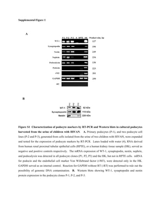
  • 1. Supplemental Figure 1 Figure S1 Characterization of podocyte markers by RT-PCR and Western blots in cultured podocytes harvested from the urine of children with HIVAN. A. Primary podocytes (P-1), and two podocyte cell lines (P-2 and P-3), generated from cells isolated from the urine of two children with HIVAN, were expanded and tested for the expression of podocyte markers by RT-PCR. Lanes loaded with water (4), RNA derived from human renal proximal tubular epithelial cells (RPTE), or a human kidney tissue sample (HK), served as negative and positive controls respectively. The mRNA expression of WT-1, synaptopodin, nestin, nephrin, and podocalyxin was detected in all podocyte clones (P1, P2, P3) and the HK, but not in RPTE cells. mRNA for podocin and the endothelial cell marker Von Willebrand factor (vWF), were detected only in the HK. GAPDH served as an internal control. Reaction for GAPDH without RT (-RT) was performed to rule out the possibility of genomic DNA contamination. B. Western blots showing WT-1, synaptopodin and nestin protein expression in the podocyte clones P-1, P-2, and P-3. A B
  • 2. Supplemental Table 1 Pairs of primers used for the characterization of human podocytes in Figure S1.
  • 3. Supplemental Figure 2 Figure S2 Sequence information corresponding to the Tat gene cloned from cultured renal epithelial cells infected with HIV-1, and used to generate the adenoviral Tat-WT vector. The upper panel shows the DNA sequence of the Tat gene cloned from cultured primary renal epithelial cells harvested from the urine a child with HIVAN, and infected with primary HIV-1 isolates corresponding to the same child, as previously described.29 In the lower panel, the protein sequence of the Tat-HIVAN gene is aligned with other Tat genes, including pcDNA3.1-Tat(101), which was subcloned from pcDNA3.1/Tat101-Flag (PEV280, NIH AIDS Research and Reference Reagent Program), and originally derived from the HIV-1 cDNA clone pCV1, and the monocyte-tropic HIV-1 isolate ADA. We used the Clustal Omega multiple sequence alignment program (http:// www.ebi.ac.uk/Tools /msa/clustalo/). The basic domain and RGD motif are indicated in brackets. The Tat sequencing procedure was repeated three times, to rule out the possibility of a sequencing error. Other Tat clones generated from the same cells, also showed a similar RGD motif deletion, suggesting that the Tat-HIVAN sequence is unlikely derived from an inactive quasispecies.
  • 4. Supplemental Figure 3 Figure S3 Translocation of Tat-HIVAN to the nucleus of cultured podocytes. Podocytes were transfected with cDNA vectors corresponding to either the wild type Tat-HIVAN, described in Figure S2, or Tat-HIVAN carrying a mutation in the basic domain (Tat-BDM1). Thirty six hours later, the cells were fixed with 4% paraformaldehyde, and stained with an anti-Flag antibody followed by anti-mouse IgG Alexa Flour 488, to detect Tat-Flag. The intracellular distribution of Tat was assessed by confocal microscopy. The left panels show the fluorescence microscopy studies, and the right panels show phase contrast microscopy. Tat- WT was detected in the cytoplasm and the nucleus of cultured podocytes, while Tat-BDM1 remained localized in the cytoplasm of these cells. These findings confirm that the wild type Tat-HIVAN can translocate to the nucleus of cultured podocytes, and demonstrate the critical role of the Tat basic domain in this process. The scale bar is 10 m.
  • 5. Supplemental Figure 4 Figure S4 The wild type Tat-HIVAN, unlike the Tat-HIVAN carrying a mutation in the basic domain, induces transactivation of the HIV-LTR in GHOST cells. GHOST cells were transiently transfected with wild type Tat-HIVAN (Tat-WT) or Tat-HIVAN carrying mutations in the basic domain (Tat-BDM1 or Tat- BDM2). Thirty six hours later, all cells were harvested for FACS analysis (upper panel) or Western blotting (lower panel). As discussed in methods, in this reporter system, the expression of GFP is under the control of the HIV-LTR and can be used as readout system to assess the transactivation activity of Tat.
  • 6. Supplemental Figure 5 Figure S5 Induction of HIV-envelope (Env) gene expression in the kidney of young HIV-Tg26 mice infected with Ad-Tat-WT. Newborn HIV-Tg26 mice (n =3 mice per group) were infected with adenoviral vectors carrying wild type Tat-HIVAN (Ad-Tat-WT) or Tat-HIVAN carrying a mutation in the basic biding domain (Ad-Tat-BDM1) as previously described. 36 Seven days later, all mice were sacrificed, and their kidneys were harvested and processed for the RT-PCR studies. The upper panel shows representative RT- PCR results corresponding to HIV-env mRNA expression in the kidney of young HIV-Tg26 mice infected with Ad-Tat-WT or Ad-Tat-BDM1. The graph summarizes the results corresponding to three mice per group (unpaired t test; ** p = 0.011).
  • 7. Supplemental Figure 6 Figure 6S Adenoviral vectors carrying WT-Tat-HIVAN precipitated the development of HIVAN in young HIV-Tg26 mice. Newborn heterozygous HIV-Tg26 mice were infected with adenoviral vectors carrying A B A B C D E F C
  • 8. either, Lac Z (rAd-LacZ), Tat-HIVAN with a mutation in the basic biding domain (rAd-Tat-BDM1), or wild type Tat-HIVAN (rAd-Tat-WT) as previously described.36 Seven days later, all mice were sacrificed, and the kidneys were processed for the RT-PCR studies. The upper panel shows representative RT-PCR results for Tat mRNA in kidney samples collected from wild type (WT) and HIV-Tg26 mice infected with rAd-LacZ, rAd- Tat-BDM1, or rAd-Tat-WT (n = 4 - 8 mice per group). The 301 bp Tat amplificaton product shown in panel A, was sequenced to confirm the expression of Tat in the kidney. Panel B shows a representative Western Blot of the immunoprecipitated Tat protein expressed in the kidney and liver of WT mice infected with rAD- Tat-WT-Flag vector. Tat was detected with an anti-Flag antibody as described in the methods section. Panel C shows representative renal sections stained with hematoxylin and eosin. All mice were infected at birth with the corresponding adenoviral vectors and sacrificed at 35 days. At this time point, fifty per cent (4/8) of the HIV-Tg26 mice infected with rAd-WT-Tat developed the complete renal histological HIVAN phenotype (F). These findings represent a significant acceleration of the timing for developing HIVAN in our HIV-Tg26 mouse colony. In contrast, none (0/8*) of the HIV-Tg26 mice infected with rAd-LacZ (D) or rAd-Tat-BDM1 (E) developed the complete renal histological HIVAN phenotype at this time point. In addition, none (0/12**) of the WT mice infected with rAd-LacZ , rAd-Tat-BDM1, or rAd-Tat-WT, showed significant renal histological lesions at 35 days of life. (Chi-square test, * p < 0.02; ** p < 0.006). (A-F, original magnification x 100)
  • 9. Supplemental Figure 7 Figure7S Circulating FGF-2 induced albuminuria in wild type and HIV-Tg26 mice. Wild type and HIV- Tg26 mice (4 weeks old), without pre-existing renal disease were infected with adenoviral viral vectors carrying either a secreted form of human recombinant FGF-2 or the Lac-Z gene (n = 5 mice per group), as described in the methods section.37 Urine samples were collected 28 days after the adenoviral infection. FGF-2 induced significant albuminuria in both WT and HIV-Tg26 mice, although these changes were more significant in HIV- Tg26 mice (* p< 0.01; ** p< 0.001, ANOVA).