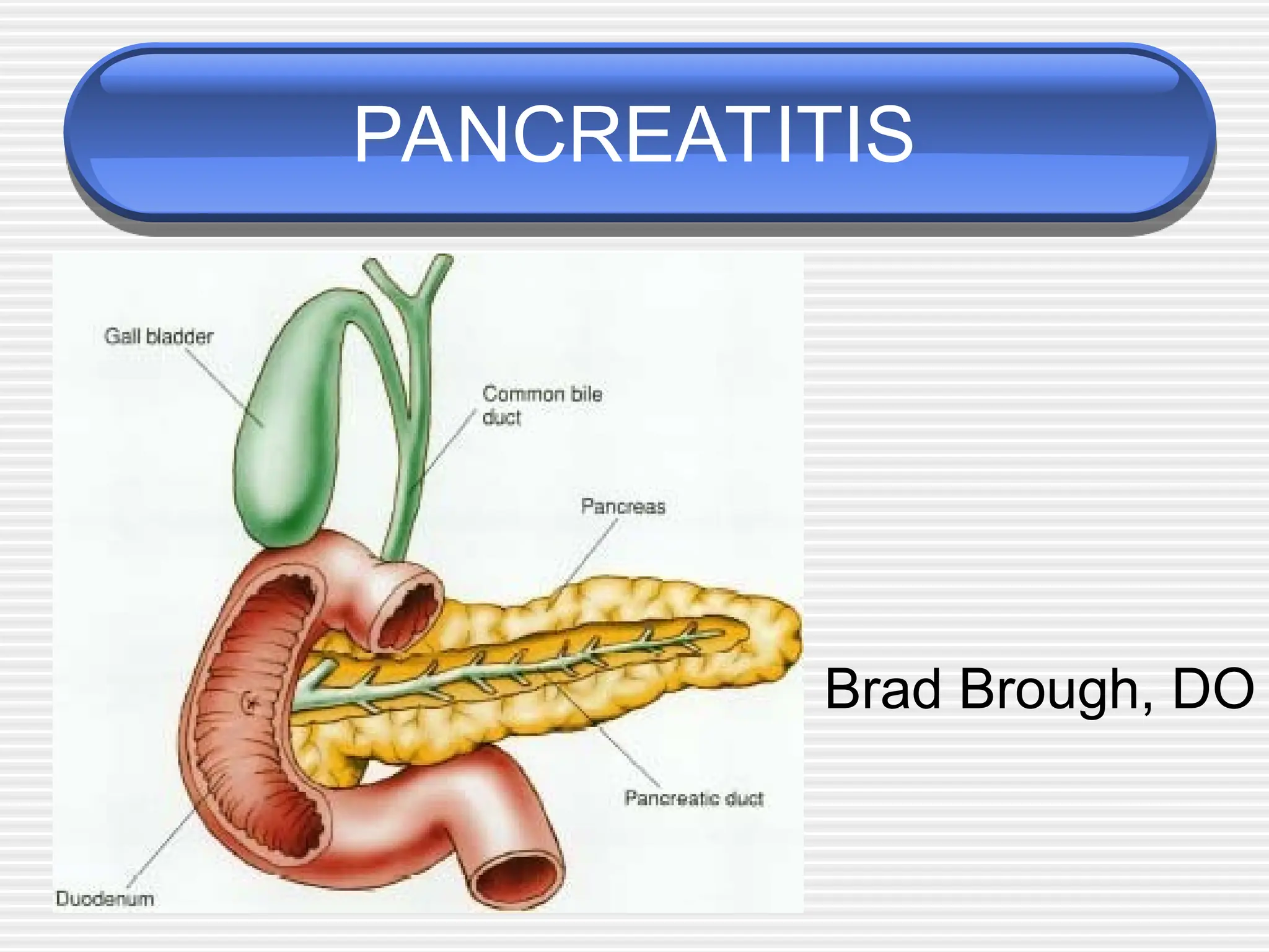
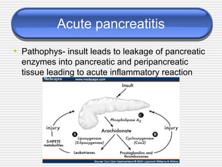
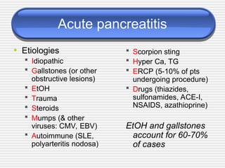
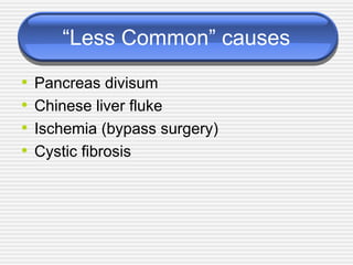
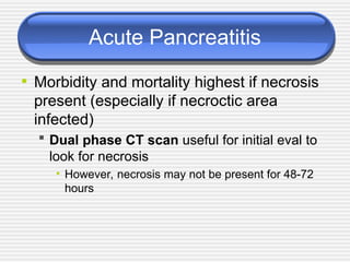
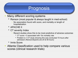
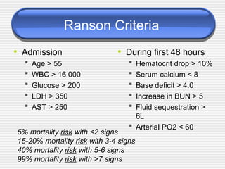
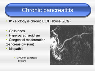
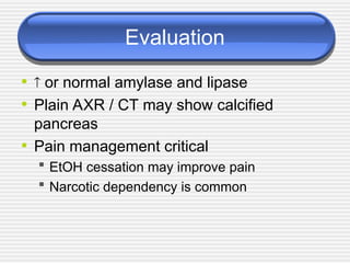
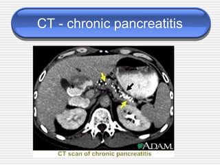
PANCREATITIS
Brad Brough, DO
Acute pancreatitis
• Pathophys- insult leads to leakage of pancreatic
enzymes into pancreatic and peripancreatic
tissue leading to acute inflammatory reaction
Acute pancreatitis
• Etiologies
 Idiopathic
 Gallstones (or other
obstructive lesions)
 EtOH
 Trauma
 Steroids
 Mumps (& other
viruses: CMV, EBV)
 Autoimmune (SLE,
polyarteritis nodosa)
 Scorpion sting
 Hyper Ca, TG
 ERCP (5-10% of pts
undergoing procedure)
 Drugs (thiazides,
sulfonamides, ACE-I,
NSAIDS, azathioprine)
EtOH and gallstones
account for 60-70%
of cases
“Less Common” causes
• Pancreas divisum
• Chinese liver fluke
• Ischemia (bypass surgery)
• Cystic fibrosis
Trivia
• What is the name of the scorpion that
causes pancreatitis?
 Hint: you won’t find it in the USA
• Tityus Trinitatis
• (Found in Central/
• South America and
• the Caribbean)
Signs & Symptoms
• Severe epigastric abdominal pain - abrupt onset
(may radiate to back)
• Nausea & Vomiting
• Weakness
• Tachycardia
• +/- Fever; +/- Hypotension or shock
 Grey Turner sign - flank discoloration due to
retroperitoneal bleed in pt. with pancreatic necrosis
(rare)
 Cullen’s sign - periumbilical discoloration (rare)
• Grey Turner sign • Cullen’s sign
Differential
• Not all inclusive, but may include:
 Biliary disease
 Intestinal obstruction
 Mesenteric Ischemia
 MI (inferior)
 AAA
 Distal aortic dissection
 PUD
Evaluation
•  amylase…Nonspecific !!!
 Amylase levels > 3x normal very suggestive of
pancreatitis
• May be normal in chronic pancreatitis!!!
 Enzyme level  severity
 False (-): acute on chronic (EtOH); HyperTG
 False (+): renal failure, other abdominal or salivary gland
process, acidemia
•  lipase
 More sensitive & specific than amylase
Evaluation
• Other inflammatory markers will be elevated
 CRP, IL-6, IL-8 (studies hoping to use these markers to aid in
detecting severity of disease)
• ALT > 3x normal  gallstone pancreatitis
 (96% specific, but only 48% sensitive)
• Depending on severity may see:
  Ca
 WBC
 BUN
  Hct
  glucose
Radiographic Evaluation
• AXR - “sentinel loop” or small bowel ileus
• US or CT may show enlarged pancreas with
stranding, abscess, fluid collections, hemorrhage,
necrosis or pseudocyst
• MRI/MRCP newest “fad”
 Decreased nephrotoxicity from gadolinium
 Better visualization of fluid collections
 MRCP allows visualization of bile ducts for stones
 Does not allow stone extraction or stent insertion
• Endoscopic US (even newer but used less)
 Useful in obese patients
CT Scan of acute pancreatitis
• CT shows
significant
swelling
and
inflammation
of the
pancreas
Gall stone pancreatitis by ERCP
Acute Pancreatitis
• Morbidity and mortality highest if necrosis
present (especially if necroctic area
infected)
 Dual phase CT scan useful for initial eval to
look for necrosis
• However, necrosis may not be present for 48-72
hours
Prognosis
• Many different scoring systems
 Ranson (most popular & always taught in med-school)
• No association found with score, and mortality or length of
hospitalization
 APACHE II
 CT severity Index
• Recent studies show this to be most predictive of adverse outcomes
 CT score > 5 associated with 15x mortality rate
 Problem is 1 CT study showing this was conducted 72 hours after
admission (Ranson/Apache are 24 & 48 hours)
 Imrie Score
• Atlanta Classification used to help compare various
scores (clinical research trials)
Ranson Criteria
• Admission
 Age > 55
 WBC > 16,000
 Glucose > 200
 LDH > 350
 AST > 250
• During first 48 hours
 Hematocrit drop > 10%
 Serum calcium < 8
 Base deficit > 4.0
 Increase in BUN > 5
 Fluid sequestration >
6L
 Arterial PO2 < 60
5% mortality risk with <2 signs
15-20% mortality risk with 3-4 signs
40% mortality risk with 5-6 signs
99% mortality risk with >7 signs
CT Severity Index
• CT Grade
 A is normal (0 points)
 B is edematous pancreas
(1 point)
 C is B plus extrapancreatic
changes (2 points)
 D is severe extrapancreatic
changes plus one fluid
collection (3 points)
 E is multiple or extensive
fluid collections (4 points)
• Necrosis score
 None (0 points)
 < 1/3 (2 points)
 > 1/3, < 1/2 (4 points)
 > 1/2 (6 points)
• TOTAL SCORE =
CT grade + Necrosis
0-1 = 0% mortality
2-3 = 3% mortality
4-6 = 6% mortality
7-10 = 17% mortality
Therapy
• Remove offending agent (if possible)
• Supportive !!!
• #1- NPO (until pain free)
 NG suction for patients with ileus or emesis
 TPN may be needed
• #2- Aggressive volume repletion with IVF
 Keep an eye on fluid balance/sequestration
and electrolyte disturbances
Therapy continued
• #3- Narcotic analgesics usually necessary for pain
relief…textbooks say Meperidine…
 NO conclusive evidence that morphine has
deleterious effect on sphincter of Oddi pressure
• #4- Urgent ERCP and biliary sphincterotomy
within 72 hours improves outcome of severe
gallstone pancreatitis
 Reduced biliary sepsis, not actual improvement of
pancreatic inflammation
• #5- Don’t forget PPI to prevent stress ulcer
Complications
• Necrotizing pancreatitis
 Significantly increases morbidity & mortality
 Usually found on CT with IV contrast
• Pseudocysts
 Suggested by persistent pain or continued high
amylase levels (may be present for 4-6 wks afterward)
 Cyst may become infected, rupture, hemorrhage or
obstruct adjacent structures
• Asymptomatic, non-enlarging pseudocysts can be watched
and followed with imaging
• Symptomatic, rapidly enlarging or complicated pseudocysts
need to be decompressed
Complications continued #2
• Infection
 Many areas for concern: abscess, pancreatic necrosis,
infected pseudocyst, cholangitis, and aspiration
pneumonia -> SEPSIS may occur
 If concerned, obtain cultures and start broad-spectrum
antimicrobials (appropriate for bowel flora)
 In the absence of fever or other clinical evidence for
infection, prophylactic antibiotics is not indicated
• Renal failure
 Severe intravascular volume depletion or acute tubular
necrosis may lead to ARF
Complications continued #3
• Pulmonary
 Atelectasis, pleural effusion, pneumonia and
ARDS can develop in severe cases
• Other
 Metabolic disturbances
• hypocalcemia, hypomagnesemia, hyperglycemia
 GI bleeds
• Stress gastritis
 Fistula formation
Prognosis
• 85-90% mild, self-limited
 Usually resolves in 3-7 days
• 10-15% severe requiring ICU admission
 Mortality may approach 50% in severe cases
Chronic pancreatitis
• Pathophys - irreversible parenchymal
destruction leading to pancreatic dysfunction
• Persistent, recurrent episodes of severe pain
• Anorexia, nausea
• Constipation, flatulence
• Steatorrhea
• Diabetes
Chronic pancreatitis
• #1- etiology is chronic EtOH abuse (90%)
• Gallstones
• Hyperparathyroidism
• Congenital malformation
(pancreas divisum)
• Idiopathic
• MRCP of pancreas
divisum
Evaluation
•  or normal amylase and lipase
• Plain AXR / CT may show calcified
pancreas
• Pain management critical
 EtOH cessation may improve pain
 Narcotic dependency is common
CT - chronic pancreatitis
Complications
• Exocrine insufficiency typically manifests
as weight loss and steatorrhea
 If steatorrhea present, a trypsinogen level < 10
is diagnostic for chronic pancreatitis
 Manage with low-fat diet and pancreatic
enzyme supplements (Pancrease, Creon)
• Endocrine insufficiency may result from
islet cell destruction which leads to
diabetes
Conclusion
• Pancreatitis is common
 YOU WILL SEE IT!!!
• 10-15% are severe = ICU admission
 Mortality may approach 50% in severe cases
• These are the cases where knowing future
complications would be great (ie finding a marker
that correlates with severity…and that’s what the
clinical researchers are attempting to do)
References
• “The Washington Manual of Medical Therapeutics.” 32nd edition. 2007.
Department of Medicine, Washington University School of Medicine.
• Caroll JK, et al. “Acute Pancreatitis: Diagnosis, Prognosis and Treament.”
American Family Physican. 2007; Vol. 75/No 10:1513-1522
• www.uptodate.com
• Leung TK et al. “Balthazar CT severity index is superior to Ranson criteria
and APACHE II scoring system in predicting acute pancreatitis outcome.”
World J Gastroenterology. 2005; 11:6049-52.
• Hatzicostas C et al. “Balthazar CT severity index is superior to Ranson
criteria and APACHE II and II scoring systems.” J Clin Gastroenterology.
2003;36:253-60
• Vriens PW et al. “CT severity index is an early prognostic tool for acute
pancreatitis.” J Am Coll Surgery. 2005; 201:497-502.

Pancreatitis_ttteeeeEDocFind_comiii_.ppt

  • 1.
  • 2.
    Acute pancreatitis • Pathophys-insult leads to leakage of pancreatic enzymes into pancreatic and peripancreatic tissue leading to acute inflammatory reaction
  • 3.
    Acute pancreatitis • Etiologies Idiopathic  Gallstones (or other obstructive lesions)  EtOH  Trauma  Steroids  Mumps (& other viruses: CMV, EBV)  Autoimmune (SLE, polyarteritis nodosa)  Scorpion sting  Hyper Ca, TG  ERCP (5-10% of pts undergoing procedure)  Drugs (thiazides, sulfonamides, ACE-I, NSAIDS, azathioprine) EtOH and gallstones account for 60-70% of cases
  • 4.
    “Less Common” causes •Pancreas divisum • Chinese liver fluke • Ischemia (bypass surgery) • Cystic fibrosis
  • 5.
    Trivia • What isthe name of the scorpion that causes pancreatitis?  Hint: you won’t find it in the USA • Tityus Trinitatis • (Found in Central/ • South America and • the Caribbean)
  • 6.
    Signs & Symptoms •Severe epigastric abdominal pain - abrupt onset (may radiate to back) • Nausea & Vomiting • Weakness • Tachycardia • +/- Fever; +/- Hypotension or shock  Grey Turner sign - flank discoloration due to retroperitoneal bleed in pt. with pancreatic necrosis (rare)  Cullen’s sign - periumbilical discoloration (rare)
  • 7.
    • Grey Turnersign • Cullen’s sign
  • 8.
    Differential • Not allinclusive, but may include:  Biliary disease  Intestinal obstruction  Mesenteric Ischemia  MI (inferior)  AAA  Distal aortic dissection  PUD
  • 9.
    Evaluation •  amylase…Nonspecific!!!  Amylase levels > 3x normal very suggestive of pancreatitis • May be normal in chronic pancreatitis!!!  Enzyme level  severity  False (-): acute on chronic (EtOH); HyperTG  False (+): renal failure, other abdominal or salivary gland process, acidemia •  lipase  More sensitive & specific than amylase
  • 10.
    Evaluation • Other inflammatorymarkers will be elevated  CRP, IL-6, IL-8 (studies hoping to use these markers to aid in detecting severity of disease) • ALT > 3x normal  gallstone pancreatitis  (96% specific, but only 48% sensitive) • Depending on severity may see:   Ca  WBC  BUN   Hct   glucose
  • 11.
    Radiographic Evaluation • AXR- “sentinel loop” or small bowel ileus • US or CT may show enlarged pancreas with stranding, abscess, fluid collections, hemorrhage, necrosis or pseudocyst • MRI/MRCP newest “fad”  Decreased nephrotoxicity from gadolinium  Better visualization of fluid collections  MRCP allows visualization of bile ducts for stones  Does not allow stone extraction or stent insertion • Endoscopic US (even newer but used less)  Useful in obese patients
  • 12.
    CT Scan ofacute pancreatitis • CT shows significant swelling and inflammation of the pancreas
  • 13.
  • 14.
    Acute Pancreatitis • Morbidityand mortality highest if necrosis present (especially if necroctic area infected)  Dual phase CT scan useful for initial eval to look for necrosis • However, necrosis may not be present for 48-72 hours
  • 15.
    Prognosis • Many differentscoring systems  Ranson (most popular & always taught in med-school) • No association found with score, and mortality or length of hospitalization  APACHE II  CT severity Index • Recent studies show this to be most predictive of adverse outcomes  CT score > 5 associated with 15x mortality rate  Problem is 1 CT study showing this was conducted 72 hours after admission (Ranson/Apache are 24 & 48 hours)  Imrie Score • Atlanta Classification used to help compare various scores (clinical research trials)
  • 16.
    Ranson Criteria • Admission Age > 55  WBC > 16,000  Glucose > 200  LDH > 350  AST > 250 • During first 48 hours  Hematocrit drop > 10%  Serum calcium < 8  Base deficit > 4.0  Increase in BUN > 5  Fluid sequestration > 6L  Arterial PO2 < 60 5% mortality risk with <2 signs 15-20% mortality risk with 3-4 signs 40% mortality risk with 5-6 signs 99% mortality risk with >7 signs
  • 17.
    CT Severity Index •CT Grade  A is normal (0 points)  B is edematous pancreas (1 point)  C is B plus extrapancreatic changes (2 points)  D is severe extrapancreatic changes plus one fluid collection (3 points)  E is multiple or extensive fluid collections (4 points) • Necrosis score  None (0 points)  < 1/3 (2 points)  > 1/3, < 1/2 (4 points)  > 1/2 (6 points) • TOTAL SCORE = CT grade + Necrosis 0-1 = 0% mortality 2-3 = 3% mortality 4-6 = 6% mortality 7-10 = 17% mortality
  • 18.
    Therapy • Remove offendingagent (if possible) • Supportive !!! • #1- NPO (until pain free)  NG suction for patients with ileus or emesis  TPN may be needed • #2- Aggressive volume repletion with IVF  Keep an eye on fluid balance/sequestration and electrolyte disturbances
  • 19.
    Therapy continued • #3-Narcotic analgesics usually necessary for pain relief…textbooks say Meperidine…  NO conclusive evidence that morphine has deleterious effect on sphincter of Oddi pressure • #4- Urgent ERCP and biliary sphincterotomy within 72 hours improves outcome of severe gallstone pancreatitis  Reduced biliary sepsis, not actual improvement of pancreatic inflammation • #5- Don’t forget PPI to prevent stress ulcer
  • 20.
    Complications • Necrotizing pancreatitis Significantly increases morbidity & mortality  Usually found on CT with IV contrast • Pseudocysts  Suggested by persistent pain or continued high amylase levels (may be present for 4-6 wks afterward)  Cyst may become infected, rupture, hemorrhage or obstruct adjacent structures • Asymptomatic, non-enlarging pseudocysts can be watched and followed with imaging • Symptomatic, rapidly enlarging or complicated pseudocysts need to be decompressed
  • 21.
    Complications continued #2 •Infection  Many areas for concern: abscess, pancreatic necrosis, infected pseudocyst, cholangitis, and aspiration pneumonia -> SEPSIS may occur  If concerned, obtain cultures and start broad-spectrum antimicrobials (appropriate for bowel flora)  In the absence of fever or other clinical evidence for infection, prophylactic antibiotics is not indicated • Renal failure  Severe intravascular volume depletion or acute tubular necrosis may lead to ARF
  • 22.
    Complications continued #3 •Pulmonary  Atelectasis, pleural effusion, pneumonia and ARDS can develop in severe cases • Other  Metabolic disturbances • hypocalcemia, hypomagnesemia, hyperglycemia  GI bleeds • Stress gastritis  Fistula formation
  • 23.
    Prognosis • 85-90% mild,self-limited  Usually resolves in 3-7 days • 10-15% severe requiring ICU admission  Mortality may approach 50% in severe cases
  • 24.
    Chronic pancreatitis • Pathophys- irreversible parenchymal destruction leading to pancreatic dysfunction • Persistent, recurrent episodes of severe pain • Anorexia, nausea • Constipation, flatulence • Steatorrhea • Diabetes
  • 25.
    Chronic pancreatitis • #1-etiology is chronic EtOH abuse (90%) • Gallstones • Hyperparathyroidism • Congenital malformation (pancreas divisum) • Idiopathic • MRCP of pancreas divisum
  • 26.
    Evaluation •  ornormal amylase and lipase • Plain AXR / CT may show calcified pancreas • Pain management critical  EtOH cessation may improve pain  Narcotic dependency is common
  • 27.
    CT - chronicpancreatitis
  • 28.
    Complications • Exocrine insufficiencytypically manifests as weight loss and steatorrhea  If steatorrhea present, a trypsinogen level < 10 is diagnostic for chronic pancreatitis  Manage with low-fat diet and pancreatic enzyme supplements (Pancrease, Creon) • Endocrine insufficiency may result from islet cell destruction which leads to diabetes
  • 29.
    Conclusion • Pancreatitis iscommon  YOU WILL SEE IT!!! • 10-15% are severe = ICU admission  Mortality may approach 50% in severe cases • These are the cases where knowing future complications would be great (ie finding a marker that correlates with severity…and that’s what the clinical researchers are attempting to do)
  • 30.
    References • “The WashingtonManual of Medical Therapeutics.” 32nd edition. 2007. Department of Medicine, Washington University School of Medicine. • Caroll JK, et al. “Acute Pancreatitis: Diagnosis, Prognosis and Treament.” American Family Physican. 2007; Vol. 75/No 10:1513-1522 • www.uptodate.com • Leung TK et al. “Balthazar CT severity index is superior to Ranson criteria and APACHE II scoring system in predicting acute pancreatitis outcome.” World J Gastroenterology. 2005; 11:6049-52. • Hatzicostas C et al. “Balthazar CT severity index is superior to Ranson criteria and APACHE II and II scoring systems.” J Clin Gastroenterology. 2003;36:253-60 • Vriens PW et al. “CT severity index is an early prognostic tool for acute pancreatitis.” J Am Coll Surgery. 2005; 201:497-502.