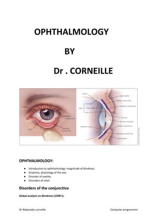
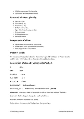
OPHTHALMOLOGY
BY
Dr . CORNEILLE
OPHTHALMOLOGY:
● Introduction to ophthalmology: magnitude of blindness.
● Anatomy, physiology of the eye.
● Disorder of eyelids.
● Disorders of orbit.
Disorders of the conjunctiva​​.
Global analysis on blindness (1990’s).
Dr Ndayisaba corneille Computer programmer
● 37 billion people are blind globally.
● 168 million people visually impaired.
Causes of blindness globally:
● Cataract (50%).
● Glaucoma. (12%).
● Trachoma.(3.5%)
● Diabetic retinopathy.
● Age related macular degeneration.
● Onchocerciasis.
● Childhood blindness
● Refractive errors.
Components of vision​​:
● Depth of vision (quantitative component)
● Width of the vision.(quantitative component).
● Colours (qualitative components)
Depth of vision:
The eye can see the object at a distance of a minimal angle of 5° but below 5° the eye loses its
visibility ie the visibility depends on the angle subtended by the object.
Assessment of vision by using Snellen’s chart​​:
A - 60 m
OMX - 36 m
Q,R,S,T, U - 24 m
A, N, O,S,P,T -18 m
X, Y,U,V,T, K - 12 m
O, X,Y,U,VX,U,V -6m ( normal vision​​)
Visual acuity, V.A = test distance/ test letter line read i.e. 6/60 =0.1
Visual acuity​​ is the ability of eye to determine the precise shape and details of the object.
Line read​​ is the line the patient stops the reading the letters.
A letter is adjusted if the patient fails to read.
Retina detects the movement of the hand and also detects light.
Dr Ndayisaba corneille Computer programmer
Categories of vision​​:
1 Normal vision​​ (6/8-6/18) according to W.H.O.
2 Visually impaired
i) Visually impaired is vision​​ of (6/24 -3/60)
ii) Severe visually impaired is vision​​ of (5/60 -3/60)
Blindness ​​ (2/60 –No perception of light.)
A blind person is any person of vision less than 3/60 (total blindness).
Trachoma​​ - cause poverty + ignorance.
Cataract cases are due to: inappropriate infrastructure and equipment for the eye.
Vision 2020:​​ is a strategy developed by W.H.O. and its partners to eliminate avoidable
blindness i.e.treatable,preventable.
Strategies:
● To provide cost effective disease intervention.
● To provide appropriate human resources for eye health.
● To provide appropriate infrastructure and equipment for eye care.
ANATOMY OF THE EYE:
The eye ball:
Is a dioptic apparatus . Its components include : content and the coat.
The coat is made up of the anterior 1/3 by the cornea . In posterior eyeball Is 2/3 layers.
Dr Ndayisaba corneille Computer programmer
The cornea:
Transparent, spherical structure.
Microscopically/ grossly:
The cornea in an adult has a horizontal diameter ​of 12mm​​ and vertical diameter of ​11mm.​​ The
cornea thickness is ​about 0.52mm​​ at the centre and about ​0.68mm​​ at periphery. Is
smaller in neonates​: 8mm horizontally and 8mm vertically​​.
Histologically:
Is made up 5 layers from outward to inward:
● Corneal epithelium
● Bowman’s membrane
● Stroma
● Descemet’s membrane
● Endothelium
The sclera:
Opaque non elastic outermost coating of the posterior 2/3 of the eye.Made up of loosely
arranged collagen fibres.
The choroid:
Pigmented,vascularised middle layer of the posterior 2/3 coating of the eye.
The Retina​​:
Innermost layer of the posterior 2/3 of the eyeball coating.
Key landmarks on the retina:
● Macula: depression on the temporal side of the retina- for light fixation.
● Optic disc/ nerve A.
● Blood vessels.
Contents of the eyeball​​:
Anteriorly- anterior chamber ; space bound anteriorly by cornea and posteriorly by iris and filled
with aqueous humour (clear,transparent fluid).
The iris:
Is a pigmented muscular tissue lying anteriorly to crystalline lens. Made up of 2 muscles:
innermost muscles are circular in nature and are called ​sphincter muscles​​- supplied by
parasympathetics​​. Outermost muscles are radial in nature and are called ​dilator
muscles- supplied by sympathetic nerve​​. The centre of the iris has got an aperture
(opening) ​called a pupil.
Posterior chamber:
Is a space lying behind the iris and behind the…… and is filled by vitreous humour.
The crystalline lens​​:
Dr Ndayisaba corneille Computer programmer
Bispherical ,elastic , transparent structure. Made up mainly of collagen fibres. Has 2 capsules in
the anterior and posterior surfaces. Lens is held in position by zonules (suspensory
ligaments) which are elastic fibres attached to the periphery of the eyeball coating.
Ciliary body​​:
3 sets of intrinsic muscles covered by epithelial lining i.e. ​horizontal muscle (outer), oblique
muscle (immediate),circular muscle (most inner).
Vitreous humour:
Innate transparent gel that fills the posterior 2/3 of the eyeball coating.
Conjunctiva:
Thin membranous epithelial lining of the anterior eyeball.
PHYSIOLOGY:
Cornea:
● Refraction- biggest refractive power because it is spherical and has interface of
tearfilm i.e. air-aqueous-cornea.
● Is transparent to ease light transmission.
Factors maintaining its transparency:
Anatomically:
● The cornea has no blood supply.
● Compact arrangement of cells + collagen fibres.
● Has got very fine nerve endings.
Physiologically:
● Cornea is kept relatively dehydrated by action of endothelial cells(active pseudo
Atphase pump)
Aqueous humour​​:
Transparent fluid filling both anterior and posterior chambers. Produced ciliary processes of
ciliary bodies by 2 mechanisms:
● Active transport mechanism.
● Ultrafiltration.
Composition of aqueous humour​​:
● Water, glucose, ions, oxygen, antioxidants like pyruvate.
● Tearfilm maintains intraocular pressure (normal 10-21 mmhg).
Iris and pupil:
2 functions​​:
Dr Ndayisaba corneille Computer programmer
● Accomodation and production of aqueous humour.
Crystalline lens:
3 functions:
● Is spherical and thus provides a refractive power of the eye.
● Transmission of light to the retina because it is transparent.
● Accommodation i.e. changing focus from distant to near.
Retina:
Neurosensory layer of the eye.Made up of 10 histological layers. Has 2 functions.
● Transduction (converting light energy into electrical stimuli)
● Initiation of the process of transmission.
Choroid:
Functions;
● Provides metabolic support to the retina
● Pigmented and absorbed light entering the retina
● Metabolism of vitamin A.
Sclera​​: maintains the eye size.
Dr Ndayisaba corneille Computer programmer
embryology of eyeembryology of eye  
Dr Ndayisaba corneille Computer programmer
Disorders of the eyelid:
Eyelids are highly vascularised. Both receive blood supply from branches of internal and external
carotid arteries. The venous drainage from the eyelids goes to the cavernous sinus.
Histologically​​, eyelids are made up of 8 layers from outward to inward.
● Skin
● Subcutaneous space
● Muscular layer
● Submuscular space
● Tarsal plate i.e. outer and inner lamellae.
● Muller muscle.
Eyelid appendages​​:
● Meibomian glands and eyelashes.
Dr Ndayisaba corneille Computer programmer
Functions of the eyelids
● Physical protection of the eyeball.
● Blinking reflex i.e.making tear flow across the eyeball. Meibomian glands produce tear film.
Disorders of the eyelids
Can be inflammation and infection.
Sty (external hordeleum):
● Is a localised inflammation of the outer lamellae of the eyelid. Commonly caused by
infection of the hair follicles of sebaceous glands caused by gram +ve organisms mainly by
staphylococci
Clinical manifestations:
● Localised, tender induration (shining swollen lesion) ending up in abscess.
RX: ​​by use of systemic antibiotics ​preferably penicillin, amoxicillin, ampicolox​​.
Simple analgesics ​like paracetamol​​.
Abscess is ​by incision and drainage (I+D)
Dr Ndayisaba corneille Computer programmer
Chalazion (external hordeleum):
● Obstruction of the meibomian glands leading to accumulation of lipid secretions in the
gland.
Clinical presentations:
● Localised, non-tender swelling of eyelids panting toward the conjunctiva side of the eyelid.
RX:​​ warm compress (2-3 days) as the initial remedy.Failure is by ​incision and curettage (I+C).
Herpes zoster ophthalmicus:
Re-activation of the primary infection of varicella zoster mainly in the ISS.
Presentations:
● Burning pain/sensation (right time for acyclovir tabs)
● Eruption
● Crustings
Dr Ndayisaba corneille Computer programmer
● Scars
Eruption of the tip on the tip of the nose​-Hutchison’s sign.
RX:​​ systemic antiviral agents i.e​. Acyclovir 800mg (10-14 days) and Acyclovir topically. Atropine​​ to
relieve pain. With pus, antibiotics are administered.Wounds should be dried, no application of
lotion.
Complications of Herpes zoster ophthalmicus:
● Keratitis
● Uveitis
● Entropion
● Post hepatic neurolysis.
Molluscum contagiosum:
● Viral infection of the eyelid. Is caused by a poxvirus. Highly contagious. It is common in
children.
● Typically presents ​with multiple /single umblicated lesions (nodules).
Dr Ndayisaba corneille Computer programmer
RX: Incision and curettage(I+C).
DISORDERS OF LID POSITION:
Ptosis:
● Drooping (lower than normal) of the upper eyelid.
Causes:
● Neurogenic i.e.those with 3​rd​
nerve palsy; those with Horner’s syndrome.
● Myogenic ptosis; those with myasthenia gravis.
● Mechanical ptosis i.e.tumours and masses.
Dr Ndayisaba corneille Computer programmer
Manifestations of Horner’s syndrome​​:
● Miosis
● Ptosis
● Anhydiosis.
Ptosis can be mild (3-4 mm), moderate (5-8 mm),severe (˃˃ 8 mm).
Problems with ptosis:
● Interferes with sight.
● Cosmetics/ disfiguring.
RX:​​ purely surgical. The approach depends on the function of elevator muscle.
Complications in children ˂˂ 6 yrs.
● Umblyopia ( permanent vision loss).
Entropion:
● Means inward turning of the eyelid.
Causes:
● Involution (weakening) i.e.aging.
● Cicatrical cause/ scaring /contractures on the conjunctival side of the eyelid.
Complication/danger:
● Trichiasis (eye lashes rubbing on the cornea).
RX: surgery. Simple mgt. tetracycline ointment for lubrication of the eye. Use of forceps to remove
eye lashes (epilation).
Ectropion:
● Means outward turning of the eye lid.
Causes:
● Involutional and cicatrical.
Complication:
Dr Ndayisaba corneille Computer programmer
Leaves the eye exposed (exposure keratopathy)
RX: lubricate the cornea using ointments. Use of shields and refer. Surgery.
Tumours of the eyelids​​:
● Benign tumours​​ i.e.papilloma and haemangiomas.
● Malignant tumours​​ i.e. basal cell carcinomas, squamous cell carcinomas and malignant
melanomas.
The orbit:
Anatomy:
Orbit is a bony structure and a socket for the eye ball.Is made up of bones and 4 walls.
Bones include​​:
● Frontal and sphenoid bones = roof of the orbit.
● Palatine and maxillary bones =floor.
● Ethmoid and maxillary bones =medial wall of orbit.
● Zygomatic bone = lateral wall of orbit.
DISORDERS OF ORBIT:
Differentials:​​ inflammation, thyroid eye disease and orbital tumours.
Inflammation​​:
Orbital cellulitis:
Causes:
● Arises from the extension of the surrounding structures.
● Arises from the surrounding sinuses.
● From tooth extraction
Dr Ndayisaba corneille Computer programmer
● Upper RTI.
Manifestations:
● Pain
● Swelling of the eyelid.
● Protrusion of the eye lid (proptosis)
● Fever ˃ 38° C
● Tender ness of eyelid
● Double vision/ diplopia.
● Chemosis (conjunctival edema)
Investigation/ assessment​​:
● Ultra sound scan of orbit.
● Blood for culture and sensitivity
● CT scan.
RX:​​ Use of systemic broad spectrum antibiotics ​preferably cephalosporins, IV quinolones, penicillin
+ gentamicin (IV) for gram –ves and gram +ves.
Complications:
● Abscess leading to osteomyelitis
● Meningitis
● Pus dislodging into cavernous sinus leading to cavernous sinus thrombosis.
Dr Ndayisaba corneille Computer programmer
Thyroid eye disease (Gravis diseas):
● Is an autoimmune disease. Can be associated with the system. Women at their 40’s are
more affected than men due to increased metabolism of glucose and disposition of fat into
the orbit pushing the eyeball out.
Manifestations:
● Proptosis
● Squinty eye / strabismus.
● Diplopia
Complications:
Pts lose their eyes from 2 mechanisms:
1 ​optic nerve compression
2 ​Exposure ophthalmopathy​​.
Investigation/assessment/ evaluation​​:
● Ulltra sound scan (fatty tissues + enlarged veins)
● Thyroxine levels are seen in orbit.
● CT scan.
RX:​​ simple mgt: underlying tissue is treated by giving antithyroid drugs i.e. iodine, systemic
steroids.Surgery by breaking the bone of the orbit. Radiation: to atrophy the tissue.Stitching of the
eyelid temporarily (tarsorrhhaphy) to prevent proptosis.
Orbital tumours​​:
Are divided into benign and malignant tumours.
Benign tumours include​​:
● Lacrimal gland tumours.
● Capillary maligioma.
● Carotid cavernous sinus.
● Dermal cyst.
Dr Ndayisaba corneille Computer programmer
Malignant tumours and primary and secondary​​. Primary malignant tumours include: lymphoma.
Secondary tumours​​:
● Retinoblastoma (cat’s glowing eye) a common cause of proptosis.
● Squamous cell carcinoma affects the orbit.
● Tumours of the nose affect the orbit.
● Metastatic tumours of the breast.
Diseases of the conjunctiva:
The conjunctiva​​:
Anatomy:
Is a thin membranous lining of the anterior segment of the eyeball. Bulbar conjunctiva lines the
surface of the sclera. Fornix conjunctiva is a loose conjunctiva. Palpebral conjunctiva lines the
posterior segment.
Histologically​​: the conjunctiva has 2 layers of epithelial cells. ​Blood vessels and lymphoid tissue​​.
Diseases:
Conjunctival inflammation​​.
Allergic conjunctiva = type I
Is chronic hypersensitivity. It is chronic / recurrent inflammation mediated by mast cells.
Clinically there 3 types of allergic reactions​​:
● Allergic Rhino conjunctivitis / seasonal allergic conjunctivitis.
● Vernal keratoconjunctivitis (very severe and can cause blindness)
● Atopic conjunctivitis (edema)
Vernal keratoconjunctivitis ( perennial ):
Manifestations:
● Red eye
● Chemosis.
Dr Ndayisaba corneille Computer programmer
● Itching.
● Tearing.
● Foreign body sensation in the eye,
Clinical findings/ examination:
● White spots around the limbus called follicles​​.
● Vegetations around the limbus lead to blindness.
● Papillae –nodular swellings on the upper tarsal of conjunctiva.
RX:​​ avoid contact with allergens (ideally) preventive measure.
● Treat the reactions/ inflammation with NSAIDS, SAIDS only at the time of inflammation +
mast cell stabilizers e.g​. sodium cromglycate and patanol​​.
Infections of the conjunctiva:
Bacterial conjunctiva​​: mainly caused +ve.
Manifestations:
● Foreign body sensation in the eye.
Dr Ndayisaba corneille Computer programmer
● Discharge – more purulent and copious discharge by Neisseria gonorrhae and mucoid watery
discharge ( chlamydia conjunctivitis)
● Painless.
RX:​​ start the pt on broad spectrum antibiotics- topical antibiotics.
Gonococcal conjunctivitis​​:
● Admission
● Drain the eye.
● Topical application after 30 min.
Viral conjunctivitis​​:
Comes in epidemics.
Manifestations:
● Photophobia.
● Red eye.
● Pain.
● Serous discharge- bilateral.
RX:​​ drop of atropine.
Pterygium:
● Wedg-shaped fibrovascular proliferation of the conjunctiva that extends onto the cornea.
Risk factors​​:
● Exposure to ultraviolet radiation (in tropical zones).
● Exposure to dust ( postulated )
Reasons for RX of pterygium​​:
● Dry eye.
● Recurrent painful red eye.
● Cosmetic problem to some people.
RX:​​ purely surgical excision.
Dr Ndayisaba corneille Computer programmer
​Pingueculum​​:
● Yellowish deposit of necrosis of conjunctiva.
● Normally localisd and enlarge slowly.
● Interferes with tear film leading to recurrent red eye.
RX:​​ give tear substitutes.
Correction: hard deposits on the palpebral conjunctiva due to shedding of epithelium cell.
Tumours:
Benign tumours:
● Lipodesmoid cyst
● Dermoid cyst
● Squamous cell carcinoma.
Presentations of squamous cell carcinoma​​:
● White / irregular pigmented surface of conjunctivitis
● Can metastatasize.
RX:​​ surgical excision with margin + absolute alcohol if detected early. Alcohol kills malignant cells.
Squamous cell carcinoma:
Dr Ndayisaba corneille Computer programmer
Lacrimal system:
Major function​​: production of tears and drainage of the eye.
Types of lacrimal glands:
1 ​Main lacrimal gland:
About 5 mm in diameter. Is located in superotemporal portion. Is a tubuloacini gland.
The main mechanism of tear production is via ​pinocytosis​​. The gland is responsible for the
production of aqueous portion of tear. Production of reflex tears is caused by irritants and emotions.
2 Accessory glands​​:
Located in the superior fornix of the conjunctiva​. Basal tear production​​ is the function of accessory
glands.
Dr Ndayisaba corneille Computer programmer
Assessment of the function of lacrimal glands​​:
Use of Schirmer test- test I​​ measures total tear production and ​test II​​ measures the basal tear
production (the most accurate).
Expectation of the test​​: 10-33mm of strip wetting in 5 min.
Diseases of the gland:
Dacryoadenitis (inflammation)
Causes:
● Viral infections
● Autoimmune
● TB. And syphilis.
Presentations:
● Painful swollen gland that causes mild proptosis which can be upward proptosis, downward
proptosis and medial proptosis.
RX: systemic​​ corticosteroids.
Dr Ndayisaba corneille Computer programmer
Tumours :​​ can be benign-called lacrimal gland adenomas.
Presentation: ​​slow progressive proptosis of the eye.(inferomedial proptosis)
Mgt:​​ total excision,
Malignant tumours(lacrimal gland adenocarcinoma​​):
● Usually develop from benign tumour.
● Pt. presents with progressive painful proptosis.
● Tumour invades the surrounding tissues.
RX:​​ excision and follow it with radiotherapy.
Drainage system​​:
Is made up of systems of tubes from lid margin upto nostril. The components include:
● Nasal lacrimal duct
● Punctum
● Canaliculus and lacrimal sac.
Mechanism of drainage is a function of ocular orbicularis.
● Contraction of ocular orbicularis forces the lacrimal sac to secret tears.
● Relaxation creates the –ve pressure in the sac leading to tear in drawing.
Tearfilm:
Made up of 3 structures/ components:
● Lipid layer/waxy –produced by meibomian gland.
● Aqueous layer
● Mucin layer.
Lipid layer​​:
● Causes the outer surface to prevent evaporation of the tear. Produced by meibomian gland.
Dr Ndayisaba corneille Computer programmer
Mucin layer of tear film:
● Produced by globlet cells of conjunctiva.it lowers the surface tension so that it reduces its
wettability.
Functions of the tear film:
● Lubrication of the eye.
● Metabolic support to the cornea.
● Contains immunoglobulins and lysosomes for defence against micro-organisms.
● Provides refractive function to the cornea.
● Removes debris or foreign bodies within the eye.
Disorders of the drainage system:
Tearing consequence of :obstruction and over production.
● Lacrimation:​​ is due to over production of tear.
● Epiphora ​​ :Is tearing due to obstruction in the lacrimal drainage system.
Causes of epiphora​​:
● Canaliculus obstruction
● Punctual occlusion
● Nasal lacrimal sac obstruction.
Presentations of epiphora​​:
● Continuous tearing.
Dr Ndayisaba corneille Computer programmer
● Eye mucoid discharge.
● Regurgitation of the pus when pressure is applied on the lacrimal sac.
● Painless swelling of lacrimal sac (chronic dacryocystitis)
Mgt:​​ adult or any body above 2 yrs.-probing and syringing.
Surgery​​- dacryocystorhinostomy (DCR).
RX of congenital nasal tearing of a child below 2 yrs.-messaging​​.
The cornea:
Disorders:
Corneal inflammation/ infection (keratitis​​)
Infective causes​​:
Bacterial keratitis follows a trauma.Bacteria do not invade cornea except Neisseria gonoccus , H.
influenzae and C. Diptheriae.
Presentations:
● Acute on set of pain in the eye.
● Red eye.
● Poor vision.
● Corneal haziness.
Examination​​:
Fluorescent corneal staining:
RX: of bacterial keratitis​​:
A )​Broad spectrum antibacterial agents​​:
1 Ceft eye drop.
2 ciprofloxacin eye drop
3 gentamicin
4 chloramphenicol eye drop in that order.
2 hourly for mild bacterial keratitis and 30 min for severe infection.
Dr Ndayisaba corneille Computer programmer
B​) Pain mgt. atropine eye drop​​.
Complications:
● Bacterial keratitis may heal with scaring leading to blindness.
● Corneal perforation
● Thin walled cornea leading to corneal herniation- staphyloma.
● Endophthalmitis.
Fungal keratitis​​:
Follows injury to the eye with vegetative matter or pts treated with herbs.
Presentations:
● Acute on set of pain in the eye.
● Red eye
● Poor vision
● Corneal haziness.
RX:​​ broad spectrum antifungal agents :
● Voriconazole eye suspension.
● Natamycin eye drop.
Dr Ndayisaba corneille Computer programmer
Viral keratitis​​:
Common causes​​:
● Herpes simplex type I.
● Varicella zoster virus.
Presentations:
● White eye.
● Little Pain.
● Reduced vision.
● Reduced corneal sensation.
RX:​​ broad spectrum agents. Acyclovir eye ointment (x5 a day 10-14/7 days.)
Protozoan infection​​:
Causes:
● Acanthamoeba. Common in people who use contact lenses.
Immunological corneal keratitis​:
Ulceration of cornea near the limbus. Typically presents ​with hanging edges​​.
RX​​: topical corticosteroids eye drop. Destroy the blood vessels around the limbus.
Dr Ndayisaba corneille Computer programmer
The uvea:
Uvea are pigmented vascularised layers of the eye.(iris, ciliary body and choroid)
Disorders are mainly inflammatory and some tumours. Inflammation of uvea is called uveitis.
Dr Ndayisaba corneille Computer programmer
Anatomical classification of the inflammation:
Anterior uveitis​​: involves the iris and or ciliary body. Can also be described to as iritis, cyclitis
or iridocyclitis (iris +ciliary body)
Causes of anterior uveitis​​:
● Range from idiopathy 30%-50% of cases to immunological cases such as juvenile idiopathic
arthritis, rheumatoid arthritis.
● May occur following infections like viral infections as in Herpes simplex type I.
● May occur following bacterial infections as in TB.
● May be a manifestation of retinoblastosus- masqueriding uveitis.
Manifestations of anterior uveitis​​:
● Acute on set of pain.
● Poor vision.
● Red eye.
On examination/ clinical findings:
● Reduced visual acuity.
● Keratic precipitates (deposits of WBC on the epithelium of cornea.)
● Flare/cloudiness in anterior chamber due to proteins.
● Pupil constriction.
● + secondary glaucoma
● Presence of cells.
RX:​​ use of topical costicosteroids. Pain is managed by use of atropine eye drop (prevents the
adhesion of eyelid on the cornea.
Complications:
● Adhesion of the iris on to the cornea leads to : anterior synachia and posterior synachia
● Glaucoma develops due anterior and posterior synachiae.
● Cataract due to interference with metabolism of the lens by inflammation.
Posterior uveitis:
Inflammation of the choroid. Always extend to the retina thus called chorioretinitis.
Causes:
Dr Ndayisaba corneille Computer programmer
● Range from idiopathy 30%-50% of cases to immunological cases such as juvenile idiopathic
arthritis, rheumatoid arthritis.
● May occur following infections like viral infections as in Herpes simplex type I.
● May occur following bacterial infections as in TB.
● May be a manifestation of retinoblastosus- masqueriding uveitis.
● May occur following protozoan infections like toxoplasmosis.
Presentations:
● Sudden reduction in vision.
● Painless.
On examination/ clinical findings​​:
● Reduced visual acuity.
● White eye.(conjunctiva).
● Cells in the vitreous (significant findings when using an ophthalmoscope)
RX:​​ use systemic corticosteroids 1 mg/kg daily. Titrate with the response i.e. increase the dose or
decrease the dose.
Complications:
● Macular edema
● Optic neuritis
● Tractional retinal detachment.
Tumours:​​ include choroidal melanoma.
The lens:
● Has got a nucleus in an adult.
Dr Ndayisaba corneille Computer programmer
● Has posterior and anterior capsules.
● Has got a cortex.
● Epithelial cells pump water actively a way.
Disorders of the lens​​:
Cataract:
Is any opacity of the lens.
Dr Ndayisaba corneille Computer programmer
Causes of cataract:
● Aging process commonest cause in adults because proteins that make up the lens get
denatured with environmental influence.
● Metabolic conditions like diabetes.
● Prolonged use of corticosteroids.
● Blunt or penetrated tauma.
● Exposure to certain types of radiation.
● Hypoparathyroidism.
● Can develop secondary to any intraocular disease e.g. uveitis.
In children:
● Can be genetic.
● Congenital abnormalties esp. intrauterine infection with toxoplasmosis. WHAT DOES THIS
‘TOOCHE’ STAND FOR?
Cataract in adults:
Presentations:
● Gradual painless loss of vision.
● Complaint of glares/ scattering of light.
● Rainbow- coloured rings or haloes.
Assessment/ evaluation:
● Visual acuity.
● Density of the cataract.
● Pupillary reaction which can be:
a) Direct light
b) Indirect light
c) Swing test.
● Ultra sound scan.
RX:​​ mainly surgery. When should surgery be done​? When the cataract interferes with the person’s
routine activities.
History of cataract surgery:
Couching ​​followed ​by intracapsular cataract extraction (I.C.C.E.) +
spectacles​​. This was followed ​by I.C.C.E. + Anterior chamber intraocular
lens(I.C.C.E.+ ACIOL )​​ This RX.is strictly recommended for the old people. It was then
followed ​by Extracapsular cataract extraction + posterior chamber
Dr Ndayisaba corneille Computer programmer
intraocular lens (E.C.C.E.+ PCIOL.)​​ This RX is mainly recommended for the young
people though recently recommended for the old people. It is followed by ​Small incision
cataract surgery (SICS) + PCIO​​L.It is the standard for cataract surgery in Uganda and
other countries. The last ​is Phacoemulsfication (Phaco) + PCIOL​​. In Phaco, a small
cut is made at the limbus and the lens is irrigated with saline. Phaco prevents astigmatism.
Small incision cataract surgery (SICS) + PCIOL
Phacoemulsfication (Phaco)
Complications of the surgery:
● Retinal detachment
Dr Ndayisaba corneille Computer programmer
● Hyphaema
● Vitreous tear
● Post operative infections.
● Postoperative endophthalmitis which can be prevented by use of iodine immediately after
surgery.
● Posterior capsule opacification.
● Cystoid macular edema.
Cataract in children:
Children are born with the potential to develop vision. The period for visual development is between
birth and 6 years​​. Above 6 years without vision, no vision at all develops. Amblyopia results- ‘lazy
eye’. ​Amblyopia is vision reduction due to a lack of visual stimulation during the critical period of
visual development in the absence of any organic lesion in the visual pathway​​.
Causes of amblyopia​​:
● Squinty of the eye.(strabismus).
● Cataract
● Ptosis.
● Refractive errors.
RX​​: surgery – lens wash out.
DX​​: leukocoria(white pupil)
Causes:
● Cataract
● Retinal detachment
● Retinocolomboma
● In children: retinoblastoma.
Disorders of lens position​​:
Sublaxation:​​ lens is in anatomical position but partially detached.
Dislocation​​: lens completely detached and moved a way from its anatomical position.
Description of the status of the eye in relation to lens:
Phakic eye​​: natural lens present.
Dr Ndayisaba corneille Computer programmer
Aphakic eye​​: no lens in the eye.it is x-terised by:
● Iridodonesis
● Black pupil
● Deep anterior chamber.
Pseudoaphakic eye​​: artificial lens present.
Correction of aphakia:
● Contact lens.
Public health eye related conditions:
Trachoma:
● Is a chronic keratoconjunctivitis caused by chlamydia trachomatis which may lead to
blindness via corneal opacification.
● Trachoma is prevalent in most parts of developing world esp. Africa, Asia,and some parts of
Latin America.In Uganda,it is common ​in Busoga, Karamoja and some parts of N. Uganda​​.
Globally about ​3 million​​ are blind with trachoma. About ​24 million​​ are at risk of blindness​.
Dr Ndayisaba corneille Computer programmer
120 million​​ are affected. Trachoma is prevalent in communities with high levels of
ignorance. It is a disease of poverty and ignorance.
Epidemiology of trachoma:
The disease shows the mild form called ​inclusion blennorrhoea​​ and severe form called
hyperendemic trachoma​​. Its epidemiology is summarised into 4 F’s and 4 D’s.
4 D’s:
● Dry
● Dusty
● Dirty
● During
4 F’s:
● Flies
● Fingers
● Feces
● Filth
The disease affects females more than men -3:1
Microbiology:
● Chlamydia trachomatis has a viral and bacterial x-tics. It is microscopic. It is an intracellular
organism.
As a bacterium:
● Multiplies by binary fission and responds to antibiotics.
Serotypes A-K.
● A-C cause trachoma
● D-k cause paratrachoma – upper respiratory tract + genital infections.
Pathophysiology:
● The main infective particle of trachoma is elementary body and has got a high affinity for
epithelial cells of cornea and the conjunctiva and after entering the epithelial cells it takes up
host cytoplasm and becomes inclusion body then bursts releasing more elementary
particles/ bodies.
Dr Ndayisaba corneille Computer programmer
o Recurrent infection leads to scaring of conjunctiva leading to
entropion-trichiasis-corneal ulceration –corneal opacification. Blindness due to
trachoma is only found in adults.
Clinical features:
W.H.O.classified clinical features into 5 stages:
Stage I:
TF- Trachomatous follicles​​. There must be at least five follicles on the upper tarsal plate + some
papillae but the blood vessels must be visible through them.
Stage II:
TI- Trachomatous inflammation intense​​. Many follicles and papillae but the conjunctival blood
vessels are not seen through them.
Dr Ndayisaba corneille Computer programmer
Stage III:
TS- Trachomatous conjunctival scaring.​​ White scars are present on the upper tarsal plate.
Stage IV:
TT- Trachomatous trichiasis.​​ Some eyelashes rubbing against the cornea.
Dr Ndayisaba corneille Computer programmer
Stage V:
CO- Corneal opacification​​.
DX ​​of trachoma is clinically based on history.
Lab DX:
Micobiological prepa ration;
● Giemsa stain
● Culture and sensitivity.
Community DX of trachoma​​:
● TT- 5% of all adults in the area with TT , the area is endemic.
● TF- 20% of the 10 yrs. Old children with TF, the area is endemic.
● 1/1000 women who are blind with CO , the area is endemic.
Control and prevention strategy​​:
It is abbreviated as ​‘SAFE’​​.
S=​​ Surgery (Bilamellar lid rotation).
A=​​ Antibiotics i.e. Azithromycin, tetracycline rifamcin to eradicate the active infection.
F=​​ Facial hygiene /washing.
Dr Ndayisaba corneille Computer programmer
E=​​ Education and environmental importance.
Onchocerciasis:
● Is a blindness condition caused by onchocerca volvulus (Nematode)
● Lives in 2 hosts i.e.human (definitive ) and an indefinitive host,simulium fly that lives around
fast flowing water inorder to get oxygen for its eggs. Black fly can fly ​about 160 Km on both
sides of the river. Adult worm has a life span ​of 14 yrs.
● Adult worm constantly produces microfilaria i.e. ​type I​​ that circulates ​in the blood​​ and it is
sucked by the black fly and ​becomes type II in its abdomen​​ .When it reaches ​the thoracic
muscles of the fly it becomes type III.
● Clinical manifestations of onchocerciasis are due to the death of the microfilaria which
results into release of toxins. Manifestations can be ocular and non ocular. Only 20% of the
pts develop ocular problem but the remaining suffer systemic problems.
Dr Ndayisaba corneille Computer programmer
STAGES
Dr Ndayisaba corneille Computer programmer
Non ocular manifestations:
● Intense puritus (itching)
● Leopard skin
● Lizard skin
● Hanging groin
● Nodules
● Epilepsy
Ocular manifestations:
● Live microfilaria on the cornea.
● Keratitis
● Chorioretinits
● Anterior uveitis
Complications arising from anterior uveitis:
● Glaucoma
● Cataract optic nerve atropy
● Corneal opacity
Prevention and control:
RX is​​ by the use of the drug ​called Ivermectin (mectizin​​) and can be used for ​prevention 125µg
/kg-800µg/kg.
Advantages of the drug:
● Wide therapeutic dose.
● Increases libido
● Given as a single dose per year and the RX lasts for 14 yrs.
Environmental prevention​​:
Spray the environment the simulium flies along the river 160 km on both sides.
Dr Ndayisaba corneille Computer programmer
Xerophthalmia:
● Is a misnoma (dry eye).
● Refers to the clinical manifestations of vitamin A deficiencies in the eye.
● Vitamin A is a fat soluble vitamin is important for epithelial lining maintenance.
● Vitamin A is essential for mucin production anywhere in the body esp. respiratory system.
● In essence out of the 100 pts suffering from Vitamin A deficiency:
50% die
25%​​ of children with Vit A deficiency recover but not fully.
25%​​ will recover fully.
Vitamin A in the body​​:
● Ingestion (not produced by the body but can be ingested.)
Dr Ndayisaba corneille Computer programmer
● Absorption (absorbed in form of retinol)
● Liver (for storage leaves the liver when bound to retinol binding protein .Adults liver stores
vitaminA but children’s liver doesnot. Vitamin A then enters the eye via epithelial lining.
Sources of vitamin A:
Animal sources​​- preformed or ready (retinol readily absorbed)
Plant sources​​-carotinoid ( can first be converted to retinol in G.I.T. before absorption and
transportation to the liver.
Rich sources of vitamin A: for a plant.
● Palm oil
● Carrots
● Green leafy vegetables
● Yellow fruits e.g. bananas ,mangoes
● Yellow sweet potatoes.
For animals​​:
● Fish liver oil.
● Animal liver
● Cheese
● Egg.
Role of vit A in the eye​​:
● Vit A is responsible for the production of outer segment of rhodopsin
● Plays a role in the corneal and conjunctival lining development.
Clinical manifestations​​:
The W.H.O. gave a category of vit.A deficiency- xerophthalmia.
Stages of vitamin A deficiency in the eye.
XN-​​ Night blindness​​ (earliest manifestation in children)
X1 A-​​ Conjunctival xerosis (dryness)
X1 B-​​ Bitot’s spot (whitish lesion on the conjunctiva)
X2 A-​​ Corneal xerosis.
Dr Ndayisaba corneille Computer programmer
X2 B-​​ Corneal ulceration ˂ 1/3 of the corneal circumference
X3 ​​– Keratomalacia (corneal melting)/ corneal ulceration ˃ 1/3 of the corneal circumference.
XF-​​ Xerophthalmic fundus degeneration.
XS​​- Xerophthalmic corneal scar.
Note: stages X1A to X2A are reversible if vit A isgiven​​.
Reasons why vit.A deficiency is more prevalent in children than in
adults​​:
Children’s liver does not have the capacity to store vit​​.A but adult’s liver can store it for a minimum
of 6 yrs.
In terms of ingestion:
● Ignorance in children
● Anorexia due to other disesases.
● Lack of vit A
In terms of absorption:
● Malabsorption
● Liver diseases
● P.E.M.of any other cause.
Children have got high metabolic demand​​ i.e.the children require 133 iµ while adults require 36 Iµ
Prevention and control of vitamin A deficiency​​:
Key strategies​​:
● Vitamin A supplementation​​:
e.g. vit ​A capsules 100,000 Iµ -˂˂ 1 yr. and 200,000 Iµ -˃˃ 1 yr​​. Given at least ​every 6 months​​ in routine
immunisation of children.
Opportunistic approach​​: any child suffering from other condition gets vit.A.
● Health education; how vit.A is prepared , what kind of food is rich in vit.A.
● Promotion of agriculture and horticulture.
● Fortification of food- addition of vitamin A into essential foods.
● Immunise all the children against immunisable diseases.
Dr Ndayisaba corneille Computer programmer
Glaucoma:
● Is the second leading cause of blindness globally.
● Blindness from it is irreversible
● Is a preventable form of blindness.
Dr Ndayisaba corneille Computer programmer
● Dfn: is a group of diseases having in common optic neuropathy with x-tic optic disc changes
and visual field constriction.
Pathophysiology:
● If there is an imbalance between aqueous humour and drainage ,pressure is built up
intraocularly. This pressure causes direct pressure on the eye structures e.g. the lens. This
pressure also causes increased pressure on the optic nerve and causes ischemia.
Classification:
Open angle glaucoma or angle closure glaucoma​​.
● An eye is said to have open angle glaucoma if there are glaucomatous damages in the eye in
presence of widely open angle of anterior chamber.
● An eye is said to have angle closure glaucoma if there are glaucomatous damages in the eye
in the presence of a closed angle of the anterior chamber.
● Primary glaucoma​​ is one where there are glaucomatous changes in the eye in the absence
of any pathology causing the changes.
● Secondary glaucoma​​ is one where there are glaucomatous damages in the eye in the
presence of pathology producing the changes.
Thus glaucoma is classified as​​:
● Primary open angle glaucoma (POAG)
● Secondary open angle glaucoma
● Primary angle closure glaucoma (PACG)
● Secondary angle closure glaucoma
● Congenital glaucoma
Primary open angle glaucoma (POAG) /chronic simple glaucoma:
Is and eye with glaucomatous changes in presence of a widely open angle of anterior chamber in
absence of any pathology causing the glaucomatous changes in the eye.
Risk factors of POAG:
● Increased intraocular pressure
● Age of 40 yrs. And above.
● Race; black race is a risk factor.
● Those with myopia.
Presentations of POAG:
Dr Ndayisaba corneille Computer programmer
● Is described as the silence thief of sight.
● Gradual painless progressive loss of vision.
Assessment/ evaluation of POAG:
● Detect the physical damage done by the glaucoma i.e. by fundoscopy/ ophthalmoscopy
Clinical findings:
✓ Increased cup to disc ratio (0.8). Normal disc ratio= 0.2-0.5.
✓ Decreased nueral rim
✓ Shift of blood vessels to one side (nasal side)
✓ Pallor of disc
✓ Notching of cup margin
2 Tonometry​​- measuring of intraocular pressure. Has two types
Applanation​​ e.g. goldmann, tonopen, perkins, pust.
Dr Ndayisaba corneille Computer programmer
Indentation ​​e.g. schiotz.
3 visual field assessment (perimetry​​). Has 2 types.
Confrontation​​: compare the pt’s visual field with yours i.e. comparing your right eye with that of the
pt. Useful when the pt. has got grossly field vision problem. All quadrants are assessed i.e. upper,
lower sides.
Automated​​:
4 ​Gonioscopy​​- measure of the depth of the angle of anterior chamber.
5 slit lamp Biomicroscopy​​ – investigates the pathology causing the glaucomaous changes.
RX of POAG:
Medical RX: categories of drug used
1 Adrenergic ββ-blockers​​ e.g. timolol
2 prostaglandin analogs​​: e.g. lantanaprose/ xalatan
3 ​Carbonic anhydrase inhibitors​​ e.g. acetazolamide
4 ​Miotics (parasympathomimetics​​) e.g. pilocarpine
5 ​Sympathomimetics ​​e.g. adrenaline drop.
Surgical options​​:
Types:
1 ​Trabeculectomy (siltration​​).
2 Laser
L=​​ light
A​​= Amplification
S=​ Stimulated
E=​​ Emission
Dr Ndayisaba corneille Computer programmer
R=​​ Radiation
3 Glaucoma implants/ bulbs for glaucoma that has failed​​.
Secondary open angle glaucoma​​:
Angle is open but there are pathologic obstructions.
Examples of secondary open angle glaucoma​​ :
● Hyphaema
● Anterior uveitis
● Pharcolytic uveitis
● Pseudo exfoliation syndrome
● Pigment dispersion syndrome
Examples of secondary angle closure glaucoma​​:
● Anterior synchia
● Posterior synchia
● Intumescent lens (swollen)
● Angle recession glaucoma.
Primary angle closure glaucoma (PACG):
There are glaucomatous changes with the closed angle in the anterior chamber but in the absence of
the pathology causing the changes.
Risk factors of PACG:
● Being of Asian origin.
● Hypermetropia.
● Being a female.
Provocative factors of PACG:
● Darkness
● Excitement and bereavement
● Looking downwards
● Pharmacological factors e.g. atropine put directly on the pupil.
Dr Ndayisaba corneille Computer programmer
Presentations of PACG:
Classified into:
Subacute attacks/ earliest attacks:
● Mild eye pain
● Bluring of vision
● Haloes
Clinical findings:
● Decreased visual acuity.
● Corneal edema
● Intraocular pressure is slightly raised (22- 33 mmHg)
● Shallow anterior chamber.
RX:​​ lower intraocular pressure by using acetazolamide and pilocarpine
Definitive RX: ​peripheral iridectomy
Acute angle closure attack​​:
Clinical presentations​​:
● Sudden on set
● Severe eye pain
● Very red eye
● Poor vision
● Nausea and vomiting
Clinical findings​​:
● Limbal injection.
● Severe corneal edema.
● Increased intraocular pressure (50- 100 mmHg)
● Iridocorneal contact.
● Pupil is semi-dilated and oval
Dr Ndayisaba corneille Computer programmer
RX:​​ in the acute phase the initial RX is to rapidly lower intraocular pressure. Drugs of choice :
osmotic agents which include manitol, glycerine, urea and isosorbide. These agents can be combined
with pilocarpine and acetazolamide.
Definitive RX​: peripheral iridectomy​​.
Childhood glaucoma​​:
Categorised into:
● Congenital/ infantile glaucoma​​- glaucoma detected from birth upto 1 yr. of age.
● Juvenile glaucoma​​: any glaucoma detected from 1 yr.and above .
Congenital glaucoma​​:
Is inherited as an autosomal gene. Affects male children than female children. Result of
developmental abnormality of the angle of the anterior chamber.
It presents with classical triad ( prominent and 3). The triad include​​:
● Lacrimation
● Photophobia
● Blepharospasm.(constriction of eyelids)
Findings on examination​​:
● Enlarged eyeball (buphthalmos) with a black centre.
● Corneal haziness
● Descemet’s membrane cracks (Haab striate)
Assessment :
Assessed under general anaesthesia. Examine the following:
● Corneal diameter is measured (9.5- 10.5mm)
● Measure intraocular pressure (12- 16 mmHg)
● Performe refraction of the eye to determine the refractive state of the eyeball.
● Do fundoscopy.
RX is mainly surgical. 3 surgical approaches​​:
● Goniotomy
● Trabeculotomy​​- make holes via trabecular mesh
● Trabeculectomy​​.
Dr Ndayisaba corneille Computer programmer
Strabismus (squints):
Is the misalignment of the eye.Normally the eyes should be facing the same direction
Classification:
1 ​manifestation​​- may be manifested as : I ​) Tropia​​ ii​) latent- phoria​​.
2 ​Direction of deviation​​ i.e. hyperdeviation ,hypodeviation, esodeviation and exodeviation. The
conditions: hypertropia , hypotropia, esotropia and exotropia.
Cuases of strabismus​​:
● Congenital in children.
● Refractive errors.
● Cranial nerve palsy iii, iv, and vi.
● Trauma.
● Thyroid eye disease.
● Orbital tumours.
● Cataract
● Retinoblastoma.
Assessment/ evaluation​​:
Considerations:
● Assess for visual acuity.
● History:
✓ Onset/ duration.
✓ Trauma
Dr Ndayisaba corneille Computer programmer
✓ Change in head position.
● Examination:
✓ Angle of deviation: corneal light and prisms.
✓ Cover- uncover test
✓ Ocular motility
✓ Refraction
✓ Complete eye examination + fundus.
Complication of strabismus:
● Amblyopia.
Mgt:
● Correct the cause i.e. if it is a refractive error
● Correct the amblyopia by patching / occluding the good eye to stimulate the affected eye.
● Surgery:
Principle​​: strengthening by resection and weakening by recession i.e. the principle operates by
strengthening the muscle and weakening the muscle.
Refractive errors
(ammetropia):
● Dioptre is a measure of refractive power of the refractive surface. In the normal eye, the rays
are focused on the retina.
Emmetropia:
Dr Ndayisaba corneille Computer programmer
● Is a state of an eye at rest where parallel rays of light from an object are brought into focus
on the retina while the ammetropia is a state of an eye at rest where parallel rays of light
cannot be brought into focus.
Types of ammetropia:
● Myopia
● Hypermetropia
● Astigmatism
● Presbyopia.
Myopia:
Is a state of an eye at rest where parallel rays of light from an object are brought into focus in front
of the retina.
Causes:
● Naturally large eyeball.
● Increased refractive power of the lens as in the diabetes.
Presentations:
● Inability to see distant objects
● No pain.
Correction of myopia:
a) Clinical refraction​​: is a procedure of determining the refractive power of an eye. Clinical
refractions are of 2 types:
Objective refraction​​- does not require the co-operation of the pt. Can be done by using a
retinoscope or autorefractor.
Subjective clinical refraction​​- requires the co-operation of the patient.
b) –ve/ diverging lens​​. Give the weakest –ve power lens to give the best correction.
Hypermetropia:
An eye is said to be hypermetropic if at rest parallel rays of light from an object are brought into
focus behind the retino.
Dr Ndayisaba corneille Computer programmer
Causes:
● Small eyeball.
Presentations:
● Long sightedness
● Pain or headache when doing near work
● Eye becomes red.
Mgt​​:
● Clinical refractions.
● +ve or converging lens. Rule: give the strongest power that gives you the best correction.
Astigmatism:
Is a state of an eye when at rest parallel rays of light from the different media cannot be brought
into single focus. It is subdivided into:
● Myopic astigmatism
● Hypermetropic astigmatism
● Compound astigmatism
Mgt:
● Clinical refractions.
● Correct with cylindrical lens.
Presbyopia:
Is the accommodation defect due to loss of elasticity of the crystalline lens from the aging process.
Correction​​:
● +ve or converging lenses.
● As for the rule of thumb: you lose + 1 dioptre at 40 yrs. And for every sequential 5 yrs.of age
you lose 0.5 dioptre.
Dr Ndayisaba corneille Computer programmer
The retina:
Histologically is made up of 10 layers from inward to outward
● Inner limiting membrane
● Nerve fibre layer
● Ganglionic cell layer
● Inner nuclear layer
● Inner plexiform layer
● Outer nuclear layer
● Outer plexiform layer
● Photoreceptor layer
● Outer limiting layer
● Retinal pigmented epithelium layer.
Disorders:
● Retinal vascular diseases:
● Diabetic retinopathy
● Hypertensive retinopathy
Dr Ndayisaba corneille Computer programmer
Diabetic retinopathy​​:
● DM is a microangiopathy due to persistent elevation of blood glucose level. There are 2
types: types I- insulin dependent and type II- non insulin dependent.
Pathophysiology:
● Persistent elevation of blood glucose levels causes viscosity of blood and damages the
endolium and pericytes of capillaries.
● Results: thromboscis . leakage of vessels . thromboscis leads to ischaemia of distal tissue and
leakage
Presentations;
● Diabetic retinopathy is divided into​: non proliferative phase and proliferative phase​​.
Non proliferative phase presents with:
● Hard exudate
● Cotton wool spots
● Microanuerism
● Dot- blot hemmorrhages
● Macular edema
● Flamed shaped hemmorrhages.
Proliferative stage (2​​nd​​
) :​​ there is growth of new cells on the retina.
Mgt:
● Control of the blood sugar levels
● Pain retinal laser photocoagulation
Dr Ndayisaba corneille Computer programmer
● Administration of intravitreal antivascular endothelial growth factor.
Complications arising from diabetic retinopathy​​:
● Vitreous haemorrhage
● Tractional retinal detachment
● Neovascular glaucoma
● Central retinal vein occlusion.
Hypertensive retinopathy:
Systemic hypertension may develop ocular manifestations.
Grades of hypertensive retinopathy:
I- Attenuated retinal vessel (copper/ silver wiring)
II- A- V nipping.
III- Splinter haemorrhage
IV- Papilloedema.
Risk factors for diabetic retinopathy:
● Duration of diabetes
● Hypertension
● Pregnancy
● Renal disease.
Mgt:
Dr Ndayisaba corneille Computer programmer
Control BP.
Retinal tumours:
Retinoblastoma;
Most common intraocular malignancy in children world wide with an incidence in 1: 10 in 20,000 life
span. Is due to a defect in the retinoblastoma suppressive gene.
Types of retinoblastoma​​:
● Sporadic type​​: accounts for 80% of cases of retinoblastoma and it is unilateral in 60% of the
cases.
● Inherited type​​: accounts for 20-40% of the retinoblastoma cases is bilateral in ---
Presentations of retinoblastoma:
● Leukocoria
● Strabismus
● Uveitis
● Orbital cellulitis
● Proptosis
RX:​ early small intraocular tumours; local focal RX is advised which is in the form of
photocoagulation or cryotherapy.
Other RX modalities:
● Focal radiotherapy
● Chemotherapy
● Beam radiotherapy
● Enucleation (remove the eye.)
Dr Ndayisaba corneille Computer programmer
The peak age of presentation of retinoblastoma ​is 18 yrs​​.,however,bilateral presents by ​12 months
and unilateral presents later ​by 24 months​​.
Retinoblastoma metastasizes to the lymphatic system, submandibular and pre-auricular lymph
nodes and the brain.
Retinal detachment​​:
Is the separation of the retina from the retinal epithelium by fluid.
Types:
Rhegmatogenous detachment:
Associated with holes/ tears on the retina.
Non rhegmatogenous detachment:
Can be tractional or exudative retinal detachment.
Rhegmatogenous detachment​​:
Causes of tears/ holes on the retina:
● Trauma
● Peripheral degeneration
● Myopia.
Presentations:
● Sudden onset of painless loss of vision
Dr Ndayisaba corneille Computer programmer
● May see floaters.
DX:​​ fundoscopy to locate the tear.
RX:​​ for small detachment ,focal RX is used i.e. pars plannaviret.
Inherited retinal diseases​​:
Retinitis pigmentosa​​:
Is an inherited retinal disease which normally presents in the 2​nd​
decade of life.
Mode​​:
● Autosomal mode
● Sex linked mode.
Presentations:
● Presents in the 2​nd​
decade of life.
● Poor night vision
● Becomes completely blind by 4​th​
decade of life.
● Disease is bilateral.
Clinical findings​​:
Classical triad are:
● Attenuated retinal blood vessels.
● Bone spicule retinal pigmentation in the periphery.
● Pale waxy optic disc.
No known RX:
Thus,counselling and visual rehabilitation are done.
Dr Ndayisaba corneille Computer programmer
Visual pathway:
Relations of the visual pathway​​:
● Optic nerve is confined within the optic canal
● The optic chiasma lies on top of the pituitary fossa. On its sides are venous cavernosa
● Optic tract which lies in the mid brain and surrounded by the brain.
Disorders :
Optic nerve:
Made up of 4 components:
● Intraocular portion (optic disc )
● Intraorbital (longest and takes a sinous course)
● Intracanalicular
● Intracranial.
Intra-orbital portion:
● Is the longest portion
● Takes the sinous course (tortuous course). This enables it to let the eyeball move freely.
● Found within the orbit.
Intra- canalicular portion:
● Found within the optic canal
● Measures 8-10 mm in length.
● Covered by the meninges which continue with those of the brain.
Dr Ndayisaba corneille Computer programmer
Intra-cranial portion;
● Found in the anterior cranial fossa.
● In optic nerve lies the ophthalmic artery that eventually forms the central retinal artery that
branches to supply the eye but the ophthalmic vein passes through superior orbital fissure.
Inflammation and infections of optic nerve​​:
Any inflammation or infection is termed ​optic neuritis​​.
Causes of optic neuritis:
● Viral infection :
Viruses include:
✓ Herpes simplex type I
✓ HIV
✓ Measles virus
✓ Herpes zoster ophthalmicus
● Immunological causes which include: multiple sclerosis and sarcoidosis
● Idiopathic (30-50%)
● Bacterial infections as in TB and syphilis
● Protozoal infections as toxoplasmosis.
Presentations of optic neuritis:
● Poor vision
● Dull pain eye movement.
Clinical findings:
● Decreased visual acuity.( mild to severe)
● Abnormal pupillary reaction(relative afferent pupil defect- only one eye.)
In papillitis (bilateral​​): swollen hyperemic optic nerve.
In retrobulbal neuritis​​ the optic nerve is normal . optic neuritis in adults are usually unilateral but it
may be bilateral in children. All the time papilloedema is bilateral most of the time optic neuritis is
unilateral.
RX of optic neuritis:
Dr Ndayisaba corneille Computer programmer
● Use of NSAID: ​ideally iv methylprednisone 1mg/kg​​ .Give oral prednisolone .Dose adjusted
depending on the response. The drug does not alter the outcome of the disease but it
quickens the outcome
● The key indicators for the outcome is V.A. at the time of presentation i.e.if pt.presents with
good V.A., the vision is going to be good but if it is bad at the time of presentation, the vision
will be poor.
Complication:
Optic atropy(sets in btn 4- 8 wks).
Papilloedema:
Refers to swollen optic nerve head due to increased intra-cranial pressure. Papilloedema is not a
diagnosis but a sign. Is always bilateral.
Causes of papilloedema(DX):
● Brain tumours
● Brain inflammation like meningitis
● Hydrocephalus
● Severe systemic hypertension
● Head injury with subdural hematomas.
RX: treat the cause.
Congenital nerve abnormalties​​:
There are a number of them and include:
● Optic nerve coloboma
● Optic nerve dysgenesis
● Morning glory syndrome.
Dr Ndayisaba corneille Computer programmer
The visual field:
Is the inverted presentation of the arrangement of the nerve fibers in the retina in space i.e.
temporal retina is responsible for nasal field of vision and vice versa.
Abnormalties:
Chiasma lesions- crossing fibres are destroyed and the ….
Pupillary pathway:
The pupillary pathway shares the common pathway with….
Pupillary pathways is divided into 2 ways:
● Input- afferent pathway
● Output- efferent pathway.
Dr Ndayisaba corneille Computer programmer
Clinical applications​​:
Pupillary reaction to light.
Assessments: 3 types:
● Direct pupillary reaction to light ;you shine light to right eye the right pupil constricts.
● Consensual/ indirect pupillary reaction; shining the light to the right eye you look at the left
pupil.
● Swinging light test; for relative afferent pupil defect.
Key stimuli in the test
● Darknest
● Accommodation
Environment for pupillary reaction assessments​​:
● Room must be darkened.
● Pt. must focus at a distance to remove the effect of accommodation.
Relationship btn pupil and vision​​:
● Pupillary reaction can be used to localised lesions in the brain(cortical diseases)
● Abnormalty of the optic nerve
Ocular and adnexal trauma:
Ocular and adnexal trauma
Dr Ndayisaba corneille Computer programmer
● Physical injury
● Mechanical
● Thermal
● Radiation
● Chemical – acids and alkali
Eye trauma is one of the leading cause of blindness
● Most eye injuries are occupational related.
● Boys are more prone to injuries than girls.
● Age group affected 3-15 yrs. And 70 yrs.+. Eye injuries are divided into mechanical and
physical
Mechanical causes of eye injuries are divided into blunt and penetrating injuries.
Blunt injuries to conjunctiva​​:
Cause:
● Subconjunctival haemorrhage which requires no RX ,just assure the pt.
● Conjunctival lacerations: small lacerations require no RX but extensive conjunctival
lacerations with loose tissue may require excision.
● Conjunctival foreign bodies; a no. of foreign bodies get dislodged on/ beneath the upper
tarsal conjunctiva .These may be metallic foreign bodies, vegetative foreign bodies ,sand or
soil. Foreign bodies on the upper tarsal conjunctiva may cause corneal abrasion and thus
require removal .Removing a foreign body from the upper tarsal conjunctiva one must evert
the upper eyelid.
Dr Ndayisaba corneille Computer programmer
The cornea:
Blunt trauma to the cornea may cause corneal abrasion. Pts.with corneal abrasion should be treated
like those with corneal ulcers. Blunt trauma may cause corneal perforation esp. at the limbus. Any
pt. presenting at the unit with no specialised care service must promptly be referred after putting a
shield on the eye.
Definitive RX​​: depends on the extent of the perforation. Small perforations suture the eye and put it
back.Extensive perforations ,suturing may initite inflammation. The eye has to be removed by
evisceration.
● Loose foreign bodies on the cornea can be removed using cotton swabs.
● Intra-corneal foreign bodies should be removed by using a microscope.
Globe injuries​​:
● Blunt injuries to the globe result into a no.of damages to intra-ocular content.
● The iris tear which leads to bleeding into anterior chamber (hyphaema)
Hyphaema:
Causes of hyphaema​​:
● Blunt and penetrating injuries.
● Bleeding disorders.
● Neovascularisation
● Use of anticoagulants e.g. warfarins
● Intra-ocular tumour(retinoblastoma).
Mgt of hypaema​​:
● It is first graded which is based on how much level of anterior chamber is field with blood
i.e.100% for total hyphaema, 75% ,25%.
˂˂ 100% hyphaema:
● Bed rest with head elevated at 45°. This prevents bleeding.Use of topical anti-inflammatory
drugs e.g. atropine.
Dr Ndayisaba corneille Computer programmer
100% hyphaema​​:
● Surgical evacuation of blood.
Complications of hyphaema​​:
● Secondary glaucoma
● Corneal endothelial staining i.e.in 100% hyphaema.
Other globe injuries​​:
Lens disorder
Intra-ocular foreign bodies- metallic and vegetative foreign bodies must be removed but glass
foreign bodies are innate and thus may be left.
Eyelid lacerations:
Dr Ndayisaba corneille Computer programmer
● Lacerations elsewhere on the eyelid
● Lacerations involving lid margin(cannot be tried by c/o)
● Lacerations involving the canalicular system (only a specialist)
● Lacerations involving tissue losss( if the laceration is ˂ 1/3 direct apposition can be applied if
it is btn 1/3- 50%).
Orbital injuries​​:
Occur in both blunt and penetrating injuries:
Blunt trauma:
When a blunt object hits the eye, iit sits on the eyeball and the pressure is then transmitted to the
weakest wall(medial wall). This trauma is called ​a blow out.
Presentations of a blow out:
● Sunken eyeball (enophthalmos)
● Squints
● Cryptations of the eyelid (escape of air)
● Loss of taste in the tongue.
Mgt.​​ depends on the extent of the fracture. In small fractures ,normally you can observe . for bigger
fractures you need to reconstruct by using the silicon plate.
​
Complications from blunt trauma:
● Hyphaema
● Retinal tear at its periphery
● Retinal edema
● Rupture of the eyeball.
Complications from penetrating injuries:
● Scar on the cornea
● Intra-ocular infections
● Sympathetic ophthalmitis
● Uveitis,glaucoma
● Retinal detachment.
Dr Ndayisaba corneille Computer programmer
Chemical injuries:
CHEMICAL INJURIES
- Chemical can be
- Solids
- Powders
- Moisture
- Steam
Vary in severity from a trivial and transient irritation of little significance to complete and
sudden loss of vision.
Domestic chemicals
- Detergents
- Disinfectants and solvents
- Cosmetic
- Breaches
Factors that determine severity of the injury
- P.H
- Volume of chemical delivered
- Duration of contact
- Inherent toxicity of particular chemical.
Modes of injury
Occur due to external contacts with chemicals following:-
- Domestic accidents with domestic chemicals
- Agricultural accidents to fertilizers insecticides etc
- Chemical laboratory accidents attacks especially with acids to disfigure faces
- Self inflicted chemical injuries seen in malingerers and psycho paths.
Types of chemicals
- Serious chemical burns are mainly from acids and alkalis
- Acids and alkalis have a high content of hydrogen and hydroxyl ions.
Dr Ndayisaba corneille Computer programmer
Alkali burns
- Most sever chemical injury
- Common alkalis include
● Lime
● Caustic potash or caustic soda
● Liquid ammonia- most harmful.
Mechanism of damage
- Alkaline burns cause increased P.H and cause carbonifation of fatty acids. This leads to
cellular and cell membrane destruction.
- Being hygroscopic​, alkalis extract water from the cells contributing to total necrosis.
- Alkalis​ combine with ​cellular lipids​ to produce soluble compounds (soap) which make
the area of contact soft and gelatinous.
- Stem cells at limbus​ get damaged leading to scarring and conjunctivazation of the cornea.
- The above mechanism results in an increased deep penetration of alkalis into tissues.
- Alkalis therefore spread widely, their actions continuing for some days and their effects
become difficult to circumscribe.
Clinical features in alkaline burns
1) Stage of acute ischemic necrosis
- Conjunctiva is edematous
● Congested
● Widespread necrosis
● Copious purulent discharge
- Wide spread corneal sloughing of epithelium, corneal edema and stromal opalescence
body become granulated
2) Stage of regeneration
- Conjunctiva and corneal epithelium regenerate
This leads to corneal vascularization and iris inflammation subsides.
3) Stage of complication
- Symblepharon formation
- Recurrent corneal ulceration
- Complicated glaucoma also develops.
Dr Ndayisaba corneille Computer programmer
Acid burns
- Less serious than alkaline burns
- Common acids
● Sulfuric acid
● Hydrochloric acid
● Nitric acid
Mechanism of damage
- Acids cause coagulation of proteins which then act as barrier to prevent deeper
penetration of acids into the tissues. Thus lesion becomes sharply demarcated.
Ocular damage
- Necrosis with sloughing of conjunctiva
- Corneal necrosis with sloughing of epithelium.
Classification of chemical burns​​.
- Depends upon severity of damage caused to limbus and cornea
- Roper -hall classification.
Grade Corneal appearance Limbal Ischemia Visual prognosis.
I Clear cornea Nil Good
II Hazy but iris details
visible
< 3
1
Good
III Opaque, iris details
occurred
-3
1
2
1
Doubtful
IV Opaque, no view of
iris and pupil
> 2
1
Poor
Treatment of chemical burns
1) Immediate and thorough irrigation of the affected eye with clean water or normal
saline tubing with at least 2 liters.
NB ​​Do not waste time with history taking. It can be done after irrigation.
2) Mechanical removal of contaminant.
- Use swab stick to remove remaining particles ie in case of lime
3) Removal of contaminated and necrotic tissues.
- Necrotic conjunctiva should be exercised.
Use cotton swabs to remove necrotic corneal epithelium.
4) Maintain favorable conditions for rapid and uncomplicated healing by applying:
Dr Ndayisaba corneille Computer programmer
- Topical atropine
- Corticosteroids
- Lubricants
5) Prevention of symblepharon
- Serial glass rodding to break fibrosis
6) Treatment of complications
- 2​0​
glaucoma with anti glaucoma drugs
- Poor corneal healing with limbal stem cell deficiency treatment with aminotic membrane
transplant with/without stem cell transplant.
- Pseudo pterygium – exicision
- Corneal opacity treatment by kerato plasty
- Kerato prosthesis is a surgical option in severely damaged eyes.
THERMAL INJURIES
- Usually caused by fire or hot fluids
- Lids are the main area of injury but in severe injuries, the conjunctiva and cornea may be
affected treatment is with atropine, steroids antibiotics and lubricants.
ELECTRICAL INJURIES
- Strong electrical current within area of eyes may cause.
● Congested conjictiva
● Punctuate or diffuse interstial opacities of the cornea
● Inflamed iris and ciliary body
● Electric cataract 2-4 months after accident.
● Multiple retina haemorrhages
● Optic nerve neuritis.
RADIATIONAL INJURIES
1) Ultraviolet radiation
Cause
- Photo opthalmia (multiple corneal epithelia erosion
- Senile contaract.
2) Intrared radiations
- Cause solar mucular burn.
3) Ionizing radiational injuries.
Caused followed radiotherapy to tumors in the vicinity of the eyes
- Common ocular lesions include
● Radiation kerato conjunctivitis
● Radiation dermatis of the lids
Dr Ndayisaba corneille Computer programmer
● Radiation contract.
Akoon Andrea​​ is responsible for ​any mistake made​​ as far as these notes are
concerned.​Dr.Onyango​​ cannot be ​blamed in any way​​.
Dr Ndayisaba corneille Computer programmer

Ophthalmology

  • 1.
    OPHTHALMOLOGY BY Dr . CORNEILLE OPHTHALMOLOGY: ●Introduction to ophthalmology: magnitude of blindness. ● Anatomy, physiology of the eye. ● Disorder of eyelids. ● Disorders of orbit. Disorders of the conjunctiva​​. Global analysis on blindness (1990’s). Dr Ndayisaba corneille Computer programmer
  • 2.
    ● 37 billionpeople are blind globally. ● 168 million people visually impaired. Causes of blindness globally: ● Cataract (50%). ● Glaucoma. (12%). ● Trachoma.(3.5%) ● Diabetic retinopathy. ● Age related macular degeneration. ● Onchocerciasis. ● Childhood blindness ● Refractive errors. Components of vision​​: ● Depth of vision (quantitative component) ● Width of the vision.(quantitative component). ● Colours (qualitative components) Depth of vision: The eye can see the object at a distance of a minimal angle of 5° but below 5° the eye loses its visibility ie the visibility depends on the angle subtended by the object. Assessment of vision by using Snellen’s chart​​: A - 60 m OMX - 36 m Q,R,S,T, U - 24 m A, N, O,S,P,T -18 m X, Y,U,V,T, K - 12 m O, X,Y,U,VX,U,V -6m ( normal vision​​) Visual acuity, V.A = test distance/ test letter line read i.e. 6/60 =0.1 Visual acuity​​ is the ability of eye to determine the precise shape and details of the object. Line read​​ is the line the patient stops the reading the letters. A letter is adjusted if the patient fails to read. Retina detects the movement of the hand and also detects light. Dr Ndayisaba corneille Computer programmer
  • 3.
    Categories of vision​​: 1Normal vision​​ (6/8-6/18) according to W.H.O. 2 Visually impaired i) Visually impaired is vision​​ of (6/24 -3/60) ii) Severe visually impaired is vision​​ of (5/60 -3/60) Blindness ​​ (2/60 –No perception of light.) A blind person is any person of vision less than 3/60 (total blindness). Trachoma​​ - cause poverty + ignorance. Cataract cases are due to: inappropriate infrastructure and equipment for the eye. Vision 2020:​​ is a strategy developed by W.H.O. and its partners to eliminate avoidable blindness i.e.treatable,preventable. Strategies: ● To provide cost effective disease intervention. ● To provide appropriate human resources for eye health. ● To provide appropriate infrastructure and equipment for eye care. ANATOMY OF THE EYE: The eye ball: Is a dioptic apparatus . Its components include : content and the coat. The coat is made up of the anterior 1/3 by the cornea . In posterior eyeball Is 2/3 layers. Dr Ndayisaba corneille Computer programmer
  • 4.
    The cornea: Transparent, sphericalstructure. Microscopically/ grossly: The cornea in an adult has a horizontal diameter ​of 12mm​​ and vertical diameter of ​11mm.​​ The cornea thickness is ​about 0.52mm​​ at the centre and about ​0.68mm​​ at periphery. Is smaller in neonates​: 8mm horizontally and 8mm vertically​​. Histologically: Is made up 5 layers from outward to inward: ● Corneal epithelium ● Bowman’s membrane ● Stroma ● Descemet’s membrane ● Endothelium The sclera: Opaque non elastic outermost coating of the posterior 2/3 of the eye.Made up of loosely arranged collagen fibres. The choroid: Pigmented,vascularised middle layer of the posterior 2/3 coating of the eye. The Retina​​: Innermost layer of the posterior 2/3 of the eyeball coating. Key landmarks on the retina: ● Macula: depression on the temporal side of the retina- for light fixation. ● Optic disc/ nerve A. ● Blood vessels. Contents of the eyeball​​: Anteriorly- anterior chamber ; space bound anteriorly by cornea and posteriorly by iris and filled with aqueous humour (clear,transparent fluid). The iris: Is a pigmented muscular tissue lying anteriorly to crystalline lens. Made up of 2 muscles: innermost muscles are circular in nature and are called ​sphincter muscles​​- supplied by parasympathetics​​. Outermost muscles are radial in nature and are called ​dilator muscles- supplied by sympathetic nerve​​. The centre of the iris has got an aperture (opening) ​called a pupil. Posterior chamber: Is a space lying behind the iris and behind the…… and is filled by vitreous humour. The crystalline lens​​: Dr Ndayisaba corneille Computer programmer
  • 5.
    Bispherical ,elastic ,transparent structure. Made up mainly of collagen fibres. Has 2 capsules in the anterior and posterior surfaces. Lens is held in position by zonules (suspensory ligaments) which are elastic fibres attached to the periphery of the eyeball coating. Ciliary body​​: 3 sets of intrinsic muscles covered by epithelial lining i.e. ​horizontal muscle (outer), oblique muscle (immediate),circular muscle (most inner). Vitreous humour: Innate transparent gel that fills the posterior 2/3 of the eyeball coating. Conjunctiva: Thin membranous epithelial lining of the anterior eyeball. PHYSIOLOGY: Cornea: ● Refraction- biggest refractive power because it is spherical and has interface of tearfilm i.e. air-aqueous-cornea. ● Is transparent to ease light transmission. Factors maintaining its transparency: Anatomically: ● The cornea has no blood supply. ● Compact arrangement of cells + collagen fibres. ● Has got very fine nerve endings. Physiologically: ● Cornea is kept relatively dehydrated by action of endothelial cells(active pseudo Atphase pump) Aqueous humour​​: Transparent fluid filling both anterior and posterior chambers. Produced ciliary processes of ciliary bodies by 2 mechanisms: ● Active transport mechanism. ● Ultrafiltration. Composition of aqueous humour​​: ● Water, glucose, ions, oxygen, antioxidants like pyruvate. ● Tearfilm maintains intraocular pressure (normal 10-21 mmhg). Iris and pupil: 2 functions​​: Dr Ndayisaba corneille Computer programmer
  • 6.
    ● Accomodation andproduction of aqueous humour. Crystalline lens: 3 functions: ● Is spherical and thus provides a refractive power of the eye. ● Transmission of light to the retina because it is transparent. ● Accommodation i.e. changing focus from distant to near. Retina: Neurosensory layer of the eye.Made up of 10 histological layers. Has 2 functions. ● Transduction (converting light energy into electrical stimuli) ● Initiation of the process of transmission. Choroid: Functions; ● Provides metabolic support to the retina ● Pigmented and absorbed light entering the retina ● Metabolism of vitamin A. Sclera​​: maintains the eye size. Dr Ndayisaba corneille Computer programmer
  • 7.
    embryology of eyeembryologyof eye   Dr Ndayisaba corneille Computer programmer
  • 8.
    Disorders of theeyelid: Eyelids are highly vascularised. Both receive blood supply from branches of internal and external carotid arteries. The venous drainage from the eyelids goes to the cavernous sinus. Histologically​​, eyelids are made up of 8 layers from outward to inward. ● Skin ● Subcutaneous space ● Muscular layer ● Submuscular space ● Tarsal plate i.e. outer and inner lamellae. ● Muller muscle. Eyelid appendages​​: ● Meibomian glands and eyelashes. Dr Ndayisaba corneille Computer programmer
  • 9.
    Functions of theeyelids ● Physical protection of the eyeball. ● Blinking reflex i.e.making tear flow across the eyeball. Meibomian glands produce tear film. Disorders of the eyelids Can be inflammation and infection. Sty (external hordeleum): ● Is a localised inflammation of the outer lamellae of the eyelid. Commonly caused by infection of the hair follicles of sebaceous glands caused by gram +ve organisms mainly by staphylococci Clinical manifestations: ● Localised, tender induration (shining swollen lesion) ending up in abscess. RX: ​​by use of systemic antibiotics ​preferably penicillin, amoxicillin, ampicolox​​. Simple analgesics ​like paracetamol​​. Abscess is ​by incision and drainage (I+D) Dr Ndayisaba corneille Computer programmer
  • 10.
    Chalazion (external hordeleum): ●Obstruction of the meibomian glands leading to accumulation of lipid secretions in the gland. Clinical presentations: ● Localised, non-tender swelling of eyelids panting toward the conjunctiva side of the eyelid. RX:​​ warm compress (2-3 days) as the initial remedy.Failure is by ​incision and curettage (I+C). Herpes zoster ophthalmicus: Re-activation of the primary infection of varicella zoster mainly in the ISS. Presentations: ● Burning pain/sensation (right time for acyclovir tabs) ● Eruption ● Crustings Dr Ndayisaba corneille Computer programmer
  • 11.
    ● Scars Eruption ofthe tip on the tip of the nose​-Hutchison’s sign. RX:​​ systemic antiviral agents i.e​. Acyclovir 800mg (10-14 days) and Acyclovir topically. Atropine​​ to relieve pain. With pus, antibiotics are administered.Wounds should be dried, no application of lotion. Complications of Herpes zoster ophthalmicus: ● Keratitis ● Uveitis ● Entropion ● Post hepatic neurolysis. Molluscum contagiosum: ● Viral infection of the eyelid. Is caused by a poxvirus. Highly contagious. It is common in children. ● Typically presents ​with multiple /single umblicated lesions (nodules). Dr Ndayisaba corneille Computer programmer
  • 12.
    RX: Incision andcurettage(I+C). DISORDERS OF LID POSITION: Ptosis: ● Drooping (lower than normal) of the upper eyelid. Causes: ● Neurogenic i.e.those with 3​rd​ nerve palsy; those with Horner’s syndrome. ● Myogenic ptosis; those with myasthenia gravis. ● Mechanical ptosis i.e.tumours and masses. Dr Ndayisaba corneille Computer programmer
  • 13.
    Manifestations of Horner’ssyndrome​​: ● Miosis ● Ptosis ● Anhydiosis. Ptosis can be mild (3-4 mm), moderate (5-8 mm),severe (˃˃ 8 mm). Problems with ptosis: ● Interferes with sight. ● Cosmetics/ disfiguring. RX:​​ purely surgical. The approach depends on the function of elevator muscle. Complications in children ˂˂ 6 yrs. ● Umblyopia ( permanent vision loss). Entropion: ● Means inward turning of the eyelid. Causes: ● Involution (weakening) i.e.aging. ● Cicatrical cause/ scaring /contractures on the conjunctival side of the eyelid. Complication/danger: ● Trichiasis (eye lashes rubbing on the cornea). RX: surgery. Simple mgt. tetracycline ointment for lubrication of the eye. Use of forceps to remove eye lashes (epilation). Ectropion: ● Means outward turning of the eye lid. Causes: ● Involutional and cicatrical. Complication: Dr Ndayisaba corneille Computer programmer
  • 14.
    Leaves the eyeexposed (exposure keratopathy) RX: lubricate the cornea using ointments. Use of shields and refer. Surgery. Tumours of the eyelids​​: ● Benign tumours​​ i.e.papilloma and haemangiomas. ● Malignant tumours​​ i.e. basal cell carcinomas, squamous cell carcinomas and malignant melanomas. The orbit: Anatomy: Orbit is a bony structure and a socket for the eye ball.Is made up of bones and 4 walls. Bones include​​: ● Frontal and sphenoid bones = roof of the orbit. ● Palatine and maxillary bones =floor. ● Ethmoid and maxillary bones =medial wall of orbit. ● Zygomatic bone = lateral wall of orbit. DISORDERS OF ORBIT: Differentials:​​ inflammation, thyroid eye disease and orbital tumours. Inflammation​​: Orbital cellulitis: Causes: ● Arises from the extension of the surrounding structures. ● Arises from the surrounding sinuses. ● From tooth extraction Dr Ndayisaba corneille Computer programmer
  • 15.
    ● Upper RTI. Manifestations: ●Pain ● Swelling of the eyelid. ● Protrusion of the eye lid (proptosis) ● Fever ˃ 38° C ● Tender ness of eyelid ● Double vision/ diplopia. ● Chemosis (conjunctival edema) Investigation/ assessment​​: ● Ultra sound scan of orbit. ● Blood for culture and sensitivity ● CT scan. RX:​​ Use of systemic broad spectrum antibiotics ​preferably cephalosporins, IV quinolones, penicillin + gentamicin (IV) for gram –ves and gram +ves. Complications: ● Abscess leading to osteomyelitis ● Meningitis ● Pus dislodging into cavernous sinus leading to cavernous sinus thrombosis. Dr Ndayisaba corneille Computer programmer
  • 16.
    Thyroid eye disease(Gravis diseas): ● Is an autoimmune disease. Can be associated with the system. Women at their 40’s are more affected than men due to increased metabolism of glucose and disposition of fat into the orbit pushing the eyeball out. Manifestations: ● Proptosis ● Squinty eye / strabismus. ● Diplopia Complications: Pts lose their eyes from 2 mechanisms: 1 ​optic nerve compression 2 ​Exposure ophthalmopathy​​. Investigation/assessment/ evaluation​​: ● Ulltra sound scan (fatty tissues + enlarged veins) ● Thyroxine levels are seen in orbit. ● CT scan. RX:​​ simple mgt: underlying tissue is treated by giving antithyroid drugs i.e. iodine, systemic steroids.Surgery by breaking the bone of the orbit. Radiation: to atrophy the tissue.Stitching of the eyelid temporarily (tarsorrhhaphy) to prevent proptosis. Orbital tumours​​: Are divided into benign and malignant tumours. Benign tumours include​​: ● Lacrimal gland tumours. ● Capillary maligioma. ● Carotid cavernous sinus. ● Dermal cyst. Dr Ndayisaba corneille Computer programmer
  • 17.
    Malignant tumours andprimary and secondary​​. Primary malignant tumours include: lymphoma. Secondary tumours​​: ● Retinoblastoma (cat’s glowing eye) a common cause of proptosis. ● Squamous cell carcinoma affects the orbit. ● Tumours of the nose affect the orbit. ● Metastatic tumours of the breast. Diseases of the conjunctiva: The conjunctiva​​: Anatomy: Is a thin membranous lining of the anterior segment of the eyeball. Bulbar conjunctiva lines the surface of the sclera. Fornix conjunctiva is a loose conjunctiva. Palpebral conjunctiva lines the posterior segment. Histologically​​: the conjunctiva has 2 layers of epithelial cells. ​Blood vessels and lymphoid tissue​​. Diseases: Conjunctival inflammation​​. Allergic conjunctiva = type I Is chronic hypersensitivity. It is chronic / recurrent inflammation mediated by mast cells. Clinically there 3 types of allergic reactions​​: ● Allergic Rhino conjunctivitis / seasonal allergic conjunctivitis. ● Vernal keratoconjunctivitis (very severe and can cause blindness) ● Atopic conjunctivitis (edema) Vernal keratoconjunctivitis ( perennial ): Manifestations: ● Red eye ● Chemosis. Dr Ndayisaba corneille Computer programmer
  • 18.
    ● Itching. ● Tearing. ●Foreign body sensation in the eye, Clinical findings/ examination: ● White spots around the limbus called follicles​​. ● Vegetations around the limbus lead to blindness. ● Papillae –nodular swellings on the upper tarsal of conjunctiva. RX:​​ avoid contact with allergens (ideally) preventive measure. ● Treat the reactions/ inflammation with NSAIDS, SAIDS only at the time of inflammation + mast cell stabilizers e.g​. sodium cromglycate and patanol​​. Infections of the conjunctiva: Bacterial conjunctiva​​: mainly caused +ve. Manifestations: ● Foreign body sensation in the eye. Dr Ndayisaba corneille Computer programmer
  • 19.
    ● Discharge –more purulent and copious discharge by Neisseria gonorrhae and mucoid watery discharge ( chlamydia conjunctivitis) ● Painless. RX:​​ start the pt on broad spectrum antibiotics- topical antibiotics. Gonococcal conjunctivitis​​: ● Admission ● Drain the eye. ● Topical application after 30 min. Viral conjunctivitis​​: Comes in epidemics. Manifestations: ● Photophobia. ● Red eye. ● Pain. ● Serous discharge- bilateral. RX:​​ drop of atropine. Pterygium: ● Wedg-shaped fibrovascular proliferation of the conjunctiva that extends onto the cornea. Risk factors​​: ● Exposure to ultraviolet radiation (in tropical zones). ● Exposure to dust ( postulated ) Reasons for RX of pterygium​​: ● Dry eye. ● Recurrent painful red eye. ● Cosmetic problem to some people. RX:​​ purely surgical excision. Dr Ndayisaba corneille Computer programmer
  • 20.
    ​Pingueculum​​: ● Yellowish depositof necrosis of conjunctiva. ● Normally localisd and enlarge slowly. ● Interferes with tear film leading to recurrent red eye. RX:​​ give tear substitutes. Correction: hard deposits on the palpebral conjunctiva due to shedding of epithelium cell. Tumours: Benign tumours: ● Lipodesmoid cyst ● Dermoid cyst ● Squamous cell carcinoma. Presentations of squamous cell carcinoma​​: ● White / irregular pigmented surface of conjunctivitis ● Can metastatasize. RX:​​ surgical excision with margin + absolute alcohol if detected early. Alcohol kills malignant cells. Squamous cell carcinoma: Dr Ndayisaba corneille Computer programmer
  • 21.
    Dr Ndayisaba corneilleComputer programmer
  • 22.
    Lacrimal system: Major function​​:production of tears and drainage of the eye. Types of lacrimal glands: 1 ​Main lacrimal gland: About 5 mm in diameter. Is located in superotemporal portion. Is a tubuloacini gland. The main mechanism of tear production is via ​pinocytosis​​. The gland is responsible for the production of aqueous portion of tear. Production of reflex tears is caused by irritants and emotions. 2 Accessory glands​​: Located in the superior fornix of the conjunctiva​. Basal tear production​​ is the function of accessory glands. Dr Ndayisaba corneille Computer programmer
  • 23.
    Assessment of thefunction of lacrimal glands​​: Use of Schirmer test- test I​​ measures total tear production and ​test II​​ measures the basal tear production (the most accurate). Expectation of the test​​: 10-33mm of strip wetting in 5 min. Diseases of the gland: Dacryoadenitis (inflammation) Causes: ● Viral infections ● Autoimmune ● TB. And syphilis. Presentations: ● Painful swollen gland that causes mild proptosis which can be upward proptosis, downward proptosis and medial proptosis. RX: systemic​​ corticosteroids. Dr Ndayisaba corneille Computer programmer
  • 24.
    Tumours :​​ canbe benign-called lacrimal gland adenomas. Presentation: ​​slow progressive proptosis of the eye.(inferomedial proptosis) Mgt:​​ total excision, Malignant tumours(lacrimal gland adenocarcinoma​​): ● Usually develop from benign tumour. ● Pt. presents with progressive painful proptosis. ● Tumour invades the surrounding tissues. RX:​​ excision and follow it with radiotherapy. Drainage system​​: Is made up of systems of tubes from lid margin upto nostril. The components include: ● Nasal lacrimal duct ● Punctum ● Canaliculus and lacrimal sac. Mechanism of drainage is a function of ocular orbicularis. ● Contraction of ocular orbicularis forces the lacrimal sac to secret tears. ● Relaxation creates the –ve pressure in the sac leading to tear in drawing. Tearfilm: Made up of 3 structures/ components: ● Lipid layer/waxy –produced by meibomian gland. ● Aqueous layer ● Mucin layer. Lipid layer​​: ● Causes the outer surface to prevent evaporation of the tear. Produced by meibomian gland. Dr Ndayisaba corneille Computer programmer
  • 25.
    Mucin layer oftear film: ● Produced by globlet cells of conjunctiva.it lowers the surface tension so that it reduces its wettability. Functions of the tear film: ● Lubrication of the eye. ● Metabolic support to the cornea. ● Contains immunoglobulins and lysosomes for defence against micro-organisms. ● Provides refractive function to the cornea. ● Removes debris or foreign bodies within the eye. Disorders of the drainage system: Tearing consequence of :obstruction and over production. ● Lacrimation:​​ is due to over production of tear. ● Epiphora ​​ :Is tearing due to obstruction in the lacrimal drainage system. Causes of epiphora​​: ● Canaliculus obstruction ● Punctual occlusion ● Nasal lacrimal sac obstruction. Presentations of epiphora​​: ● Continuous tearing. Dr Ndayisaba corneille Computer programmer
  • 26.
    ● Eye mucoiddischarge. ● Regurgitation of the pus when pressure is applied on the lacrimal sac. ● Painless swelling of lacrimal sac (chronic dacryocystitis) Mgt:​​ adult or any body above 2 yrs.-probing and syringing. Surgery​​- dacryocystorhinostomy (DCR). RX of congenital nasal tearing of a child below 2 yrs.-messaging​​. The cornea: Disorders: Corneal inflammation/ infection (keratitis​​) Infective causes​​: Bacterial keratitis follows a trauma.Bacteria do not invade cornea except Neisseria gonoccus , H. influenzae and C. Diptheriae. Presentations: ● Acute on set of pain in the eye. ● Red eye. ● Poor vision. ● Corneal haziness. Examination​​: Fluorescent corneal staining: RX: of bacterial keratitis​​: A )​Broad spectrum antibacterial agents​​: 1 Ceft eye drop. 2 ciprofloxacin eye drop 3 gentamicin 4 chloramphenicol eye drop in that order. 2 hourly for mild bacterial keratitis and 30 min for severe infection. Dr Ndayisaba corneille Computer programmer
  • 27.
    B​) Pain mgt.atropine eye drop​​. Complications: ● Bacterial keratitis may heal with scaring leading to blindness. ● Corneal perforation ● Thin walled cornea leading to corneal herniation- staphyloma. ● Endophthalmitis. Fungal keratitis​​: Follows injury to the eye with vegetative matter or pts treated with herbs. Presentations: ● Acute on set of pain in the eye. ● Red eye ● Poor vision ● Corneal haziness. RX:​​ broad spectrum antifungal agents : ● Voriconazole eye suspension. ● Natamycin eye drop. Dr Ndayisaba corneille Computer programmer
  • 28.
    Viral keratitis​​: Common causes​​: ●Herpes simplex type I. ● Varicella zoster virus. Presentations: ● White eye. ● Little Pain. ● Reduced vision. ● Reduced corneal sensation. RX:​​ broad spectrum agents. Acyclovir eye ointment (x5 a day 10-14/7 days.) Protozoan infection​​: Causes: ● Acanthamoeba. Common in people who use contact lenses. Immunological corneal keratitis​: Ulceration of cornea near the limbus. Typically presents ​with hanging edges​​. RX​​: topical corticosteroids eye drop. Destroy the blood vessels around the limbus. Dr Ndayisaba corneille Computer programmer
  • 29.
    The uvea: Uvea arepigmented vascularised layers of the eye.(iris, ciliary body and choroid) Disorders are mainly inflammatory and some tumours. Inflammation of uvea is called uveitis. Dr Ndayisaba corneille Computer programmer
  • 30.
    Anatomical classification ofthe inflammation: Anterior uveitis​​: involves the iris and or ciliary body. Can also be described to as iritis, cyclitis or iridocyclitis (iris +ciliary body) Causes of anterior uveitis​​: ● Range from idiopathy 30%-50% of cases to immunological cases such as juvenile idiopathic arthritis, rheumatoid arthritis. ● May occur following infections like viral infections as in Herpes simplex type I. ● May occur following bacterial infections as in TB. ● May be a manifestation of retinoblastosus- masqueriding uveitis. Manifestations of anterior uveitis​​: ● Acute on set of pain. ● Poor vision. ● Red eye. On examination/ clinical findings: ● Reduced visual acuity. ● Keratic precipitates (deposits of WBC on the epithelium of cornea.) ● Flare/cloudiness in anterior chamber due to proteins. ● Pupil constriction. ● + secondary glaucoma ● Presence of cells. RX:​​ use of topical costicosteroids. Pain is managed by use of atropine eye drop (prevents the adhesion of eyelid on the cornea. Complications: ● Adhesion of the iris on to the cornea leads to : anterior synachia and posterior synachia ● Glaucoma develops due anterior and posterior synachiae. ● Cataract due to interference with metabolism of the lens by inflammation. Posterior uveitis: Inflammation of the choroid. Always extend to the retina thus called chorioretinitis. Causes: Dr Ndayisaba corneille Computer programmer
  • 31.
    ● Range fromidiopathy 30%-50% of cases to immunological cases such as juvenile idiopathic arthritis, rheumatoid arthritis. ● May occur following infections like viral infections as in Herpes simplex type I. ● May occur following bacterial infections as in TB. ● May be a manifestation of retinoblastosus- masqueriding uveitis. ● May occur following protozoan infections like toxoplasmosis. Presentations: ● Sudden reduction in vision. ● Painless. On examination/ clinical findings​​: ● Reduced visual acuity. ● White eye.(conjunctiva). ● Cells in the vitreous (significant findings when using an ophthalmoscope) RX:​​ use systemic corticosteroids 1 mg/kg daily. Titrate with the response i.e. increase the dose or decrease the dose. Complications: ● Macular edema ● Optic neuritis ● Tractional retinal detachment. Tumours:​​ include choroidal melanoma. The lens: ● Has got a nucleus in an adult. Dr Ndayisaba corneille Computer programmer
  • 32.
    ● Has posteriorand anterior capsules. ● Has got a cortex. ● Epithelial cells pump water actively a way. Disorders of the lens​​: Cataract: Is any opacity of the lens. Dr Ndayisaba corneille Computer programmer
  • 33.
    Causes of cataract: ●Aging process commonest cause in adults because proteins that make up the lens get denatured with environmental influence. ● Metabolic conditions like diabetes. ● Prolonged use of corticosteroids. ● Blunt or penetrated tauma. ● Exposure to certain types of radiation. ● Hypoparathyroidism. ● Can develop secondary to any intraocular disease e.g. uveitis. In children: ● Can be genetic. ● Congenital abnormalties esp. intrauterine infection with toxoplasmosis. WHAT DOES THIS ‘TOOCHE’ STAND FOR? Cataract in adults: Presentations: ● Gradual painless loss of vision. ● Complaint of glares/ scattering of light. ● Rainbow- coloured rings or haloes. Assessment/ evaluation: ● Visual acuity. ● Density of the cataract. ● Pupillary reaction which can be: a) Direct light b) Indirect light c) Swing test. ● Ultra sound scan. RX:​​ mainly surgery. When should surgery be done​? When the cataract interferes with the person’s routine activities. History of cataract surgery: Couching ​​followed ​by intracapsular cataract extraction (I.C.C.E.) + spectacles​​. This was followed ​by I.C.C.E. + Anterior chamber intraocular lens(I.C.C.E.+ ACIOL )​​ This RX.is strictly recommended for the old people. It was then followed ​by Extracapsular cataract extraction + posterior chamber Dr Ndayisaba corneille Computer programmer
  • 34.
    intraocular lens (E.C.C.E.+PCIOL.)​​ This RX is mainly recommended for the young people though recently recommended for the old people. It is followed by ​Small incision cataract surgery (SICS) + PCIO​​L.It is the standard for cataract surgery in Uganda and other countries. The last ​is Phacoemulsfication (Phaco) + PCIOL​​. In Phaco, a small cut is made at the limbus and the lens is irrigated with saline. Phaco prevents astigmatism. Small incision cataract surgery (SICS) + PCIOL Phacoemulsfication (Phaco) Complications of the surgery: ● Retinal detachment Dr Ndayisaba corneille Computer programmer
  • 35.
    ● Hyphaema ● Vitreoustear ● Post operative infections. ● Postoperative endophthalmitis which can be prevented by use of iodine immediately after surgery. ● Posterior capsule opacification. ● Cystoid macular edema. Cataract in children: Children are born with the potential to develop vision. The period for visual development is between birth and 6 years​​. Above 6 years without vision, no vision at all develops. Amblyopia results- ‘lazy eye’. ​Amblyopia is vision reduction due to a lack of visual stimulation during the critical period of visual development in the absence of any organic lesion in the visual pathway​​. Causes of amblyopia​​: ● Squinty of the eye.(strabismus). ● Cataract ● Ptosis. ● Refractive errors. RX​​: surgery – lens wash out. DX​​: leukocoria(white pupil) Causes: ● Cataract ● Retinal detachment ● Retinocolomboma ● In children: retinoblastoma. Disorders of lens position​​: Sublaxation:​​ lens is in anatomical position but partially detached. Dislocation​​: lens completely detached and moved a way from its anatomical position. Description of the status of the eye in relation to lens: Phakic eye​​: natural lens present. Dr Ndayisaba corneille Computer programmer
  • 36.
    Aphakic eye​​: nolens in the eye.it is x-terised by: ● Iridodonesis ● Black pupil ● Deep anterior chamber. Pseudoaphakic eye​​: artificial lens present. Correction of aphakia: ● Contact lens. Public health eye related conditions: Trachoma: ● Is a chronic keratoconjunctivitis caused by chlamydia trachomatis which may lead to blindness via corneal opacification. ● Trachoma is prevalent in most parts of developing world esp. Africa, Asia,and some parts of Latin America.In Uganda,it is common ​in Busoga, Karamoja and some parts of N. Uganda​​. Globally about ​3 million​​ are blind with trachoma. About ​24 million​​ are at risk of blindness​. Dr Ndayisaba corneille Computer programmer
  • 37.
    120 million​​ areaffected. Trachoma is prevalent in communities with high levels of ignorance. It is a disease of poverty and ignorance. Epidemiology of trachoma: The disease shows the mild form called ​inclusion blennorrhoea​​ and severe form called hyperendemic trachoma​​. Its epidemiology is summarised into 4 F’s and 4 D’s. 4 D’s: ● Dry ● Dusty ● Dirty ● During 4 F’s: ● Flies ● Fingers ● Feces ● Filth The disease affects females more than men -3:1 Microbiology: ● Chlamydia trachomatis has a viral and bacterial x-tics. It is microscopic. It is an intracellular organism. As a bacterium: ● Multiplies by binary fission and responds to antibiotics. Serotypes A-K. ● A-C cause trachoma ● D-k cause paratrachoma – upper respiratory tract + genital infections. Pathophysiology: ● The main infective particle of trachoma is elementary body and has got a high affinity for epithelial cells of cornea and the conjunctiva and after entering the epithelial cells it takes up host cytoplasm and becomes inclusion body then bursts releasing more elementary particles/ bodies. Dr Ndayisaba corneille Computer programmer
  • 38.
    o Recurrent infectionleads to scaring of conjunctiva leading to entropion-trichiasis-corneal ulceration –corneal opacification. Blindness due to trachoma is only found in adults. Clinical features: W.H.O.classified clinical features into 5 stages: Stage I: TF- Trachomatous follicles​​. There must be at least five follicles on the upper tarsal plate + some papillae but the blood vessels must be visible through them. Stage II: TI- Trachomatous inflammation intense​​. Many follicles and papillae but the conjunctival blood vessels are not seen through them. Dr Ndayisaba corneille Computer programmer
  • 39.
    Stage III: TS- Trachomatousconjunctival scaring.​​ White scars are present on the upper tarsal plate. Stage IV: TT- Trachomatous trichiasis.​​ Some eyelashes rubbing against the cornea. Dr Ndayisaba corneille Computer programmer
  • 40.
    Stage V: CO- Cornealopacification​​. DX ​​of trachoma is clinically based on history. Lab DX: Micobiological prepa ration; ● Giemsa stain ● Culture and sensitivity. Community DX of trachoma​​: ● TT- 5% of all adults in the area with TT , the area is endemic. ● TF- 20% of the 10 yrs. Old children with TF, the area is endemic. ● 1/1000 women who are blind with CO , the area is endemic. Control and prevention strategy​​: It is abbreviated as ​‘SAFE’​​. S=​​ Surgery (Bilamellar lid rotation). A=​​ Antibiotics i.e. Azithromycin, tetracycline rifamcin to eradicate the active infection. F=​​ Facial hygiene /washing. Dr Ndayisaba corneille Computer programmer
  • 41.
    E=​​ Education andenvironmental importance. Onchocerciasis: ● Is a blindness condition caused by onchocerca volvulus (Nematode) ● Lives in 2 hosts i.e.human (definitive ) and an indefinitive host,simulium fly that lives around fast flowing water inorder to get oxygen for its eggs. Black fly can fly ​about 160 Km on both sides of the river. Adult worm has a life span ​of 14 yrs. ● Adult worm constantly produces microfilaria i.e. ​type I​​ that circulates ​in the blood​​ and it is sucked by the black fly and ​becomes type II in its abdomen​​ .When it reaches ​the thoracic muscles of the fly it becomes type III. ● Clinical manifestations of onchocerciasis are due to the death of the microfilaria which results into release of toxins. Manifestations can be ocular and non ocular. Only 20% of the pts develop ocular problem but the remaining suffer systemic problems. Dr Ndayisaba corneille Computer programmer
  • 42.
    STAGES Dr Ndayisaba corneilleComputer programmer
  • 43.
    Non ocular manifestations: ●Intense puritus (itching) ● Leopard skin ● Lizard skin ● Hanging groin ● Nodules ● Epilepsy Ocular manifestations: ● Live microfilaria on the cornea. ● Keratitis ● Chorioretinits ● Anterior uveitis Complications arising from anterior uveitis: ● Glaucoma ● Cataract optic nerve atropy ● Corneal opacity Prevention and control: RX is​​ by the use of the drug ​called Ivermectin (mectizin​​) and can be used for ​prevention 125µg /kg-800µg/kg. Advantages of the drug: ● Wide therapeutic dose. ● Increases libido ● Given as a single dose per year and the RX lasts for 14 yrs. Environmental prevention​​: Spray the environment the simulium flies along the river 160 km on both sides. Dr Ndayisaba corneille Computer programmer
  • 44.
    Xerophthalmia: ● Is amisnoma (dry eye). ● Refers to the clinical manifestations of vitamin A deficiencies in the eye. ● Vitamin A is a fat soluble vitamin is important for epithelial lining maintenance. ● Vitamin A is essential for mucin production anywhere in the body esp. respiratory system. ● In essence out of the 100 pts suffering from Vitamin A deficiency: 50% die 25%​​ of children with Vit A deficiency recover but not fully. 25%​​ will recover fully. Vitamin A in the body​​: ● Ingestion (not produced by the body but can be ingested.) Dr Ndayisaba corneille Computer programmer
  • 45.
    ● Absorption (absorbedin form of retinol) ● Liver (for storage leaves the liver when bound to retinol binding protein .Adults liver stores vitaminA but children’s liver doesnot. Vitamin A then enters the eye via epithelial lining. Sources of vitamin A: Animal sources​​- preformed or ready (retinol readily absorbed) Plant sources​​-carotinoid ( can first be converted to retinol in G.I.T. before absorption and transportation to the liver. Rich sources of vitamin A: for a plant. ● Palm oil ● Carrots ● Green leafy vegetables ● Yellow fruits e.g. bananas ,mangoes ● Yellow sweet potatoes. For animals​​: ● Fish liver oil. ● Animal liver ● Cheese ● Egg. Role of vit A in the eye​​: ● Vit A is responsible for the production of outer segment of rhodopsin ● Plays a role in the corneal and conjunctival lining development. Clinical manifestations​​: The W.H.O. gave a category of vit.A deficiency- xerophthalmia. Stages of vitamin A deficiency in the eye. XN-​​ Night blindness​​ (earliest manifestation in children) X1 A-​​ Conjunctival xerosis (dryness) X1 B-​​ Bitot’s spot (whitish lesion on the conjunctiva) X2 A-​​ Corneal xerosis. Dr Ndayisaba corneille Computer programmer
  • 46.
    X2 B-​​ Cornealulceration ˂ 1/3 of the corneal circumference X3 ​​– Keratomalacia (corneal melting)/ corneal ulceration ˃ 1/3 of the corneal circumference. XF-​​ Xerophthalmic fundus degeneration. XS​​- Xerophthalmic corneal scar. Note: stages X1A to X2A are reversible if vit A isgiven​​. Reasons why vit.A deficiency is more prevalent in children than in adults​​: Children’s liver does not have the capacity to store vit​​.A but adult’s liver can store it for a minimum of 6 yrs. In terms of ingestion: ● Ignorance in children ● Anorexia due to other disesases. ● Lack of vit A In terms of absorption: ● Malabsorption ● Liver diseases ● P.E.M.of any other cause. Children have got high metabolic demand​​ i.e.the children require 133 iµ while adults require 36 Iµ Prevention and control of vitamin A deficiency​​: Key strategies​​: ● Vitamin A supplementation​​: e.g. vit ​A capsules 100,000 Iµ -˂˂ 1 yr. and 200,000 Iµ -˃˃ 1 yr​​. Given at least ​every 6 months​​ in routine immunisation of children. Opportunistic approach​​: any child suffering from other condition gets vit.A. ● Health education; how vit.A is prepared , what kind of food is rich in vit.A. ● Promotion of agriculture and horticulture. ● Fortification of food- addition of vitamin A into essential foods. ● Immunise all the children against immunisable diseases. Dr Ndayisaba corneille Computer programmer
  • 47.
    Glaucoma: ● Is thesecond leading cause of blindness globally. ● Blindness from it is irreversible ● Is a preventable form of blindness. Dr Ndayisaba corneille Computer programmer
  • 48.
    ● Dfn: isa group of diseases having in common optic neuropathy with x-tic optic disc changes and visual field constriction. Pathophysiology: ● If there is an imbalance between aqueous humour and drainage ,pressure is built up intraocularly. This pressure causes direct pressure on the eye structures e.g. the lens. This pressure also causes increased pressure on the optic nerve and causes ischemia. Classification: Open angle glaucoma or angle closure glaucoma​​. ● An eye is said to have open angle glaucoma if there are glaucomatous damages in the eye in presence of widely open angle of anterior chamber. ● An eye is said to have angle closure glaucoma if there are glaucomatous damages in the eye in the presence of a closed angle of the anterior chamber. ● Primary glaucoma​​ is one where there are glaucomatous changes in the eye in the absence of any pathology causing the changes. ● Secondary glaucoma​​ is one where there are glaucomatous damages in the eye in the presence of pathology producing the changes. Thus glaucoma is classified as​​: ● Primary open angle glaucoma (POAG) ● Secondary open angle glaucoma ● Primary angle closure glaucoma (PACG) ● Secondary angle closure glaucoma ● Congenital glaucoma Primary open angle glaucoma (POAG) /chronic simple glaucoma: Is and eye with glaucomatous changes in presence of a widely open angle of anterior chamber in absence of any pathology causing the glaucomatous changes in the eye. Risk factors of POAG: ● Increased intraocular pressure ● Age of 40 yrs. And above. ● Race; black race is a risk factor. ● Those with myopia. Presentations of POAG: Dr Ndayisaba corneille Computer programmer
  • 49.
    ● Is describedas the silence thief of sight. ● Gradual painless progressive loss of vision. Assessment/ evaluation of POAG: ● Detect the physical damage done by the glaucoma i.e. by fundoscopy/ ophthalmoscopy Clinical findings: ✓ Increased cup to disc ratio (0.8). Normal disc ratio= 0.2-0.5. ✓ Decreased nueral rim ✓ Shift of blood vessels to one side (nasal side) ✓ Pallor of disc ✓ Notching of cup margin 2 Tonometry​​- measuring of intraocular pressure. Has two types Applanation​​ e.g. goldmann, tonopen, perkins, pust. Dr Ndayisaba corneille Computer programmer
  • 50.
    Indentation ​​e.g. schiotz. 3visual field assessment (perimetry​​). Has 2 types. Confrontation​​: compare the pt’s visual field with yours i.e. comparing your right eye with that of the pt. Useful when the pt. has got grossly field vision problem. All quadrants are assessed i.e. upper, lower sides. Automated​​: 4 ​Gonioscopy​​- measure of the depth of the angle of anterior chamber. 5 slit lamp Biomicroscopy​​ – investigates the pathology causing the glaucomaous changes. RX of POAG: Medical RX: categories of drug used 1 Adrenergic ββ-blockers​​ e.g. timolol 2 prostaglandin analogs​​: e.g. lantanaprose/ xalatan 3 ​Carbonic anhydrase inhibitors​​ e.g. acetazolamide 4 ​Miotics (parasympathomimetics​​) e.g. pilocarpine 5 ​Sympathomimetics ​​e.g. adrenaline drop. Surgical options​​: Types: 1 ​Trabeculectomy (siltration​​). 2 Laser L=​​ light A​​= Amplification S=​ Stimulated E=​​ Emission Dr Ndayisaba corneille Computer programmer
  • 51.
    R=​​ Radiation 3 Glaucomaimplants/ bulbs for glaucoma that has failed​​. Secondary open angle glaucoma​​: Angle is open but there are pathologic obstructions. Examples of secondary open angle glaucoma​​ : ● Hyphaema ● Anterior uveitis ● Pharcolytic uveitis ● Pseudo exfoliation syndrome ● Pigment dispersion syndrome Examples of secondary angle closure glaucoma​​: ● Anterior synchia ● Posterior synchia ● Intumescent lens (swollen) ● Angle recession glaucoma. Primary angle closure glaucoma (PACG): There are glaucomatous changes with the closed angle in the anterior chamber but in the absence of the pathology causing the changes. Risk factors of PACG: ● Being of Asian origin. ● Hypermetropia. ● Being a female. Provocative factors of PACG: ● Darkness ● Excitement and bereavement ● Looking downwards ● Pharmacological factors e.g. atropine put directly on the pupil. Dr Ndayisaba corneille Computer programmer
  • 52.
    Presentations of PACG: Classifiedinto: Subacute attacks/ earliest attacks: ● Mild eye pain ● Bluring of vision ● Haloes Clinical findings: ● Decreased visual acuity. ● Corneal edema ● Intraocular pressure is slightly raised (22- 33 mmHg) ● Shallow anterior chamber. RX:​​ lower intraocular pressure by using acetazolamide and pilocarpine Definitive RX: ​peripheral iridectomy Acute angle closure attack​​: Clinical presentations​​: ● Sudden on set ● Severe eye pain ● Very red eye ● Poor vision ● Nausea and vomiting Clinical findings​​: ● Limbal injection. ● Severe corneal edema. ● Increased intraocular pressure (50- 100 mmHg) ● Iridocorneal contact. ● Pupil is semi-dilated and oval Dr Ndayisaba corneille Computer programmer
  • 53.
    RX:​​ in theacute phase the initial RX is to rapidly lower intraocular pressure. Drugs of choice : osmotic agents which include manitol, glycerine, urea and isosorbide. These agents can be combined with pilocarpine and acetazolamide. Definitive RX​: peripheral iridectomy​​. Childhood glaucoma​​: Categorised into: ● Congenital/ infantile glaucoma​​- glaucoma detected from birth upto 1 yr. of age. ● Juvenile glaucoma​​: any glaucoma detected from 1 yr.and above . Congenital glaucoma​​: Is inherited as an autosomal gene. Affects male children than female children. Result of developmental abnormality of the angle of the anterior chamber. It presents with classical triad ( prominent and 3). The triad include​​: ● Lacrimation ● Photophobia ● Blepharospasm.(constriction of eyelids) Findings on examination​​: ● Enlarged eyeball (buphthalmos) with a black centre. ● Corneal haziness ● Descemet’s membrane cracks (Haab striate) Assessment : Assessed under general anaesthesia. Examine the following: ● Corneal diameter is measured (9.5- 10.5mm) ● Measure intraocular pressure (12- 16 mmHg) ● Performe refraction of the eye to determine the refractive state of the eyeball. ● Do fundoscopy. RX is mainly surgical. 3 surgical approaches​​: ● Goniotomy ● Trabeculotomy​​- make holes via trabecular mesh ● Trabeculectomy​​. Dr Ndayisaba corneille Computer programmer
  • 54.
    Strabismus (squints): Is themisalignment of the eye.Normally the eyes should be facing the same direction Classification: 1 ​manifestation​​- may be manifested as : I ​) Tropia​​ ii​) latent- phoria​​. 2 ​Direction of deviation​​ i.e. hyperdeviation ,hypodeviation, esodeviation and exodeviation. The conditions: hypertropia , hypotropia, esotropia and exotropia. Cuases of strabismus​​: ● Congenital in children. ● Refractive errors. ● Cranial nerve palsy iii, iv, and vi. ● Trauma. ● Thyroid eye disease. ● Orbital tumours. ● Cataract ● Retinoblastoma. Assessment/ evaluation​​: Considerations: ● Assess for visual acuity. ● History: ✓ Onset/ duration. ✓ Trauma Dr Ndayisaba corneille Computer programmer
  • 55.
    ✓ Change inhead position. ● Examination: ✓ Angle of deviation: corneal light and prisms. ✓ Cover- uncover test ✓ Ocular motility ✓ Refraction ✓ Complete eye examination + fundus. Complication of strabismus: ● Amblyopia. Mgt: ● Correct the cause i.e. if it is a refractive error ● Correct the amblyopia by patching / occluding the good eye to stimulate the affected eye. ● Surgery: Principle​​: strengthening by resection and weakening by recession i.e. the principle operates by strengthening the muscle and weakening the muscle. Refractive errors (ammetropia): ● Dioptre is a measure of refractive power of the refractive surface. In the normal eye, the rays are focused on the retina. Emmetropia: Dr Ndayisaba corneille Computer programmer
  • 56.
    ● Is astate of an eye at rest where parallel rays of light from an object are brought into focus on the retina while the ammetropia is a state of an eye at rest where parallel rays of light cannot be brought into focus. Types of ammetropia: ● Myopia ● Hypermetropia ● Astigmatism ● Presbyopia. Myopia: Is a state of an eye at rest where parallel rays of light from an object are brought into focus in front of the retina. Causes: ● Naturally large eyeball. ● Increased refractive power of the lens as in the diabetes. Presentations: ● Inability to see distant objects ● No pain. Correction of myopia: a) Clinical refraction​​: is a procedure of determining the refractive power of an eye. Clinical refractions are of 2 types: Objective refraction​​- does not require the co-operation of the pt. Can be done by using a retinoscope or autorefractor. Subjective clinical refraction​​- requires the co-operation of the patient. b) –ve/ diverging lens​​. Give the weakest –ve power lens to give the best correction. Hypermetropia: An eye is said to be hypermetropic if at rest parallel rays of light from an object are brought into focus behind the retino. Dr Ndayisaba corneille Computer programmer
  • 57.
    Causes: ● Small eyeball. Presentations: ●Long sightedness ● Pain or headache when doing near work ● Eye becomes red. Mgt​​: ● Clinical refractions. ● +ve or converging lens. Rule: give the strongest power that gives you the best correction. Astigmatism: Is a state of an eye when at rest parallel rays of light from the different media cannot be brought into single focus. It is subdivided into: ● Myopic astigmatism ● Hypermetropic astigmatism ● Compound astigmatism Mgt: ● Clinical refractions. ● Correct with cylindrical lens. Presbyopia: Is the accommodation defect due to loss of elasticity of the crystalline lens from the aging process. Correction​​: ● +ve or converging lenses. ● As for the rule of thumb: you lose + 1 dioptre at 40 yrs. And for every sequential 5 yrs.of age you lose 0.5 dioptre. Dr Ndayisaba corneille Computer programmer
  • 58.
    The retina: Histologically ismade up of 10 layers from inward to outward ● Inner limiting membrane ● Nerve fibre layer ● Ganglionic cell layer ● Inner nuclear layer ● Inner plexiform layer ● Outer nuclear layer ● Outer plexiform layer ● Photoreceptor layer ● Outer limiting layer ● Retinal pigmented epithelium layer. Disorders: ● Retinal vascular diseases: ● Diabetic retinopathy ● Hypertensive retinopathy Dr Ndayisaba corneille Computer programmer
  • 59.
    Diabetic retinopathy​​: ● DMis a microangiopathy due to persistent elevation of blood glucose level. There are 2 types: types I- insulin dependent and type II- non insulin dependent. Pathophysiology: ● Persistent elevation of blood glucose levels causes viscosity of blood and damages the endolium and pericytes of capillaries. ● Results: thromboscis . leakage of vessels . thromboscis leads to ischaemia of distal tissue and leakage Presentations; ● Diabetic retinopathy is divided into​: non proliferative phase and proliferative phase​​. Non proliferative phase presents with: ● Hard exudate ● Cotton wool spots ● Microanuerism ● Dot- blot hemmorrhages ● Macular edema ● Flamed shaped hemmorrhages. Proliferative stage (2​​nd​​ ) :​​ there is growth of new cells on the retina. Mgt: ● Control of the blood sugar levels ● Pain retinal laser photocoagulation Dr Ndayisaba corneille Computer programmer
  • 60.
    ● Administration ofintravitreal antivascular endothelial growth factor. Complications arising from diabetic retinopathy​​: ● Vitreous haemorrhage ● Tractional retinal detachment ● Neovascular glaucoma ● Central retinal vein occlusion. Hypertensive retinopathy: Systemic hypertension may develop ocular manifestations. Grades of hypertensive retinopathy: I- Attenuated retinal vessel (copper/ silver wiring) II- A- V nipping. III- Splinter haemorrhage IV- Papilloedema. Risk factors for diabetic retinopathy: ● Duration of diabetes ● Hypertension ● Pregnancy ● Renal disease. Mgt: Dr Ndayisaba corneille Computer programmer
  • 61.
    Control BP. Retinal tumours: Retinoblastoma; Mostcommon intraocular malignancy in children world wide with an incidence in 1: 10 in 20,000 life span. Is due to a defect in the retinoblastoma suppressive gene. Types of retinoblastoma​​: ● Sporadic type​​: accounts for 80% of cases of retinoblastoma and it is unilateral in 60% of the cases. ● Inherited type​​: accounts for 20-40% of the retinoblastoma cases is bilateral in --- Presentations of retinoblastoma: ● Leukocoria ● Strabismus ● Uveitis ● Orbital cellulitis ● Proptosis RX:​ early small intraocular tumours; local focal RX is advised which is in the form of photocoagulation or cryotherapy. Other RX modalities: ● Focal radiotherapy ● Chemotherapy ● Beam radiotherapy ● Enucleation (remove the eye.) Dr Ndayisaba corneille Computer programmer
  • 62.
    The peak ageof presentation of retinoblastoma ​is 18 yrs​​.,however,bilateral presents by ​12 months and unilateral presents later ​by 24 months​​. Retinoblastoma metastasizes to the lymphatic system, submandibular and pre-auricular lymph nodes and the brain. Retinal detachment​​: Is the separation of the retina from the retinal epithelium by fluid. Types: Rhegmatogenous detachment: Associated with holes/ tears on the retina. Non rhegmatogenous detachment: Can be tractional or exudative retinal detachment. Rhegmatogenous detachment​​: Causes of tears/ holes on the retina: ● Trauma ● Peripheral degeneration ● Myopia. Presentations: ● Sudden onset of painless loss of vision Dr Ndayisaba corneille Computer programmer
  • 63.
    ● May seefloaters. DX:​​ fundoscopy to locate the tear. RX:​​ for small detachment ,focal RX is used i.e. pars plannaviret. Inherited retinal diseases​​: Retinitis pigmentosa​​: Is an inherited retinal disease which normally presents in the 2​nd​ decade of life. Mode​​: ● Autosomal mode ● Sex linked mode. Presentations: ● Presents in the 2​nd​ decade of life. ● Poor night vision ● Becomes completely blind by 4​th​ decade of life. ● Disease is bilateral. Clinical findings​​: Classical triad are: ● Attenuated retinal blood vessels. ● Bone spicule retinal pigmentation in the periphery. ● Pale waxy optic disc. No known RX: Thus,counselling and visual rehabilitation are done. Dr Ndayisaba corneille Computer programmer
  • 64.
    Visual pathway: Relations ofthe visual pathway​​: ● Optic nerve is confined within the optic canal ● The optic chiasma lies on top of the pituitary fossa. On its sides are venous cavernosa ● Optic tract which lies in the mid brain and surrounded by the brain. Disorders : Optic nerve: Made up of 4 components: ● Intraocular portion (optic disc ) ● Intraorbital (longest and takes a sinous course) ● Intracanalicular ● Intracranial. Intra-orbital portion: ● Is the longest portion ● Takes the sinous course (tortuous course). This enables it to let the eyeball move freely. ● Found within the orbit. Intra- canalicular portion: ● Found within the optic canal ● Measures 8-10 mm in length. ● Covered by the meninges which continue with those of the brain. Dr Ndayisaba corneille Computer programmer
  • 65.
    Intra-cranial portion; ● Foundin the anterior cranial fossa. ● In optic nerve lies the ophthalmic artery that eventually forms the central retinal artery that branches to supply the eye but the ophthalmic vein passes through superior orbital fissure. Inflammation and infections of optic nerve​​: Any inflammation or infection is termed ​optic neuritis​​. Causes of optic neuritis: ● Viral infection : Viruses include: ✓ Herpes simplex type I ✓ HIV ✓ Measles virus ✓ Herpes zoster ophthalmicus ● Immunological causes which include: multiple sclerosis and sarcoidosis ● Idiopathic (30-50%) ● Bacterial infections as in TB and syphilis ● Protozoal infections as toxoplasmosis. Presentations of optic neuritis: ● Poor vision ● Dull pain eye movement. Clinical findings: ● Decreased visual acuity.( mild to severe) ● Abnormal pupillary reaction(relative afferent pupil defect- only one eye.) In papillitis (bilateral​​): swollen hyperemic optic nerve. In retrobulbal neuritis​​ the optic nerve is normal . optic neuritis in adults are usually unilateral but it may be bilateral in children. All the time papilloedema is bilateral most of the time optic neuritis is unilateral. RX of optic neuritis: Dr Ndayisaba corneille Computer programmer
  • 66.
    ● Use ofNSAID: ​ideally iv methylprednisone 1mg/kg​​ .Give oral prednisolone .Dose adjusted depending on the response. The drug does not alter the outcome of the disease but it quickens the outcome ● The key indicators for the outcome is V.A. at the time of presentation i.e.if pt.presents with good V.A., the vision is going to be good but if it is bad at the time of presentation, the vision will be poor. Complication: Optic atropy(sets in btn 4- 8 wks). Papilloedema: Refers to swollen optic nerve head due to increased intra-cranial pressure. Papilloedema is not a diagnosis but a sign. Is always bilateral. Causes of papilloedema(DX): ● Brain tumours ● Brain inflammation like meningitis ● Hydrocephalus ● Severe systemic hypertension ● Head injury with subdural hematomas. RX: treat the cause. Congenital nerve abnormalties​​: There are a number of them and include: ● Optic nerve coloboma ● Optic nerve dysgenesis ● Morning glory syndrome. Dr Ndayisaba corneille Computer programmer
  • 67.
    The visual field: Isthe inverted presentation of the arrangement of the nerve fibers in the retina in space i.e. temporal retina is responsible for nasal field of vision and vice versa. Abnormalties: Chiasma lesions- crossing fibres are destroyed and the …. Pupillary pathway: The pupillary pathway shares the common pathway with…. Pupillary pathways is divided into 2 ways: ● Input- afferent pathway ● Output- efferent pathway. Dr Ndayisaba corneille Computer programmer
  • 68.
    Clinical applications​​: Pupillary reactionto light. Assessments: 3 types: ● Direct pupillary reaction to light ;you shine light to right eye the right pupil constricts. ● Consensual/ indirect pupillary reaction; shining the light to the right eye you look at the left pupil. ● Swinging light test; for relative afferent pupil defect. Key stimuli in the test ● Darknest ● Accommodation Environment for pupillary reaction assessments​​: ● Room must be darkened. ● Pt. must focus at a distance to remove the effect of accommodation. Relationship btn pupil and vision​​: ● Pupillary reaction can be used to localised lesions in the brain(cortical diseases) ● Abnormalty of the optic nerve Ocular and adnexal trauma: Ocular and adnexal trauma Dr Ndayisaba corneille Computer programmer
  • 69.
    ● Physical injury ●Mechanical ● Thermal ● Radiation ● Chemical – acids and alkali Eye trauma is one of the leading cause of blindness ● Most eye injuries are occupational related. ● Boys are more prone to injuries than girls. ● Age group affected 3-15 yrs. And 70 yrs.+. Eye injuries are divided into mechanical and physical Mechanical causes of eye injuries are divided into blunt and penetrating injuries. Blunt injuries to conjunctiva​​: Cause: ● Subconjunctival haemorrhage which requires no RX ,just assure the pt. ● Conjunctival lacerations: small lacerations require no RX but extensive conjunctival lacerations with loose tissue may require excision. ● Conjunctival foreign bodies; a no. of foreign bodies get dislodged on/ beneath the upper tarsal conjunctiva .These may be metallic foreign bodies, vegetative foreign bodies ,sand or soil. Foreign bodies on the upper tarsal conjunctiva may cause corneal abrasion and thus require removal .Removing a foreign body from the upper tarsal conjunctiva one must evert the upper eyelid. Dr Ndayisaba corneille Computer programmer
  • 70.
    The cornea: Blunt traumato the cornea may cause corneal abrasion. Pts.with corneal abrasion should be treated like those with corneal ulcers. Blunt trauma may cause corneal perforation esp. at the limbus. Any pt. presenting at the unit with no specialised care service must promptly be referred after putting a shield on the eye. Definitive RX​​: depends on the extent of the perforation. Small perforations suture the eye and put it back.Extensive perforations ,suturing may initite inflammation. The eye has to be removed by evisceration. ● Loose foreign bodies on the cornea can be removed using cotton swabs. ● Intra-corneal foreign bodies should be removed by using a microscope. Globe injuries​​: ● Blunt injuries to the globe result into a no.of damages to intra-ocular content. ● The iris tear which leads to bleeding into anterior chamber (hyphaema) Hyphaema: Causes of hyphaema​​: ● Blunt and penetrating injuries. ● Bleeding disorders. ● Neovascularisation ● Use of anticoagulants e.g. warfarins ● Intra-ocular tumour(retinoblastoma). Mgt of hypaema​​: ● It is first graded which is based on how much level of anterior chamber is field with blood i.e.100% for total hyphaema, 75% ,25%. ˂˂ 100% hyphaema: ● Bed rest with head elevated at 45°. This prevents bleeding.Use of topical anti-inflammatory drugs e.g. atropine. Dr Ndayisaba corneille Computer programmer
  • 71.
    100% hyphaema​​: ● Surgicalevacuation of blood. Complications of hyphaema​​: ● Secondary glaucoma ● Corneal endothelial staining i.e.in 100% hyphaema. Other globe injuries​​: Lens disorder Intra-ocular foreign bodies- metallic and vegetative foreign bodies must be removed but glass foreign bodies are innate and thus may be left. Eyelid lacerations: Dr Ndayisaba corneille Computer programmer
  • 72.
    ● Lacerations elsewhereon the eyelid ● Lacerations involving lid margin(cannot be tried by c/o) ● Lacerations involving the canalicular system (only a specialist) ● Lacerations involving tissue losss( if the laceration is ˂ 1/3 direct apposition can be applied if it is btn 1/3- 50%). Orbital injuries​​: Occur in both blunt and penetrating injuries: Blunt trauma: When a blunt object hits the eye, iit sits on the eyeball and the pressure is then transmitted to the weakest wall(medial wall). This trauma is called ​a blow out. Presentations of a blow out: ● Sunken eyeball (enophthalmos) ● Squints ● Cryptations of the eyelid (escape of air) ● Loss of taste in the tongue. Mgt.​​ depends on the extent of the fracture. In small fractures ,normally you can observe . for bigger fractures you need to reconstruct by using the silicon plate. ​ Complications from blunt trauma: ● Hyphaema ● Retinal tear at its periphery ● Retinal edema ● Rupture of the eyeball. Complications from penetrating injuries: ● Scar on the cornea ● Intra-ocular infections ● Sympathetic ophthalmitis ● Uveitis,glaucoma ● Retinal detachment. Dr Ndayisaba corneille Computer programmer
  • 73.
    Chemical injuries: CHEMICAL INJURIES -Chemical can be - Solids - Powders - Moisture - Steam Vary in severity from a trivial and transient irritation of little significance to complete and sudden loss of vision. Domestic chemicals - Detergents - Disinfectants and solvents - Cosmetic - Breaches Factors that determine severity of the injury - P.H - Volume of chemical delivered - Duration of contact - Inherent toxicity of particular chemical. Modes of injury Occur due to external contacts with chemicals following:- - Domestic accidents with domestic chemicals - Agricultural accidents to fertilizers insecticides etc - Chemical laboratory accidents attacks especially with acids to disfigure faces - Self inflicted chemical injuries seen in malingerers and psycho paths. Types of chemicals - Serious chemical burns are mainly from acids and alkalis - Acids and alkalis have a high content of hydrogen and hydroxyl ions. Dr Ndayisaba corneille Computer programmer
  • 74.
    Alkali burns - Mostsever chemical injury - Common alkalis include ● Lime ● Caustic potash or caustic soda ● Liquid ammonia- most harmful. Mechanism of damage - Alkaline burns cause increased P.H and cause carbonifation of fatty acids. This leads to cellular and cell membrane destruction. - Being hygroscopic​, alkalis extract water from the cells contributing to total necrosis. - Alkalis​ combine with ​cellular lipids​ to produce soluble compounds (soap) which make the area of contact soft and gelatinous. - Stem cells at limbus​ get damaged leading to scarring and conjunctivazation of the cornea. - The above mechanism results in an increased deep penetration of alkalis into tissues. - Alkalis therefore spread widely, their actions continuing for some days and their effects become difficult to circumscribe. Clinical features in alkaline burns 1) Stage of acute ischemic necrosis - Conjunctiva is edematous ● Congested ● Widespread necrosis ● Copious purulent discharge - Wide spread corneal sloughing of epithelium, corneal edema and stromal opalescence body become granulated 2) Stage of regeneration - Conjunctiva and corneal epithelium regenerate This leads to corneal vascularization and iris inflammation subsides. 3) Stage of complication - Symblepharon formation - Recurrent corneal ulceration - Complicated glaucoma also develops. Dr Ndayisaba corneille Computer programmer
  • 75.
    Acid burns - Lessserious than alkaline burns - Common acids ● Sulfuric acid ● Hydrochloric acid ● Nitric acid Mechanism of damage - Acids cause coagulation of proteins which then act as barrier to prevent deeper penetration of acids into the tissues. Thus lesion becomes sharply demarcated. Ocular damage - Necrosis with sloughing of conjunctiva - Corneal necrosis with sloughing of epithelium. Classification of chemical burns​​. - Depends upon severity of damage caused to limbus and cornea - Roper -hall classification. Grade Corneal appearance Limbal Ischemia Visual prognosis. I Clear cornea Nil Good II Hazy but iris details visible < 3 1 Good III Opaque, iris details occurred -3 1 2 1 Doubtful IV Opaque, no view of iris and pupil > 2 1 Poor Treatment of chemical burns 1) Immediate and thorough irrigation of the affected eye with clean water or normal saline tubing with at least 2 liters. NB ​​Do not waste time with history taking. It can be done after irrigation. 2) Mechanical removal of contaminant. - Use swab stick to remove remaining particles ie in case of lime 3) Removal of contaminated and necrotic tissues. - Necrotic conjunctiva should be exercised. Use cotton swabs to remove necrotic corneal epithelium. 4) Maintain favorable conditions for rapid and uncomplicated healing by applying: Dr Ndayisaba corneille Computer programmer
  • 76.
    - Topical atropine -Corticosteroids - Lubricants 5) Prevention of symblepharon - Serial glass rodding to break fibrosis 6) Treatment of complications - 2​0​ glaucoma with anti glaucoma drugs - Poor corneal healing with limbal stem cell deficiency treatment with aminotic membrane transplant with/without stem cell transplant. - Pseudo pterygium – exicision - Corneal opacity treatment by kerato plasty - Kerato prosthesis is a surgical option in severely damaged eyes. THERMAL INJURIES - Usually caused by fire or hot fluids - Lids are the main area of injury but in severe injuries, the conjunctiva and cornea may be affected treatment is with atropine, steroids antibiotics and lubricants. ELECTRICAL INJURIES - Strong electrical current within area of eyes may cause. ● Congested conjictiva ● Punctuate or diffuse interstial opacities of the cornea ● Inflamed iris and ciliary body ● Electric cataract 2-4 months after accident. ● Multiple retina haemorrhages ● Optic nerve neuritis. RADIATIONAL INJURIES 1) Ultraviolet radiation Cause - Photo opthalmia (multiple corneal epithelia erosion - Senile contaract. 2) Intrared radiations - Cause solar mucular burn. 3) Ionizing radiational injuries. Caused followed radiotherapy to tumors in the vicinity of the eyes - Common ocular lesions include ● Radiation kerato conjunctivitis ● Radiation dermatis of the lids Dr Ndayisaba corneille Computer programmer
  • 77.
    ● Radiation contract. AkoonAndrea​​ is responsible for ​any mistake made​​ as far as these notes are concerned.​Dr.Onyango​​ cannot be ​blamed in any way​​. Dr Ndayisaba corneille Computer programmer
  • 78.
    Dr Ndayisaba corneilleComputer programmer