1) The document describes an OCT catheter and its modes for high resolution and survey imaging of coronary arteries. It also details the pullback types and user interface.
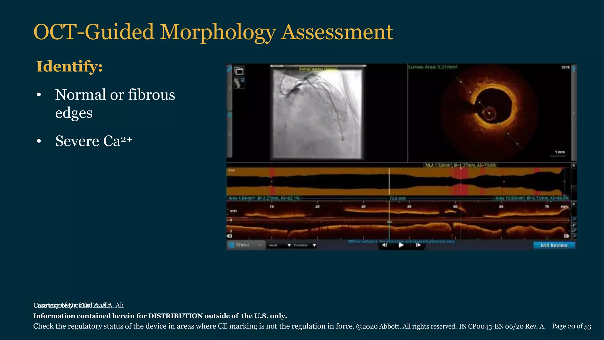
2) OCT can be used to assess lesion morphology, length, and diameter to guide percutaneous coronary intervention planning and stent optimization. Morphology is assessed to determine lesion preparation needs while length and diameter measurements inform stent and balloon sizing.
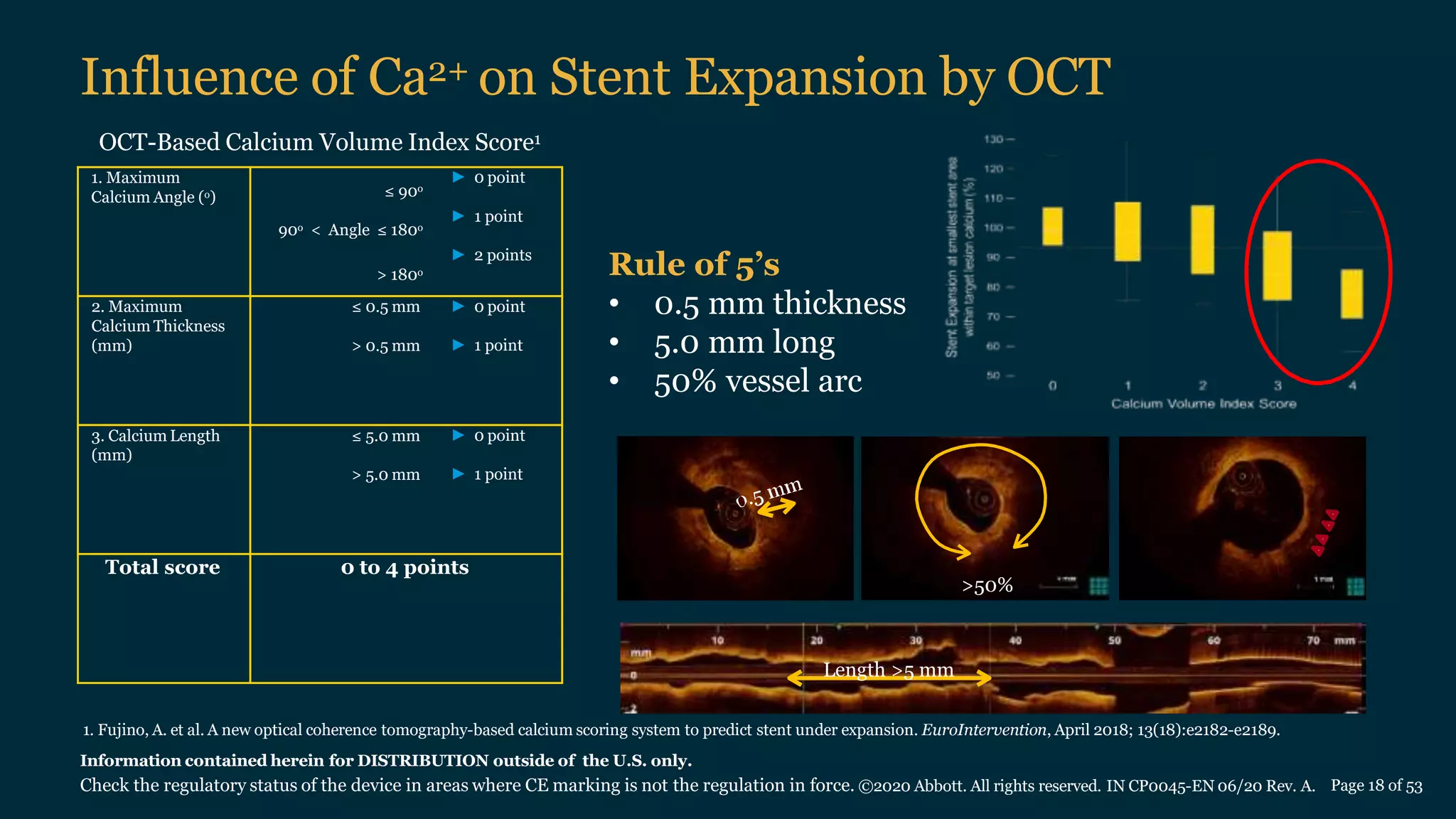
3) Pre-PCI OCT imaging helps determine lesion characteristics to strategize the optimal PCI approach, while post-PCI OCT allows optimization and assessment of stent apposition and expansion.