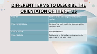
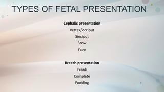
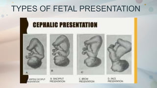
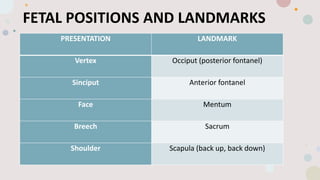
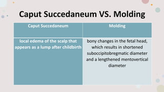
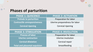
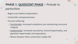
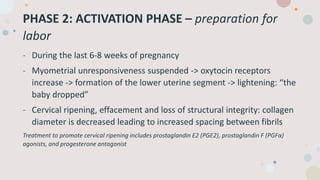
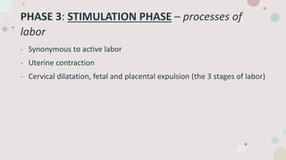
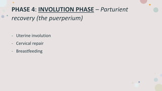
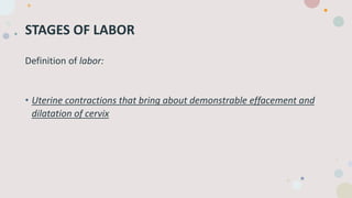
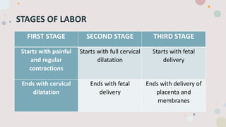
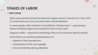
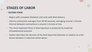
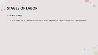
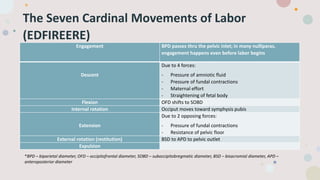
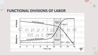
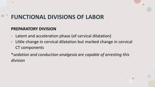
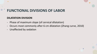
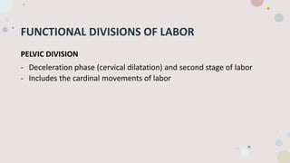
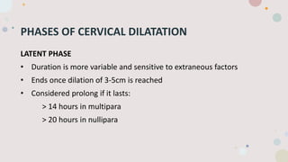
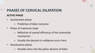
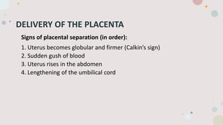
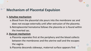
2. DIFFERENT TERMS TO DESCRIBE THE
ORIENTATON OF THE FETUS
FETAL LIE Relation of the fetal long axis to that of the
mother
FETAL PRESENTATION Portion of the body that is the foremost within
the birth canal
FETAL ATTITUDE Posture or habitus
FETAL POSITION Relationship of the fetal presenting part to the
right or left of the birth canal
3. TYPES OF FETAL PRESENTATION
Cephalic presentation
Vertex/occiput
Sinciput
Brow
Face
Breech presentation
Frank
Complete
Footling
5. FETAL POSITIONS AND LANDMARKS
PRESENTATION LANDMARK
Vertex Occiput (posterior fontanel)
Sinciput Anterior fontanel
Face Mentum
Breech Sacrum
Shoulder Scapula (back up, back down)
6. Caput Succedaneum VS. Molding
Caput Succedaneum Molding
local edema of the scalp that
appears as a lump after childbirth
bony changes in the fetal head,
which results in shortened
suboccipitobregmatic diameter
and a lengthened mentovertical
diameter
8. Phases of parturition
PHASE 1: QUISCENCE PHASE 2: ACTIVATION
Prelude to parturition Preparation for labor
Contractile unresponsiveness
Cervical ripening
Uterine preparedness for labor
Cervical ripening
PHASE 3: STIMULATION PHASE 4: INVOLUTION
Process of labor Preparation for labor
Uterine contraction
Cervical dilatation
Fetal and placental expulsion
Uterine involution
Cervical repair
breastfeeding
9. PHASE 1: QUIESCENT PHASE – Prelude to
parturition
- Begins even before implantation
- Contractile unresponsiveness
- Cervical softening:
o Functionally: increased compliance yet maintaining structural
integrity
o Anatomically: increased vascularity, stromal hypertrophy, and
glandular hypertrophy and hyperplasia
o Where Braxton-Hicks contraction maybe felt
10. PHASE 2: ACTIVATION PHASE – preparation for
labor
- During the last 6-8 weeks of pregnancy
- Myometrial unresponsiveness suspended -> oxytocin receptors
increase -> formation of the lower uterine segment -> lightening: “the
baby dropped”
- Cervical ripening, effacement and loss of structural integrity: collagen
diameter is decreased leading to increased spacing between fibrils
Treatment to promote cervical ripening includes prostaglandin E2 (PGE2), prostaglandin F (PGFα)
agonists, and progesterone antagonist
11. PHASE 3: STIMULATION PHASE – processes of
labor
- Synonymous to active labor
- Uterine contraction
- Cervical dilatation, fetal and placental expulsion (the 3 stages of labor)
13. STAGES OF LABOR
Definition of labor:
• Uterine contractions that bring about demonstrable effacement and
dilatation of cervix
14. STAGES OF LABOR
FIRST STAGE SECOND STAGE THIRD STAGE
Starts with painful
and regular
contractions
Starts with full cervical
dilatation
Starts with fetal
delivery
Ends with cervical
dilatation
Ends with fetal
delivery
Ends with delivery of
placenta and
membranes
15. STAGES OF LABOR
• FIRST STAGE
- Starts when painful contractions become regular (every 5 minutes for 1 hour OR ≥
12 contractions per hour) and ends with cervical dilatation
- In some people, labor initiation is heralded by “bloody show” – spontaneous
release of blood-tinged mucus plug from the cervical canal
- Ferguson reflex – mechanical stretching of the cervix enhances uterine activity
- Contractions are painful possibly because of:
o Hypoxia of the myometrium
o Compression of the nerve ganglia
o Cervical stretching during dilatation
16. STAGES OF LABOR
• SECOND STAGE
- Begins with complete dilatation and ends with fetal delivery
- Uterine contraction averages from 30-90 seconds, averaging around 1 minute.
Interval between contractions is around 1 minute or less.
- The most important force in fetal expulsion is produced by maternal
intraabdominal pressure
- Station describes the descent of the fetal biparietal diameter in relation to a line
drawn between 2 maternal ischial spines
17. STAGES OF LABOR
• THIRD STAGE
- Starts with fetal delivery and ends with expulsion of placenta and membranes
18. The Seven Cardinal Movements of Labor
(EDFIREERE)
Engagement BPD passes thru the pelvic inlet; in many nulliparas,
engagement happens even before labor begins
Descent
Due to 4 forces:
- Pressure of amniotic fluid
- Pressure of fundal contractions
- Maternal effort
- Straightening of fetal body
Flexion OFD shifts to SOBD
Internal rotation Occiput moves toward symphysis pubis
Extension
Due to 2 opposing forces:
- Pressure of fundal contractions
- Resistance of pelvic floor
External rotation (restitution) BSD to APD to pelvic outlet
Expulsion
*BPD – biparietal diameter, OFD – occipitofrontal diameter, SOBD – suboccipitobregmatic diameter, BSD – bisacromial diameter, APD –
anteroposterior diameter
20. FUNCTIONAL DIVISIONS OF LABOR
PREPARATORY DIVISION
- Latent and acceleration phase (of cervical dilatation)
- Little change in cervical dilatation but marked change in cervical
CT components
*sedation and conduction analgesia are capable of arresting this
division
21. FUNCTIONAL DIVISIONS OF LABOR
DILATATION DIVISION
- Phase of maximum slope (of cervical dilatation)
- Occurs most commonly after 6 cm dilatation (Zhang curve, 2010)
- Unaffected by sedation
22. FUNCTIONAL DIVISIONS OF LABOR
PELVIC DIVISION
- Deceleration phase (cervical dilatation) and second stage of labor
- Includes the cardinal movements of labor
23. PHASES OF CERVICAL DILATATION
LATENT PHASE
• Duration is more variable and sensitive to extraneous factors
• Ends once dilation of 3-5cm is reached
• Considered prolong if it lasts:
> 14 hours in multipara
> 20 hours in nullipara
24. PHASES OF CERVICAL DILATATION
ACTIVE PHASE
• Acceleration phase
o Predictive of labor outcome
• Phase of maximum slope
o Reflective of overall efficiency of the contractile
mechanism
o Usually the descent in nulliparas occur here
• Deceleration phase
o Heralds entry into the pelvic division of labor
25. DELIVERY OF THE PLACENTA
Signs of placental separation (in order):
1. Uterus becomes globular and firmer (Calkin’s sign)
2. Sudden gush of blood
3. Uterus rises in the abdomen
4. Lengthening of the umbilical cord
26. Mechanism of Placental Expulsion
1. Schultze mechanism
a.Blood from the placental site pours into the membrane sac and
does not escape externally until after extrusion of the placenta.
b.Retroplacental hematoma follows the placenta or is found within
the inverted sac.
2. Duncan mechanism
a.Placenta separates first at the periphery and the blood collects
between the membranes and the uterine wall and the escapes
the vagina.
b.Placenta descends sideways, maternal surface appears first
27. References:
Cunningham F. Gary, Leveno Kenneth, Bloom Steven, Spong Catherine, Dashe Jodi, Hoffman Barbara, Casey
Brian, and Sheffield Jeanne. (2014). “Williams Obstetrics, 24th edition.” McGraw-Hill Education. USA.