Introduction
• Most ofthe chest x-rays you will see
will be normal
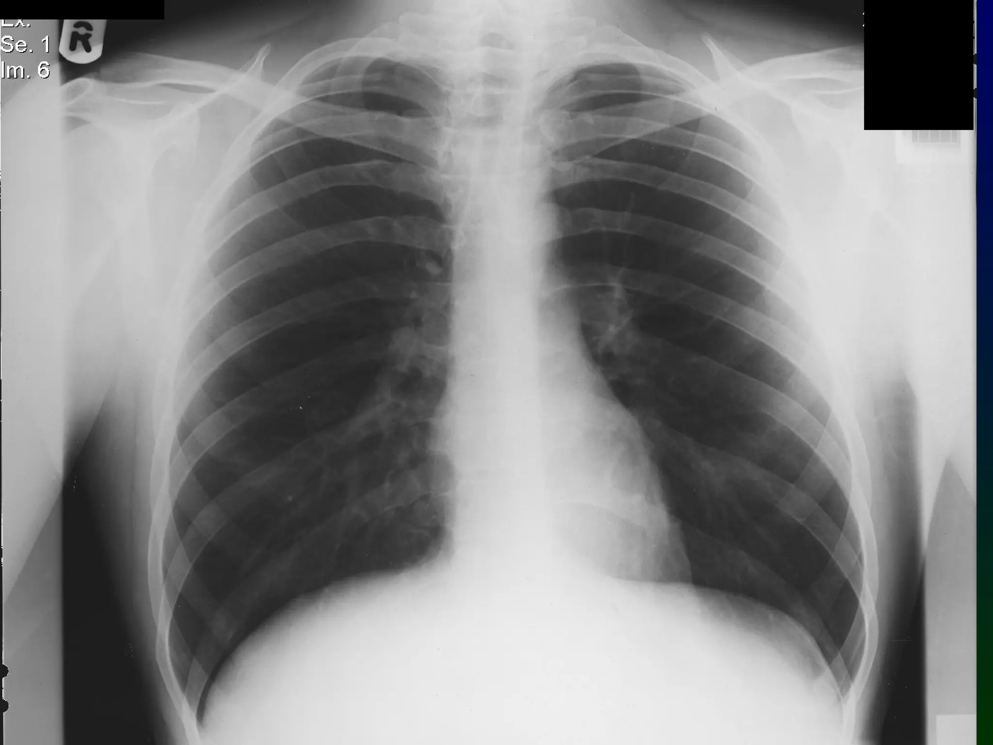
• In order to recognise abnormality, you
need to know what a normal CXR looks
like
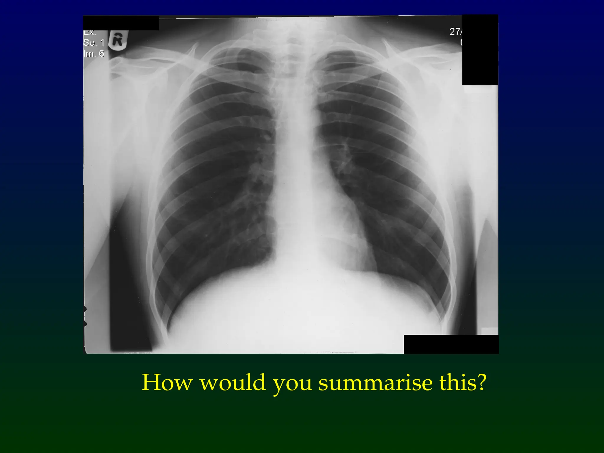
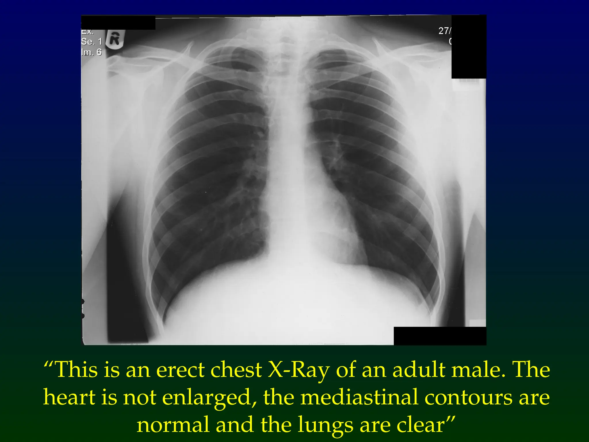
The CXR on the next slide is normal. How
would you interpret it?
4.
General Principles
• Havea systematic approach
• Interpret the CXR in conjunction with
the clinical findings
• Always compare with previous CXR if
available to assess for change
• Ask yourself “does my interpretation
make sense?”
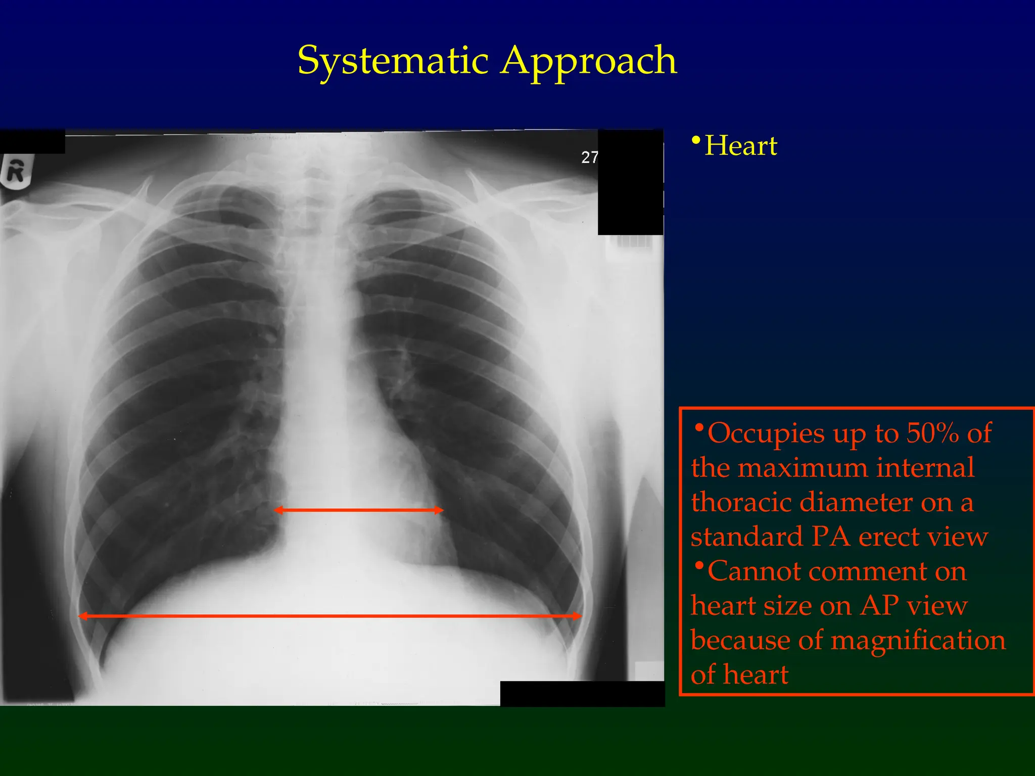
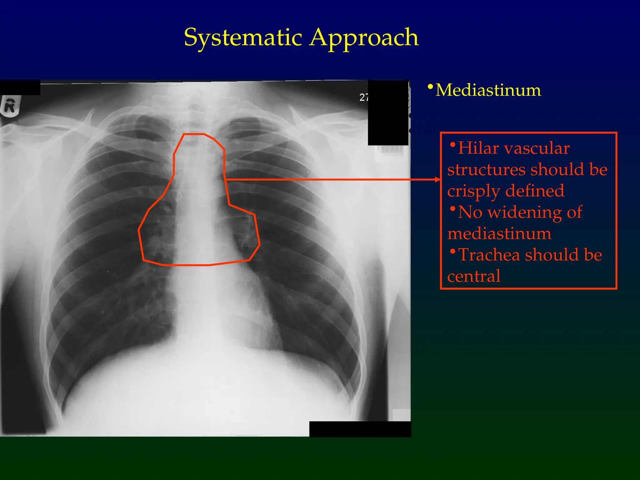
•Heart
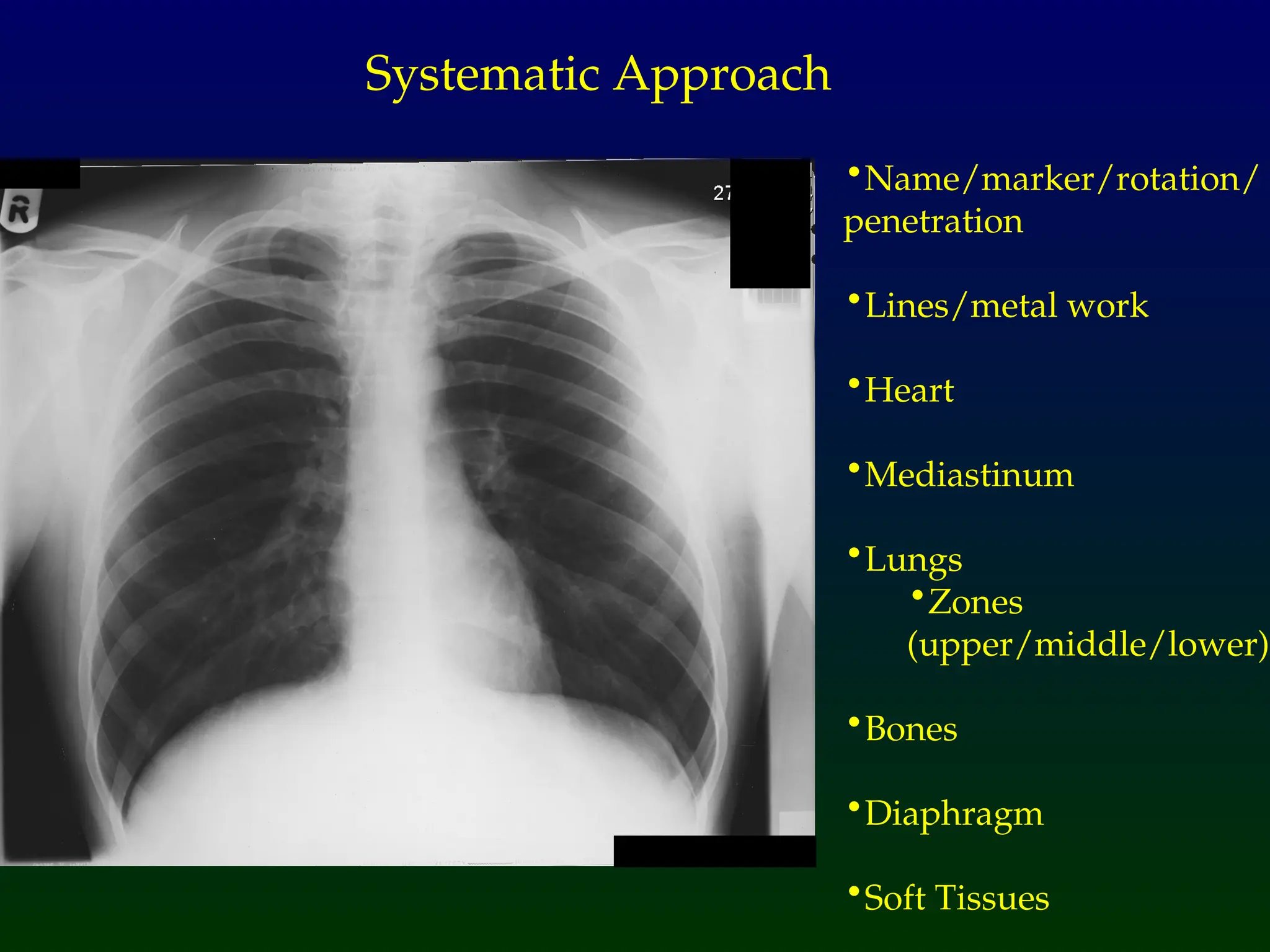
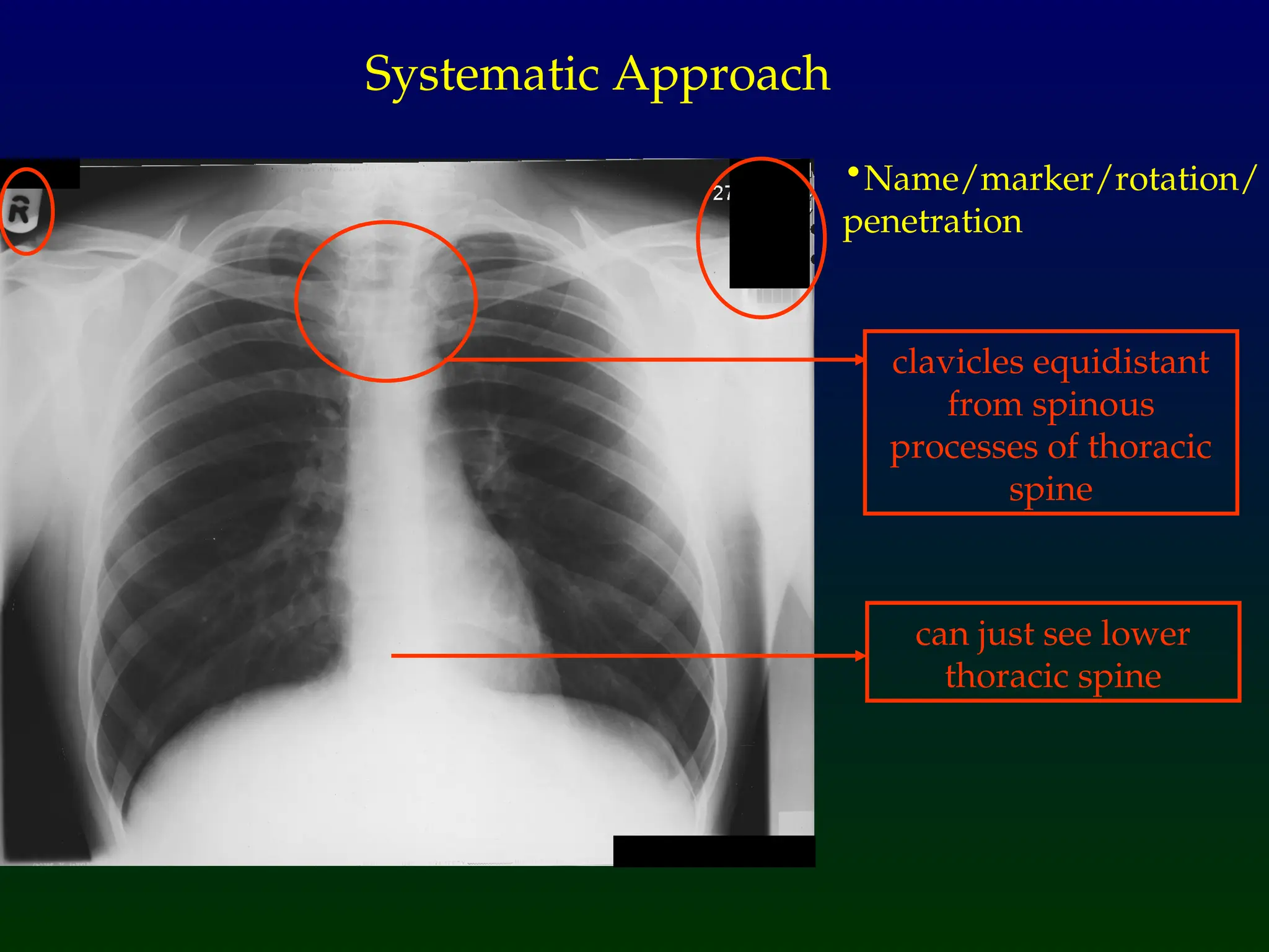
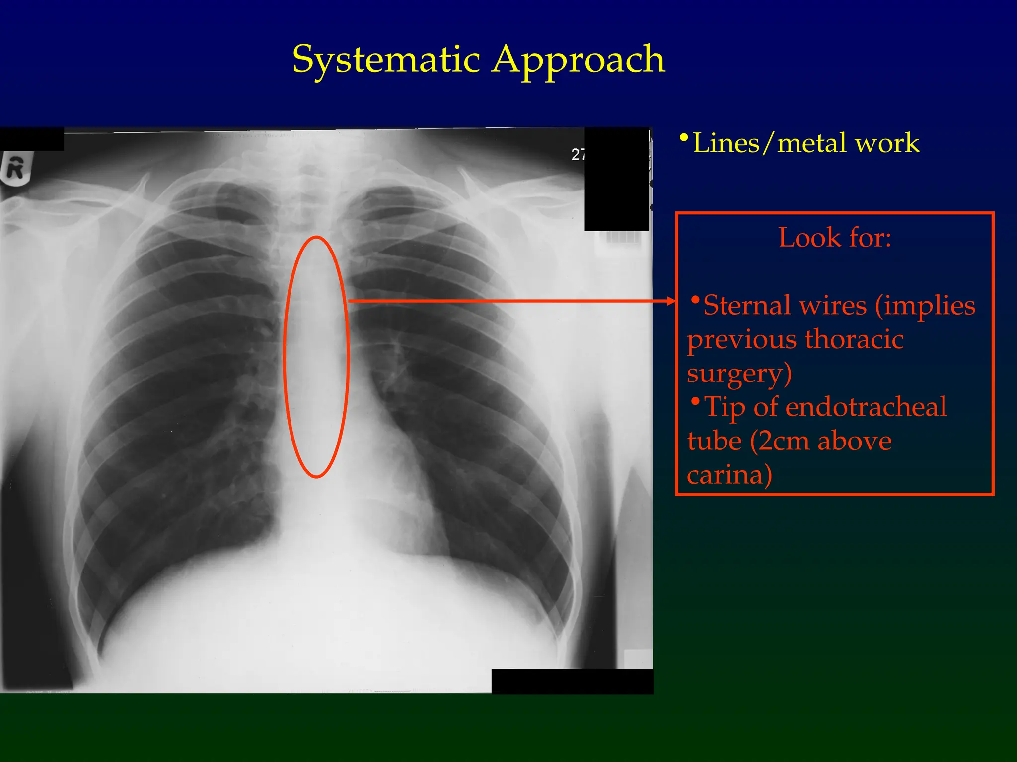
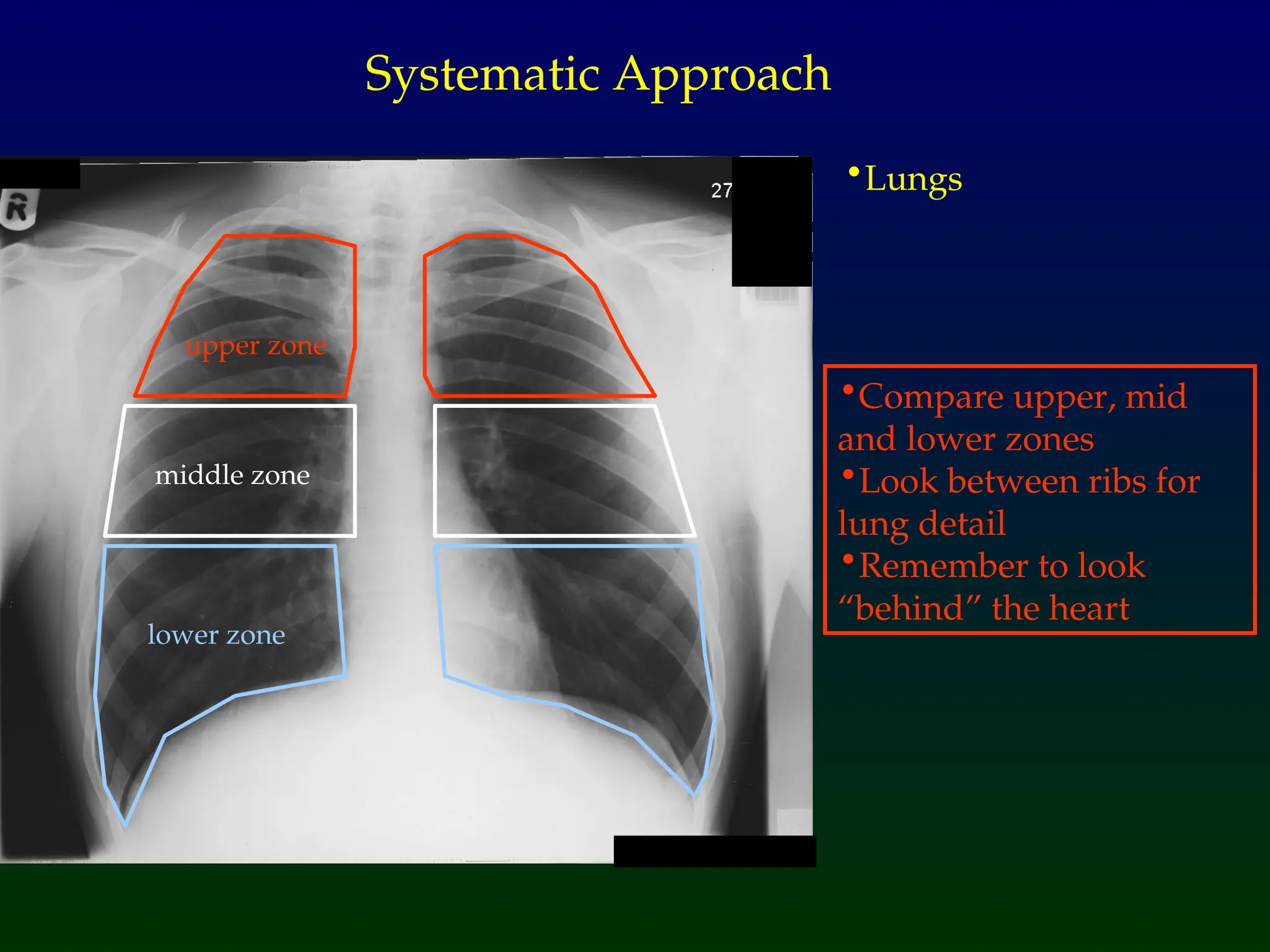
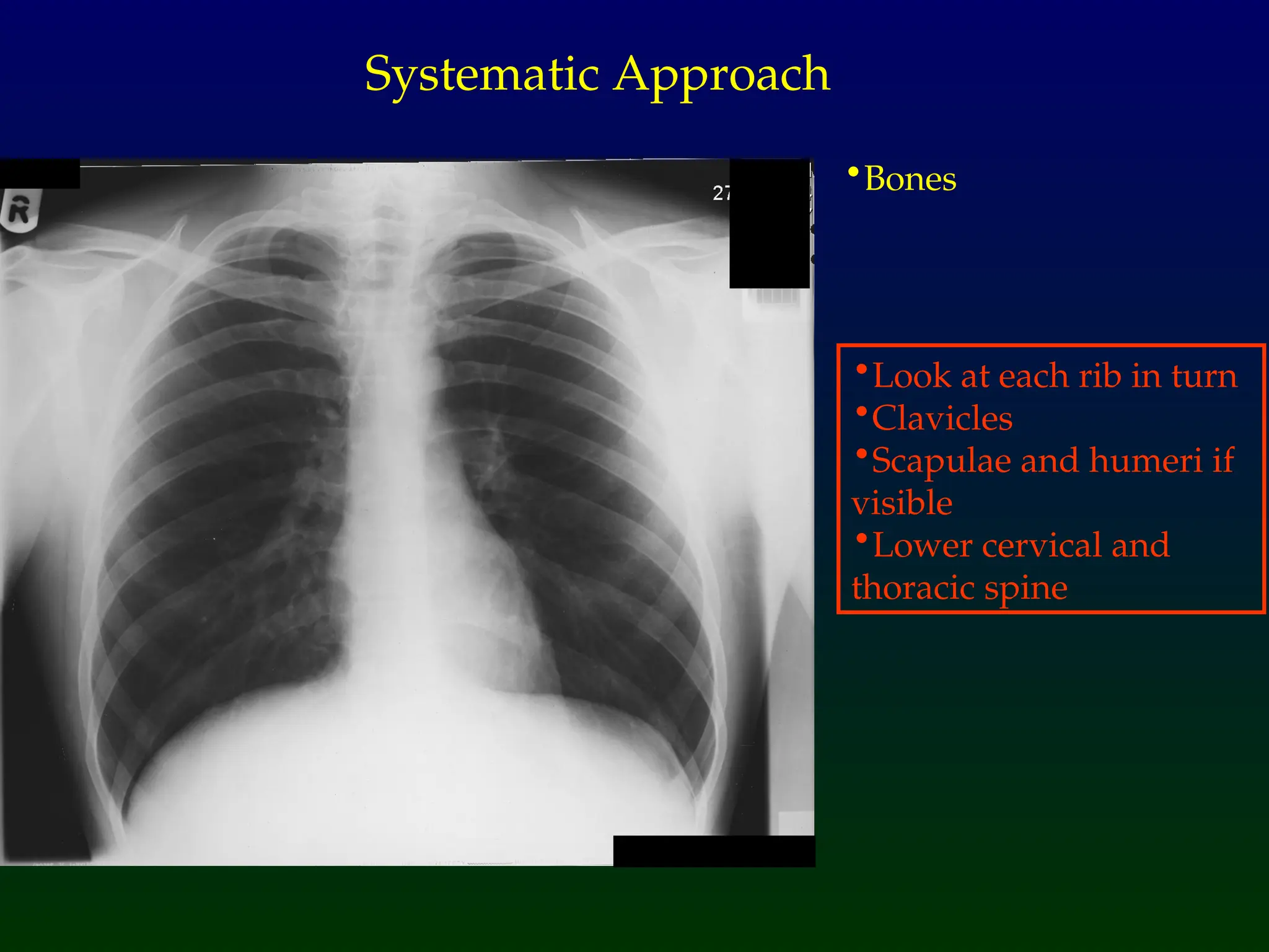
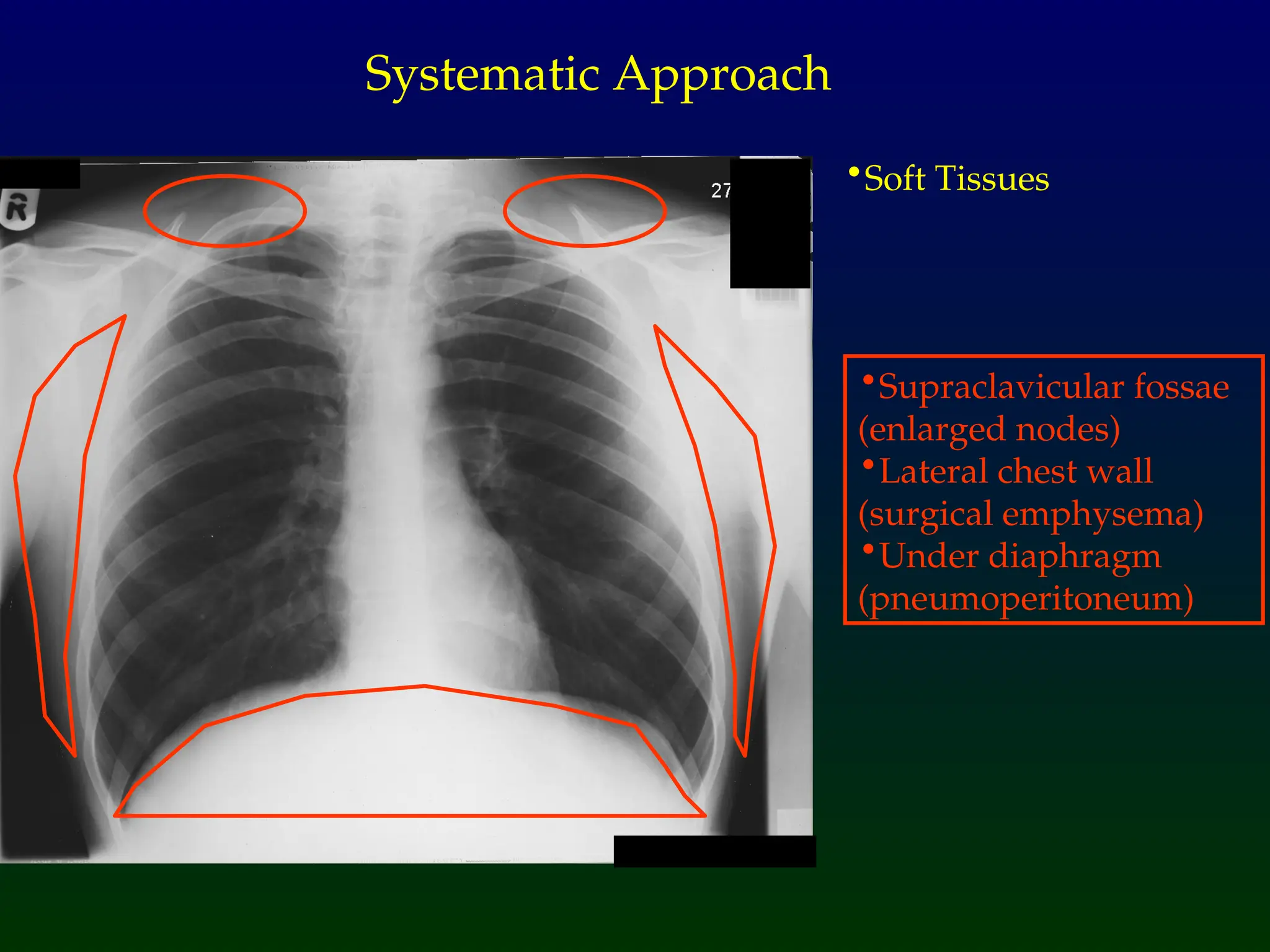
Systematic Approach
•Occupies upto 50% of
the maximum internal
thoracic diameter on a
standard PA erect view
•Cannot comment on
heart size on AP view
because of magnification
of heart
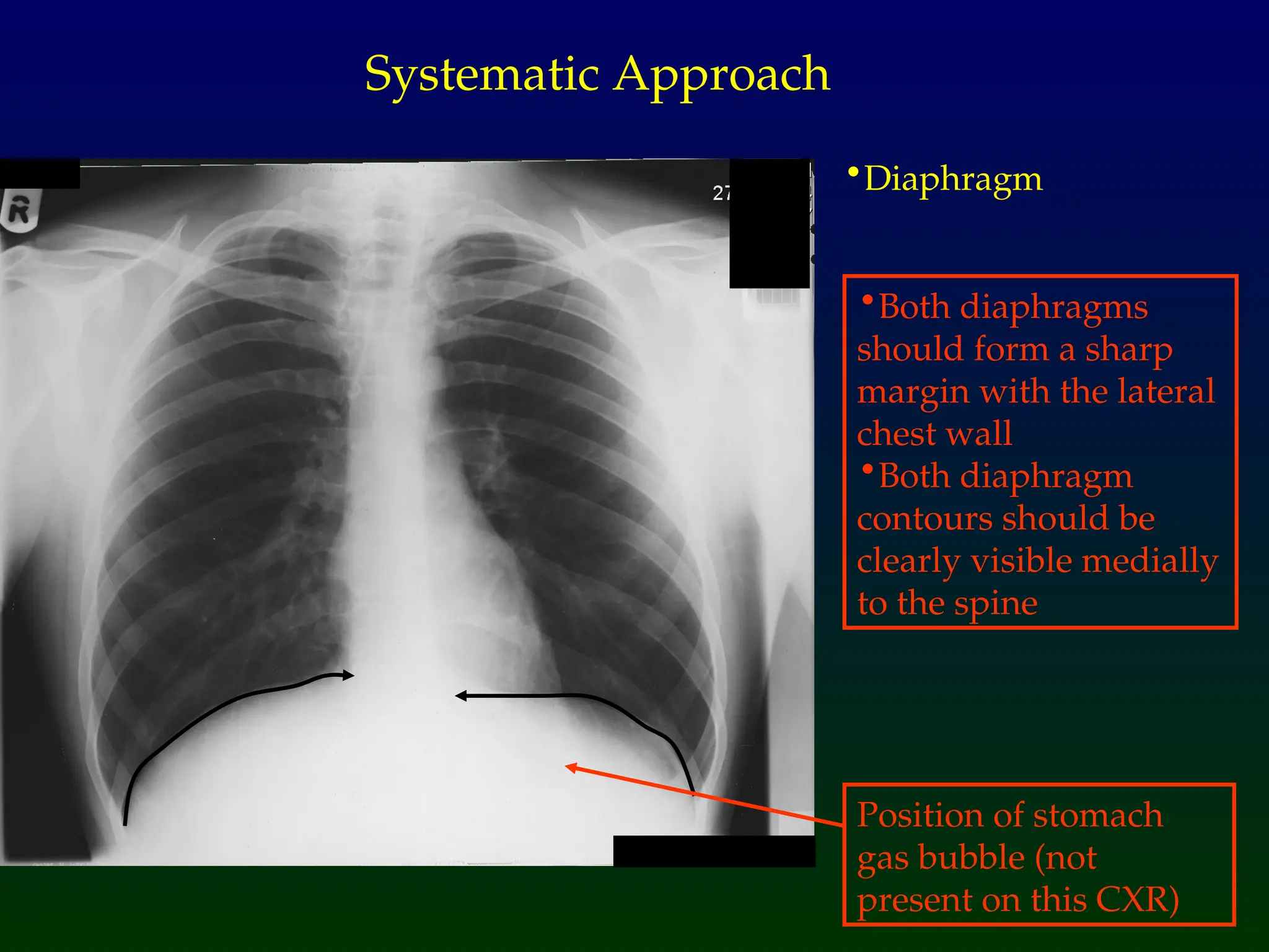
•Diaphragm
Systematic Approach
•Both diaphragms
shouldform a sharp
margin with the lateral
chest wall
•Both diaphragm
contours should be
clearly visible medially
to the spine
Position of stomach
gas bubble (not
present on this CXR)


![Radiological_diagnosis_of_TB_ECHO_MOH[1].pptx](https://cdn.slidesharecdn.com/ss_thumbnails/radiologicaldiagnosisoftbechomoh1-240905083452-eb26e5f9-thumbnail.jpg?width=640&height=640&fit=bounds)


