Download to read offline
![Fan et al. Page 9
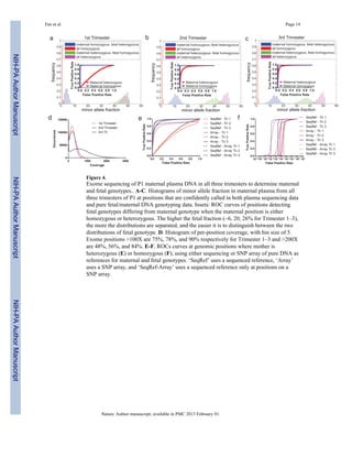
homozygous, ε/2 for group 2 SNPs of which fetus is heterozygous and mother is
homozygous, 1-ε/2 for group 3 SNPs at which fetus is homozygous and mother is
heterozygous, and ½ for group 3 SNPs at which both mother and fetus are heterozygous,
where ε is the fetal DNA fraction. We used the median of the distribution of minor allele
fraction for group 2 SNPs to provide an estimate of fetal DNA fraction.
Supplementary Material
Refer to Web version on PubMed Central for supplementary material.
Acknowledgments
The authors would like to thank Elizabeth Kogut and staff of the Division of Perinatal Genetics and the General
Clinical Research Center of Stanford University for coordination of patient recruitment; Ron Wong for initial
sample processing of clinical samples; Norma Neff, Gary Mantalas, Ben Passarelli, and Winston Koh for their help
in sequencing library preparation and data analysis.
References
1. Mandel P, Metais P. Les acides nucleiques du plasma sanguin chez l’homme. C R Acad Sci Paris.
1948; 142:241–243.
2. Lo YM, et al. Quantitative analysis of fetal DNA in maternal plasma and serum: implications for
noninvasive prenatal diagnosis. Am J Hum Genet. 1998; 62:768–775. [PubMed: 9529358]
3. Bodurtha J, Strauss JF 3rd. Genomics and perinatal care. N Engl J Med. 2012; 366:64–73. doi:
10.1056/NEJMra1105043. [PubMed: 22216843]
4. Fan HC, Blumenfeld YJ, Chitkara U, Hudgins L, Quake SR. Noninvasive diagnosis of fetal
aneuploidy by shotgun sequencing DNA from maternal blood. Proc Natl Acad Sci U S A. 2008;
105:16266–16271. [PubMed: 18838674]
5. Sehnert AJ, et al. Optimal detection of fetal chromosomal abnormalities by massively parallel DNA
sequencing of cell-free fetal DNA from maternal blood. Clinical chemistry. 2011; 57:1042–1049.
doi: 10.1373/clinchem.2011.165910. [PubMed: 21519036]
6. Bianchi DW, et al. Genome-Wide Fetal Aneuploidy Detection by Maternal Plasma DNA
Sequencing. Obstetrics and gynecology. 2012 doi: 10.1097/AOG.0b013e31824fb482.
7. Palomaki GE, et al. DNA sequencing of maternal plasma reliably identifies trisomy 18 and trisomy
13 as well as Down syndrome: an international collaborative study. Genetics in medicine : official
journal of the American College of Medical Genetics. 2012; 14:296–305. doi: 10.1038/gim.
2011.73. [PubMed: 22281937]
8. Palomaki GE, et al. DNA sequencing of maternal plasma to detect Down syndrome: an international
clinical validation study. Genet Med. 2011; 13:913–920. doi: 10.1097/GIM.0b013e3182368a0e.
[PubMed: 22005709]
9. Ehrich M, et al. Noninvasive detection of fetal trisomy 21 by sequencing of DNA in maternal blood:
a study in a clinical setting. Am J Obstet Gynecol. 2011; 204:205, e201–e211. doi:
S0002-9378(11)00018-4 [pii] 10.1016/j.ajog.2010.12.060. [PubMed: 21310373]
10. Chiu RW, et al. Non-invasive prenatal assessment of trisomy 21 by multiplexed maternal plasma
DNA sequencing: large scale validity study. Bmj. 2011; 342:c7401. [PubMed: 21224326]
11. Lo YM, et al. Maternal plasma DNA sequencing reveals the genome-wide genetic and mutational
profile of the fetus. Sci Transl Med. 2010; 2:61ra91. doi: 2/61/61ra91 [pii] 10.1126/scitranslmed.
3001720.
12. Fan HC, Quake SR. In principle method for noninvasive determination of the fetal genome.
Available from Nature Proceedings. 2010 doi: <http://dx.doi.org/10.1038/npre.2010.5373.1>.
13. Macintyre S, Sooman A. Non-paternity and prenatal genetic screening. Lancet. 1991; 338:869–
871. doi: 01406736(91)91513-T [pii]. [PubMed: 1681226]
14. Bellis MA, Hughes K, Hughes S, Ashton JR. Measuring paternal discrepancy and its public health
consequences. J Epidemiol Community Health. 2005; 59:749–754. doi: 59/9/749 [pii] 10.1136/
jech.2005.036517. [PubMed: 16100312]
Nature. Author manuscript; available in PMC 2013 February 01.
NIH-PA Author Manuscript NIH-PA Author Manuscript NIH-PA Author Manuscript](https://image.slidesharecdn.com/nihms379831stephenquake-141021231411-conversion-gate01/85/Nihms379831-stephen-quake-9-320.jpg)
![Fan et al. Page 10
15. Fan HC, Wang J, Potanina A, Quake SR. Whole-genome molecular haplotyping of single cells.
Nat Biotechnol. 2011; 29:51–57. doi: nbt.1739 [pii] 10.1038/nbt.1739. [PubMed: 21170043]
16. Consortium TGP. A map of human genome variation from population-scale sequencing. Nature.
2010; 467:1061–1073. doi: nature09534 [pii] 10.1038/nature09534. [PubMed: 20981092]
17. Marchini J, et al. A comparison of phasing algorithms for trios and unrelated individuals. Am J
Hum Genet. 2006; 78:437–450. [PubMed: 16465620]
18. White RA 3rd, Blainey PC, Fan HC, Quake SR. Digital PCR provides sensitive and absolute
calibration for high throughput sequencing. BMC Genomics. 2009; 10:116. [PubMed: 19298667]
19. Clark MJ, et al. Performance comparison of exome DNA sequencing technologies. Nat Biotechnol.
2011; 29:908–914. doi: nbt.1975 [pii] 10.1038/nbt.1975. [PubMed: 21947028]
20. Kinde I, Wu J, Papadopoulos N, Kinzler KW, Vogelstein B. Detection and quantification of rare
mutations with massively parallel sequencing. Proc Natl Acad Sci U S A. 2011; 108:9530–9535.
doi: 1105422108 [pii]10.1073/pnas.1105422108. [PubMed: 21586637]
Nature. Author manuscript; available in PMC 2013 February 01.
NIH-PA Author Manuscript NIH-PA Author Manuscript NIH-PA Author Manuscript](https://image.slidesharecdn.com/nihms379831stephenquake-141021231411-conversion-gate01/85/Nihms379831-stephen-quake-10-320.jpg)
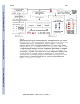

This document describes a new noninvasive method for sequencing the entire fetal genome using cell-free DNA found in a pregnant woman's blood. The method works by counting parental haplotypes - combinations of maternal and paternal chromosomes passed to the fetus. Since a small percentage of cell-free DNA comes from the fetus, haplotypes inherited by the fetus can be identified by which have a higher count. Researchers tested this method on two pregnancies and were able to determine the fetal genomes without any invasive procedures. This noninvasive prenatal testing could allow comprehensive screening for genetic diseases.


