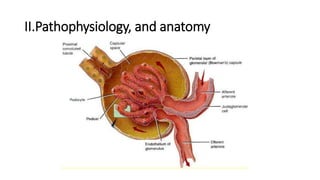
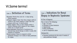
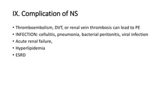
NEPHROTIC SYNDROME
SINGI
OUTLINE
• I.DEFINITION
• II. Pathophysiology,and anatomy
• III. Presentation
• IV. Investigation
• V. Classification of nephrotic syndrome
• VI. Some terms!
• VII. Management using General measures!
• VII. Treatment Goal
• IX. Complication of NS
• X.REFERENCE
I.DEFINITION
• Nephrotic syndrome is a syndrome caused by alteration in the
glomerular capillary wall.
• It consists of heavy proteinuria, hypoalbuminemia and often
associated with edema and hyperlipidemia
II.Pathophysiology, and anatomy
Plasma is filtered through the glomerular
barrier
• Components of plasma cross the three layers of the glomerular barrier during
filtration
• Capillary endothelium (cells have openings, fenestrae, of 70-100 nm D)
• Basement membrane, heparin sulphate proteoglycans (net negative charge)
• Epithelium of Bowman’s Capsule ( Podocytes–filtration slits allow size <60kD)
• The ability of a molecule to cross the membrane depends on size, charge, and
shape
• Glomerular filtrate therefore contains all molecules not contained by the
glomerular barrier - it is NOT URINE YET!
Pathophysiology cont’’’
• Increased permeability of glomerular to large molecules, mostly
albumin lead to proteinuria
• Proteinuria causes fall in serum albumin, if liver fails to compensate
by synthesis, plasma albumin decline, leading to edema formation.
• Edema. May be due to retention of sodium induced by renal disease
(overfill hypothesis)
• or secondary Sodium retention: hypoalbuminemia, low oncotic
pressure, fluid move to interstitial space, renal hypo perfusion,
activation of renin angiotensin system (underfill hypothesis)
III. Presentation
Clinical presentation
• edema (periorbital, scrotal, labial region then anasarca), Morning:
periorbital, Day time: extremity edema
• tiredness, leukonychia
• Hypertension, acute renal failure,
• Allergy in 50% with MCNS
Labs
• Proteinuria > 3g/24hrs, spot urine ACR>300mg/mmol, or urine protein to
urine creat ratio >2g/g, dipstick hematuria&3+.4+ proteinuria
• Hypoalbuminia, serum albumin <25g/dl
• Microscopic hematuria
• Hyperlipidemia, total cholesterol often > 10 mmol
• Protein electrophoresis: if no albumin, PARAPROTEIN, multiple myeloma
IV. Other investigation
• Exclude infection: Midstream urine, microscopy, culture & sensitivity
• HIV
• ESR, CRP, Glucose
• FBC and coagulation studies
• U&Es, and eGFR
• HBV&HCV, and LFT to exclude liver pathology
• Chest X-Ray and renal ultrasound, Doppler
• Renal u/s
• Renal biopsy
V. Classification of nephrotic syndrome
• Nephrotic syndrome:
• idiopathic 90% : MCN, FSGS, MN
• or secondary: Infection(HIV, HBV&HCV, malaria, syphilis )
• Drug (Gold, interferon, heroin, lithium, NSAID)
• Malignancies (leukemia, lymphoma)
• miscellaneous (diabetes, SLE…)
Idiopathic nephrotic syndrome
A.Minimal change nephrotic syndrome:
• nephrotic syndrome without hematuria
• Consist of 95% of nephrotic syndrome in children and 25% in adults
• Renal biopsy shows normal appearance, EM: fusion of foot processes
• Normal renal function usually.
• Spontaneous remission, treat only if edema and proteinuria
• B. Focal segmental glomerulosclerosis (FSGS)
• Nephrotic syndrome with microscopic hematuria, often HTN&
worsening renal impairment.
• Primary FSGS: unknown cause. on biopsy renal scleroses are seen,
interstitial fibrosis and focal tubal atrophy are common, mesangial
hypercellularity may be present
• Secondary FSGS: Occur as a result of loss of functioning nephrons
• C. Membranous glomerulonephritis
• Asymptomatic proteinuria with or without renal impairment,
microscopic hematuria and hypertension, it usually affect adults,
mostly men.
• Can have spontaneous remission
• Treatment for those with renal impairment
• Men, old age, heavy proteinuria are main progression risks
VI.Some terms!
VII. Management using General measures!
1.Initial treatment: stop Na+ intake, thiazide diuretic
or Furosemide(40-120mg/day + amiloride 5mg/day
2.Protein intake should be reduced, high induce proteinuria
3.Albumin infusion: only in diuretic resistance, or oliguria/uremia
without damage to glomerular. In MCNS: Some people need albumin +
diuretic
4.Hypercoagulable state: prophylactic anti coagulant, if RVT permant
anti coagulant
5.infection: due to loss of immunoglobulin in urine, Early detection and
aggressive treatment no prophylaxis
6.Lipid abonormalities, if high cholesterol, give atorvastatin…
7.ACEI/AIIRA: Decrease proteinuria by ↓BP, Glomerular Filtr pressure
Treatment cont’’’
• MCNS
• Start prednisolone 1 mg/kg/day (Max 80mg/day) 4-6 weeks
• Once remission reduce prednisolone by 30% every 4 to 6 weeks and stop
steroids in at least 12 weeks
• Relapse may occur and is treated with prednisolone
• Frequent relapses: can be due to steroid dependence or steroid resistant
Nephrotic syndrome,
cyclophosphamide 7.5 to 15 mg/day for 8 to 12 weeks,
cyclosporine 3-5 mg/kg/day target blood level 80-150ng/ml 8-12 weeks
•
FSGS
•
• Overt NS/Progressing renal impairment: drop in eGFR >15% in 1 year or >
10% in 2 consecutive years.
• Prednisolone 0.5-2 mg/kg/day should be continue for 6 months before
diagnosing steroid resistance
• cyclosporine aiming 150-300ng/ml in blood may be used but relapse after
discontinuation
• Cyclophosphamide 1-1.5 mg/kg/day with 1mg/kg/day steroid for 3-6
months, followed by a maintenance of prednisolone and azathioprine may
decrease proteinuria and slow down progession.
• Despite treatment 50% progress to ESRD in 10 years.
MGN
• There is spontaneous remission in 40%, hold treatment for 6 months
• Only people with severe proteinuria and progressing renal
impairment should be treated
• A) prednisolone 1mg/kg/day alternating daily with cyclophosphamide
1.5 to 2.5 mg/kg/day for 6-12 months
• Or
• Chlorambicil 0.2mg/kg/day in months 2, 4, and 6 and prednisolone
0.4 mg/kg/day in months 1, 3, and 5
VIII. Treatment Goal
• BP: <130/80 OR <120/75 If proteinuria or diabetes
• proteinuria,<1g/24 hours
IX. Complication of NS
• Thromboembolism, DVT, or renal vein thrombosis can lead to PE
• INFECTION: cellulitis, pneumonia, bacterial peritonitis, viral infection
• Acute renal failure,
• Hyperlipidemia
• ESRD
X.REFERENCE
• Kumar & Clarks medical management and therapeutics 1rst Ed 2011
• Uptodate
• The nephrotic syndrome,Pediatrics in Review Vol.30 No.3 March 2009
• www.ncbi.nlm.nih.gov/Nephrotic syndrome in adults
• Kumar & Clark’s clinical medicine 8th Ed 2012
THANK YOU

Nephrotic syndrom

  • 1.
  • 2.
    OUTLINE • I.DEFINITION • II.Pathophysiology,and anatomy • III. Presentation • IV. Investigation • V. Classification of nephrotic syndrome • VI. Some terms! • VII. Management using General measures! • VII. Treatment Goal • IX. Complication of NS • X.REFERENCE
  • 3.
    I.DEFINITION • Nephrotic syndromeis a syndrome caused by alteration in the glomerular capillary wall. • It consists of heavy proteinuria, hypoalbuminemia and often associated with edema and hyperlipidemia
  • 4.
  • 5.
    Plasma is filteredthrough the glomerular barrier • Components of plasma cross the three layers of the glomerular barrier during filtration • Capillary endothelium (cells have openings, fenestrae, of 70-100 nm D) • Basement membrane, heparin sulphate proteoglycans (net negative charge) • Epithelium of Bowman’s Capsule ( Podocytes–filtration slits allow size <60kD) • The ability of a molecule to cross the membrane depends on size, charge, and shape • Glomerular filtrate therefore contains all molecules not contained by the glomerular barrier - it is NOT URINE YET!
  • 6.
    Pathophysiology cont’’’ • Increasedpermeability of glomerular to large molecules, mostly albumin lead to proteinuria • Proteinuria causes fall in serum albumin, if liver fails to compensate by synthesis, plasma albumin decline, leading to edema formation. • Edema. May be due to retention of sodium induced by renal disease (overfill hypothesis) • or secondary Sodium retention: hypoalbuminemia, low oncotic pressure, fluid move to interstitial space, renal hypo perfusion, activation of renin angiotensin system (underfill hypothesis)
  • 7.
    III. Presentation Clinical presentation •edema (periorbital, scrotal, labial region then anasarca), Morning: periorbital, Day time: extremity edema • tiredness, leukonychia • Hypertension, acute renal failure, • Allergy in 50% with MCNS Labs • Proteinuria > 3g/24hrs, spot urine ACR>300mg/mmol, or urine protein to urine creat ratio >2g/g, dipstick hematuria&3+.4+ proteinuria • Hypoalbuminia, serum albumin <25g/dl • Microscopic hematuria • Hyperlipidemia, total cholesterol often > 10 mmol • Protein electrophoresis: if no albumin, PARAPROTEIN, multiple myeloma
  • 8.
    IV. Other investigation •Exclude infection: Midstream urine, microscopy, culture & sensitivity • HIV • ESR, CRP, Glucose • FBC and coagulation studies • U&Es, and eGFR • HBV&HCV, and LFT to exclude liver pathology • Chest X-Ray and renal ultrasound, Doppler • Renal u/s • Renal biopsy
  • 9.
    V. Classification ofnephrotic syndrome • Nephrotic syndrome: • idiopathic 90% : MCN, FSGS, MN • or secondary: Infection(HIV, HBV&HCV, malaria, syphilis ) • Drug (Gold, interferon, heroin, lithium, NSAID) • Malignancies (leukemia, lymphoma) • miscellaneous (diabetes, SLE…)
  • 10.
    Idiopathic nephrotic syndrome A.Minimalchange nephrotic syndrome: • nephrotic syndrome without hematuria • Consist of 95% of nephrotic syndrome in children and 25% in adults • Renal biopsy shows normal appearance, EM: fusion of foot processes • Normal renal function usually. • Spontaneous remission, treat only if edema and proteinuria
  • 11.
    • B. Focalsegmental glomerulosclerosis (FSGS) • Nephrotic syndrome with microscopic hematuria, often HTN& worsening renal impairment. • Primary FSGS: unknown cause. on biopsy renal scleroses are seen, interstitial fibrosis and focal tubal atrophy are common, mesangial hypercellularity may be present • Secondary FSGS: Occur as a result of loss of functioning nephrons
  • 12.
    • C. Membranousglomerulonephritis • Asymptomatic proteinuria with or without renal impairment, microscopic hematuria and hypertension, it usually affect adults, mostly men. • Can have spontaneous remission • Treatment for those with renal impairment • Men, old age, heavy proteinuria are main progression risks
  • 13.
  • 14.
    VII. Management usingGeneral measures! 1.Initial treatment: stop Na+ intake, thiazide diuretic or Furosemide(40-120mg/day + amiloride 5mg/day 2.Protein intake should be reduced, high induce proteinuria 3.Albumin infusion: only in diuretic resistance, or oliguria/uremia without damage to glomerular. In MCNS: Some people need albumin + diuretic 4.Hypercoagulable state: prophylactic anti coagulant, if RVT permant anti coagulant 5.infection: due to loss of immunoglobulin in urine, Early detection and aggressive treatment no prophylaxis 6.Lipid abonormalities, if high cholesterol, give atorvastatin… 7.ACEI/AIIRA: Decrease proteinuria by ↓BP, Glomerular Filtr pressure
  • 15.
    Treatment cont’’’ • MCNS •Start prednisolone 1 mg/kg/day (Max 80mg/day) 4-6 weeks • Once remission reduce prednisolone by 30% every 4 to 6 weeks and stop steroids in at least 12 weeks • Relapse may occur and is treated with prednisolone • Frequent relapses: can be due to steroid dependence or steroid resistant Nephrotic syndrome, cyclophosphamide 7.5 to 15 mg/day for 8 to 12 weeks, cyclosporine 3-5 mg/kg/day target blood level 80-150ng/ml 8-12 weeks •
  • 16.
  • 17.
    • Overt NS/Progressingrenal impairment: drop in eGFR >15% in 1 year or > 10% in 2 consecutive years. • Prednisolone 0.5-2 mg/kg/day should be continue for 6 months before diagnosing steroid resistance • cyclosporine aiming 150-300ng/ml in blood may be used but relapse after discontinuation • Cyclophosphamide 1-1.5 mg/kg/day with 1mg/kg/day steroid for 3-6 months, followed by a maintenance of prednisolone and azathioprine may decrease proteinuria and slow down progession. • Despite treatment 50% progress to ESRD in 10 years.
  • 18.
    MGN • There isspontaneous remission in 40%, hold treatment for 6 months • Only people with severe proteinuria and progressing renal impairment should be treated • A) prednisolone 1mg/kg/day alternating daily with cyclophosphamide 1.5 to 2.5 mg/kg/day for 6-12 months • Or • Chlorambicil 0.2mg/kg/day in months 2, 4, and 6 and prednisolone 0.4 mg/kg/day in months 1, 3, and 5
  • 19.
    VIII. Treatment Goal •BP: <130/80 OR <120/75 If proteinuria or diabetes • proteinuria,<1g/24 hours
  • 20.
    IX. Complication ofNS • Thromboembolism, DVT, or renal vein thrombosis can lead to PE • INFECTION: cellulitis, pneumonia, bacterial peritonitis, viral infection • Acute renal failure, • Hyperlipidemia • ESRD
  • 21.
    X.REFERENCE • Kumar &Clarks medical management and therapeutics 1rst Ed 2011 • Uptodate • The nephrotic syndrome,Pediatrics in Review Vol.30 No.3 March 2009 • www.ncbi.nlm.nih.gov/Nephrotic syndrome in adults • Kumar & Clark’s clinical medicine 8th Ed 2012
  • 22.