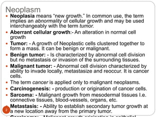
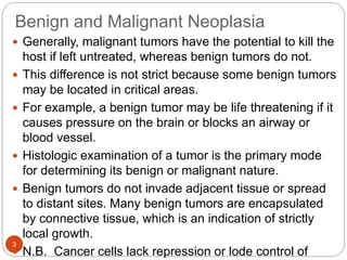
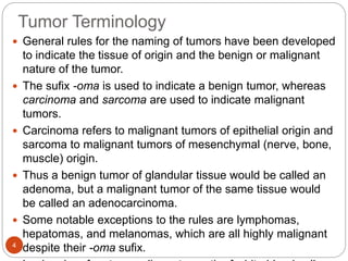
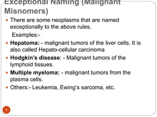
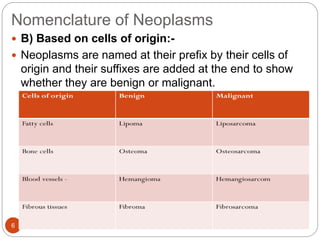
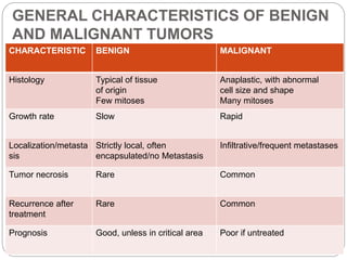
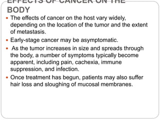
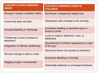
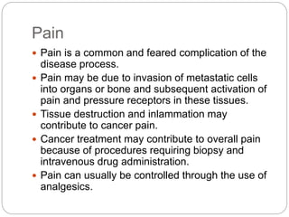
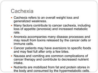
OJORA K. A
NEOPLASIA
Neoplasm
2
 Neoplasia means “new growth.” In common use, the term
implies an abnormality of cellular growth and may be used
interchangeably with the term tumor.
 Aberrant cellular growth:- An alteration in normal cell
growth
 Tumor: - A growth of Neoplastic cells clustered together to
form a mass. It can be benign or malignant.
 Benign tumor: - Is characterized by abnormal cell division
but no metastasis or invasion of the surrounding tissues.
 Malignant tumor: - Abnormal cell division characterized by
ability to invade locally, metastasize and reoccur. It is cancer
cells.
 The term cancer is applied only to malignant neoplasms.
 Carcinogenesis: - production or origination of cancer cells.
 Sarcoma: - Malignant growth from mesodermal tissues I.e.
connective tissues, blood-vessels, organs, etc.
 Metastasis: - Ability to establish secondary tumor growth at
a new location away from the primary tumor.
Benign and Malignant Neoplasia
3
 Generally, malignant tumors have the potential to kill the
host if left untreated, whereas benign tumors do not.
 This difference is not strict because some benign tumors
may be located in critical areas.
 For example, a benign tumor may be life threatening if it
causes pressure on the brain or blocks an airway or
blood vessel.
 Histologic examination of a tumor is the primary mode
for determining its benign or malignant nature.
 Benign tumors do not invade adjacent tissue or spread
to distant sites. Many benign tumors are encapsulated
by connective tissue, which is an indication of strictly
local growth.
 N.B. Cancer cells lack repression or lode control of
Tumor Terminology
4
 General rules for the naming of tumors have been developed
to indicate the tissue of origin and the benign or malignant
nature of the tumor.
 The sufix -oma is used to indicate a benign tumor, whereas
carcinoma and sarcoma are used to indicate malignant
tumors.
 Carcinoma refers to malignant tumors of epithelial origin and
sarcoma to malignant tumors of mesenchymal (nerve, bone,
muscle) origin.
 Thus a benign tumor of glandular tissue would be called an
adenoma, but a malignant tumor of the same tissue would
be called an adenocarcinoma.
 Some notable exceptions to the rules are lymphomas,
hepatomas, and melanomas, which are all highly malignant
despite their -oma sufix.
Exceptional Naming (Malignant
Misnomers)
5
 There are some neoplasms that are named
exceptionally to the above rules.
Examples:-
 Hepatoma: - malignant tumors of the liver cells. It is
also called Hepato-cellular carcinoma
 Hodgkin’s disease: - Malignant tumors of the
lymphoid tissues.
 Multiple myeloma: - malignant tumors from the
plasma cells.
 Others:- Leukemia, Ewing’s sarcoma, etc.
Nomenclature of Neoplasms
6
 B) Based on cells of origin:-
 Neoplasms are named at their prefix by their cells of
origin and their suffixes are added at the end to show
whether they are benign or malignant.
GENERAL CHARACTERISTICS OF BENIGN
AND MALIGNANT TUMORS
7
CHARACTERISTIC BENIGN MALIGNANT
Histology Typical of tissue
of origin
Few mitoses
Anaplastic, with abnormal
cell size and shape
Many mitoses
Growth rate Slow Rapid
Localization/metasta
sis
Strictly local, often
encapsulated/no Metastasis
Infiltrative/frequent metastases
Tumor necrosis Rare Common
Recurrence after
treatment
Rare Common
Prognosis Good, unless in critical area Poor if untreated
EPIDEMIOLOGY AND CANCER RISK
FACTORS
 Cancer accounts for approximately 25% of all deaths,
which makes it the second leading cause of death in the
United States.
 Most cancer deaths (77%) occur in persons older than
55 years.
 The American Cancer Society (ACS) estimates that men
have almost a 1 in 2 lifetime risk of developing cancer
and women have slightly higher than a 1 in 3 risk.
Tobacco Use
 Lung cancer remains the leading cause of cancer death in
both men and women, accounting for 30% of all cancer
deaths.
 Lung cancer has one of the worst survival rates of all
cancers—only 15%.
 In addition to lung cancer, tobacco use has been linked
with cancer of the pancreas, bladder, kidney, mouth,
esophagus, and cervix.
 Carcinogens can be grouped into two major types: those
that cause genetic damage (initiators) and those that
promote growth of the tumor (promoters).
 Tobacco smoke contains hundreds of compounds, many
of which have known genotoxicity (e.g., polycyclic
aromatic hydrocarbons, nicotine derivatives) and probably
serve as initiators.
 Tobacco smoke also contains promoters, which spur the
Nutrition
 The scientific study of nutrition and cancer is complex,
and it is not clear how single nutrients, combinations of
nutrients, overnutrition and energy imbalance, or the
amount and distribution of body fat affect a person’s risk
for specific cancers.
 Fat-Several epidemiologic studies performed in the
1970s and early 1980s suggested a relationship
between high-fat diets and the development of breast,
colon, and prostate cancer. In some studies, however,
higher fat intake was found to be protective against
some cancers.
 Alcohol-Alcohol intake has been linked to a number of
cancers, including breast, esophageal, laryngeal, and
liver cancer. Alcohol may exert its cancer-promoting
effects through impairment of the liver’s ability to
metabolize harmful substances and endogenous
 Antioxidants
 Until recently, the emphasis of cancer prevention has
been on the identification and avoidance of cancer-
causing agents.
 However, increasing interest has been shown in finding
substances with cancer-protective properties.
 The fact that DNA damage is an important step in
cancer initiation, coupled with the knowledge that
oxygen free radicals can impart this damage, led to the
idea that antioxidants may have protective effects for
cancer.
 The specific agents tested in clinical trials included -
carotene, vitamin E, vitamin C, selenium, retinol, zinc,
ribolavin, and molybdenum
CARCINOGENESIS
 The steps of carcinogenesis have been labeled
initiation, promotion, and progression.
 Initiation
 Initiating events are thought to be the genetic
mutations that inappropriately activate proto-
oncogenes and inactivate tumor suppressor genes.
 However, the genetic mutations are not evident until
the mutant cell proliferates.
 Proliferation is a requirement for cancer development,
and nonproliferating cells are unlikely to cause
cancer. It has been suggested that several mutations
may be necessary to achieve full malignancy.
 Promotion
 Promotion is the stage during which the mutant cell
proliferates.
 The transition from initiation to promotion may involve the
activation of another oncogene or the inactivation of a tumor
suppressor gene that has kept proliferation in check.
 Nonmutating factors may also be important in promoting
cellular proliferation.
 Nutritional factors and infection may provide a stimulus for
cellular proliferation.
 The relationship between estrogen hormones and breast,
ovarian, and uterine cancer is an important example.
 Epidemiologic studies indicate that the greater the number of
menstrual cycles experienced, the higher the risk of these
types of cancer developing.
 Women with early menarche, late irst pregnancy, lack of
breast feeding, and late menopause have a greater risk of
developing breast, uterine, and ovarian cancer.
 This enhanced susceptibility is thought to occur in part
Progression
 Progression is the stage during which the mutant,
proliferating cells begin to exhibit malignant behavior.
 The mutations suffered during initiation are not
suficient to cause all the biochemical changes
necessary for malignant behavior.
 The proliferating cells are genetically unstable and
undergo chance mutations that give them a growth
advantage.
 Clones of mutant cells exhibit a wide variation in
phenotype.
 Phenotype refers to the cell’s traits, such as
morphology, metabolism, and biochemical
composition.
Commonly encountered carcinogens
are:
15
 Chemical carcinogens
 Many chemical agents are capable of causing
Neoplasms in either humans or animals. Chemical
carcinogens are grouped as:-
a. Polycyclic aromatic hydrocarbons:- They are
common carcinogens; present in tobacco smoke or
automobile exhaust, .Usually cause cancer of lips, oral-
cavity, lungs, neck, pancreases,
b. Aromatic amines: Commonly found in
insecticides,certain foods and Naphthalene, Usually
related with cancer of the bladder.
c. Alkylating agents: They can cause cancer when given
in large dose. Are usually used as therapeutic agents.
example:-Nitrogen mustard - Mustard gas
16
 Physical carcinogenic agents
 Ionizing radiation is a recognized cause of cellular
mutations. Damage to DNA may be direct or indirect.
A long latent period often exists between exposure
and development of clinical disease.
 Example:-leukemia and skin cancers became very
common long years later in Hiroshima and
Nagasaki,Japan;after atomic bomb detonation
17
 Viral carcinogens (oncogenic viruses)
 Viruses are thought to cause some human and
Animals malignant neoplasms. Current evidence
shows that viruses alter the genome of the infected
cells, which then alter the offspring of the host cells.
Some of the oncognic viruses are:-
1. EBV (Epstein-Barr virus) associated with Burkett’s
lymphoma.
2. HPV (Human-papilloma-virus) associated with
cervical cancer and skin-papilloma.
 Other Factors in carcinogenesis
 Epidemiologic studies have revealed other factors in
the occurrence of neoplasms besides chemical,
physical and viral-carcinogens. Some of these factors
are dietary habits, sexuality, and other personal habits
METASTASIS
18
 Metastasis is the process whereby cancer cells escape
their tissue of origin and initiate new colonies of cancer in
distant sites.
 For tumor cells to gain access to the blood or lymphatic
circulation, they must first escape the basement membrane
of the tissue of origin, move through the extracellular space,
and penetrate the basement membrane of the vessel.
 This process is thought to involve loss of cell-to-cell
adhesion and binding to matrix components such as
laminin via specific laminin receptors on the tumor cell,
followed by release of enzymes such as proteases and
collagenases that digest the basement membrane.
 The cancer cell then squeezes through the rift by ameboid
movement.
 The process is repeated at the vessel basement membrane
to access the blood or lymphatic vessel.
 When the cell reaches the tissue to be colonized, it must
again traverse the basement membranes by using similar
mechanisms considerably from its origin.
Angiogenesis
 Tumors cannot enlarge more than about 2 mm in diameter
unless they grow blood vessels into the tumor mass to
provide oxygen and nutrients.
 Angiogenesis is the process of forming new blood vessels.
 Most tumors do not induce angiogenesis until late in the
stage of cancer development and so remain small and
nonvascularized for years.
 The triggers that spur the cancer to begin angiogenesis are
not completely understood.
 Tumor cells may begin to produce angiogenic factors such as
vascular endothelial growth factor (VEGF) in response to
hypoxia or other signals.
 VEGF stimulates proliferation of vascular endothelial cells,
which then migrate to the tumor and orchestrate blood vessel
development.
 Metastatic tumors must also initiate angiogenesis in their new
locations or they will not survive.
Grading and Staging of Tumors
 Grading and staging of tumors are done to predict the clinical
behavior of a malignant tumor and to guide therapy.
 Grading refers to the histologic characterization of tumor cells and
is basically a determination of the degree of anaplasia.
 Most grading systems classify tumors into three or four classes of
increasing degrees of malignancy.
 A greater degree of anaplasia indicates a greater malignant
potential.
 The correlation between the grade of the tumor and its biological
behavior is not perfect.
 Some low-grade tumors have proved to be quite malignant.
 The choice of treatment modality is usually inluenced more by the
stage of the tumor than by its histologic grade.
 Staging describes the location and pattern of spread of a tumor
within the host.
 Factors such as tumor size, extent of local growth, lymph node and
organ involvement, and presence of distant metastases are
considered.
 Several staging systems exist; however, the international TNM
(tumor, node,metastasis) system is used extensively as a general
EFFECTS OF CANCER ON THE
BODY
 The effects of cancer on the host vary widely,
depending on the location of the tumor and the extent
of metastasis.
 Early-stage cancer may be asymptomatic.
 As the tumor increases in size and spreads through
the body, a number of symptoms typically become
apparent, including pain, cachexia, immune
suppression, and infection.
 Once treatment has begun, patients may also suffer
hair loss and sloughing of mucosal membranes.
CANCER’S SEVEN WARNING
SIGNS
Change in bowel or bladder habits
A sore that does not heal
Unusual bleeding or discharge
Thickening or lump in breast or
elsewhere
Indigestion or dificulty swallowing
Obvious change in wart or mole
Nagging cough or hoarseness
CANCER’S WARNING SIGNS IN
CHILDREN
Continued, unexplained weight loss
Headaches with vomiting in the morning
Increased swelling or persistent pain in
bones or joints
Lump or mass in abdomen, neck, or
elsewhere
Development of whitish appearance in pupil
of the eye
Recurrent fevers not caused by infections
Excessive bleeding or bruising
Noticeable paleness or prolonged tiredness
Pain
 Pain is a common and feared complication of the
disease process.
 Pain may be due to invasion of metastatic cells
into organs or bone and subsequent activation of
pain and pressure receptors in these tissues.
 Tissue destruction and inlammation may
contribute to cancer pain.
 Cancer treatment may contribute to overall pain
because of procedures requiring biopsy and
intravenous drug administration.
 Pain can usually be controlled through the use of
analgesics.
Cachexia
 Cachexia refers to an overall weight loss and
generalized weakness.
 Many factors contribute to cancer cachexia, including
loss of appetite (anorexia) and increased metabolic
rate.
 Anorexia accompanies many disease processes and
may result from toxins released by the cancer cells or
immune cells.
 Cancer patients may have aversions to specific foods
and may feel full after only a few bites.
 Nausea and vomiting are common complications of
cancer therapy and contribute to decreased nutrient
intake.
 Nutrients are mobilized from fat and protein stores in
the body and consumed by the hypermetabolic cells.
Immune suppression
 Individuals with cancer often demonstrate deicits in
immune system competence.
 Cancer cells secrete substances that suppress the
immune system.
 Individuals with cancer may have reduced populations
of T and B cells and may respond poorly to injected
antigens.
 The mechanisms by which cancer cells depress
immune responses are not well understood, but the
prognosis for cancer recovery is poorer when the
immune system is depressed.
 Immune cells, including cytotoxic T cells and natural
killer (NK) cells, actively detect and destroy cancer
cells.
 Bone marrow suppression contributes to the anemia,
leukopenia, and thrombocytopenia that often
accompany cancer.
 Bone marrow suppression may be due to invasion
and destruction of blood-forming cells in the bone
marrow, poor nutrition, and chemotherapeutic drugs.
 Anemia, leukopenia, and thrombocytopenia may be
managed by administration of blood products
containing red blood cells, white blood cells, and
platelets, respectively.
 In fact, blood replacement therapy is used more often
in cancer patients than in patients with any other
medical condition.
 When chemotherapy is terminated, stem cells in the
bone marrow generally recover and the production of
 Hair loss and the sloughing of mucosal
membranes are complications of radiation
therapy and chemotherapy.
 Treatment is designed to kill the rapidly
proliferating cancer cells, but normal cells with
high growth rates such as mucosal epithelia and
hair follicle cells are also damaged.
 Damaged mucosa is a primary source of cancer
pain and anorexia, and may provide a portal for
the invasion of organisms from the skin or
gastrointestinal tract.
Paraneoplastic syndromes
 Paraneoplastic syndromes are symptom complexes that
cannot be explained by obvious tumor properties and occur in
10% to 15% of patients with cancer.
 Many of the syndromes are associated with excessive
production of hormones or cytokines by the tumor.
 Common paraneoplastic syndromes include (1)
hypercalcemia, (2) Cushing syndrome secondary to excess
adrenocorticotropic hormone (ACTH) secretion, and (3)
hyponatremia and water overload secondary to excess
antidiuretic hormone (SIADH, syndrome of inappropriate
ADH) secretion.
 Small cell carcinoma of the lung is commonly the culprit for
excess ACTH and ADH syndromes. Hypercalcemia (elevated
concentration of serum calcium) is a paraneoplastic syndrome
associated with abnormal production of parathyroid hormone–
related protein (PTHrP) by the tumor cells.
 Unexplained hypercalcemia is regarded as evidence of
 If left untreated, cancer has the potential to kill the
host. The cause of death is multifactorial.
 Infection, hemorrhage, and organ failure are the
primary causes of cancer death.
 The failure of cancer-ridden organs such as the
liver, kidney, brain, and lung results in the loss of
life- sustaining functions.
CANCER THERAPY
 Surgery
 The majority of patients with solid tumors are treated
surgically, which can be curative in some localized
cancers. The main beneit of surgery is removal of a
tumor with minimal damage to other body cells.
 The surgeon generally removes a margin of normal-
appearing tissue around the resected tumor to ensure
complete tumor removal.
 Lymph nodes are subjected to biopsy and also removed
if evidence of metastasis is present.
 Surgical resection of some tumors can be tricky if vital
structures such as neurons or blood vessels are
involved.
Radiation Therapy
 Ionizing radiation is used for two principal reasons: to
kill tumor cells that are not resectable because of
location in a vital or inaccessible area and to kill
tumor cells that may have escaped the surgeon’s
scalpel and remain undetected in the local area.
 Radiation kills cells by damaging their nuclear DNA.
 Cells that are rapidly cycling are more susceptible to
radiation death because there is little time for DNA
repair.
 Radiation may not kill cells directly; rather, it may
initiate apoptosis.
Drug Therapy
 Chemotherapy generally refers to the systemic
administration of anticancer chemicals as treatment for
cancers that are known or suspected to be
disseminated in the body.
 Unlike surgery or radiation therapy, which is locally or
regionally applied, parenterally administered
chemotherapeutic drugs can ind their cancer cell
targets in areas throughout the entire body.
 Chemotherapeutic agents are not selective for tumor
cells, and a certain amount of normal cell death also
occurs.
 Rapidly dividing cells, particularly those of the bone
marrow, intestinal epithelia, and hair follicles, are most
Immunotherapy
 Harnessing the power of the immune system to
ight cancer is a particularly appealing idea
because of the potential for speciicity.
 Current modes of immunomodulation primarily
involve the use of interferons, interleukins, and
monoclonal antibodies.
 These therapies are generally used as adjuncts
to surgery, irradiation, and chemotherapy.
Stem Cell Transplantation
 Transplantation of hematologic stem cells is used to manage
lifethreatening disorders in which the patient’s bone marrow
is incapable of manufacturing white blood cells, red blood
cells, or platelets.
 Most often, nonfunctional marrow is a consequence of the
high-dosechemotherapy and radiation used to manage
hematologic malignancies such as leukemia and lymphoma.
 Stem cell transplantation also has been applied to other
malignancies (e.g., breast cancer) and to nonmalignant
disorders (e.g., aplastic anemia, sickle cell anemia, and
thalassemia).
 Stem cells can be harvested from aspirates of bone marrow
or from the donor’s peripheral bloodstream.
 Bone marrow is rich in stem cells, but the peripheral blood is
poor.
 The stem cell donor can be a tissue-matched individual
(allogeneic), an identical twin (syngeneic), or the patient in
question (autologous).
 A closer match between donor and recipient is associated
Sample questions
 The different between benign and malignant
tumor.
 Difference between staging and grading of
malignant tumor
 List the types of carcinogens with examples
 List the different type of cancer therapy,.

neoplasm2. pptx

  • 1.
  • 2.
    Neoplasm 2  Neoplasia means“new growth.” In common use, the term implies an abnormality of cellular growth and may be used interchangeably with the term tumor.  Aberrant cellular growth:- An alteration in normal cell growth  Tumor: - A growth of Neoplastic cells clustered together to form a mass. It can be benign or malignant.  Benign tumor: - Is characterized by abnormal cell division but no metastasis or invasion of the surrounding tissues.  Malignant tumor: - Abnormal cell division characterized by ability to invade locally, metastasize and reoccur. It is cancer cells.  The term cancer is applied only to malignant neoplasms.  Carcinogenesis: - production or origination of cancer cells.  Sarcoma: - Malignant growth from mesodermal tissues I.e. connective tissues, blood-vessels, organs, etc.  Metastasis: - Ability to establish secondary tumor growth at a new location away from the primary tumor.
  • 3.
    Benign and MalignantNeoplasia 3  Generally, malignant tumors have the potential to kill the host if left untreated, whereas benign tumors do not.  This difference is not strict because some benign tumors may be located in critical areas.  For example, a benign tumor may be life threatening if it causes pressure on the brain or blocks an airway or blood vessel.  Histologic examination of a tumor is the primary mode for determining its benign or malignant nature.  Benign tumors do not invade adjacent tissue or spread to distant sites. Many benign tumors are encapsulated by connective tissue, which is an indication of strictly local growth.  N.B. Cancer cells lack repression or lode control of
  • 4.
    Tumor Terminology 4  Generalrules for the naming of tumors have been developed to indicate the tissue of origin and the benign or malignant nature of the tumor.  The sufix -oma is used to indicate a benign tumor, whereas carcinoma and sarcoma are used to indicate malignant tumors.  Carcinoma refers to malignant tumors of epithelial origin and sarcoma to malignant tumors of mesenchymal (nerve, bone, muscle) origin.  Thus a benign tumor of glandular tissue would be called an adenoma, but a malignant tumor of the same tissue would be called an adenocarcinoma.  Some notable exceptions to the rules are lymphomas, hepatomas, and melanomas, which are all highly malignant despite their -oma sufix.
  • 5.
    Exceptional Naming (Malignant Misnomers) 5 There are some neoplasms that are named exceptionally to the above rules. Examples:-  Hepatoma: - malignant tumors of the liver cells. It is also called Hepato-cellular carcinoma  Hodgkin’s disease: - Malignant tumors of the lymphoid tissues.  Multiple myeloma: - malignant tumors from the plasma cells.  Others:- Leukemia, Ewing’s sarcoma, etc.
  • 6.
    Nomenclature of Neoplasms 6 B) Based on cells of origin:-  Neoplasms are named at their prefix by their cells of origin and their suffixes are added at the end to show whether they are benign or malignant.
  • 7.
    GENERAL CHARACTERISTICS OFBENIGN AND MALIGNANT TUMORS 7 CHARACTERISTIC BENIGN MALIGNANT Histology Typical of tissue of origin Few mitoses Anaplastic, with abnormal cell size and shape Many mitoses Growth rate Slow Rapid Localization/metasta sis Strictly local, often encapsulated/no Metastasis Infiltrative/frequent metastases Tumor necrosis Rare Common Recurrence after treatment Rare Common Prognosis Good, unless in critical area Poor if untreated
  • 8.
    EPIDEMIOLOGY AND CANCERRISK FACTORS  Cancer accounts for approximately 25% of all deaths, which makes it the second leading cause of death in the United States.  Most cancer deaths (77%) occur in persons older than 55 years.  The American Cancer Society (ACS) estimates that men have almost a 1 in 2 lifetime risk of developing cancer and women have slightly higher than a 1 in 3 risk.
  • 9.
    Tobacco Use  Lungcancer remains the leading cause of cancer death in both men and women, accounting for 30% of all cancer deaths.  Lung cancer has one of the worst survival rates of all cancers—only 15%.  In addition to lung cancer, tobacco use has been linked with cancer of the pancreas, bladder, kidney, mouth, esophagus, and cervix.  Carcinogens can be grouped into two major types: those that cause genetic damage (initiators) and those that promote growth of the tumor (promoters).  Tobacco smoke contains hundreds of compounds, many of which have known genotoxicity (e.g., polycyclic aromatic hydrocarbons, nicotine derivatives) and probably serve as initiators.  Tobacco smoke also contains promoters, which spur the
  • 10.
    Nutrition  The scientificstudy of nutrition and cancer is complex, and it is not clear how single nutrients, combinations of nutrients, overnutrition and energy imbalance, or the amount and distribution of body fat affect a person’s risk for specific cancers.  Fat-Several epidemiologic studies performed in the 1970s and early 1980s suggested a relationship between high-fat diets and the development of breast, colon, and prostate cancer. In some studies, however, higher fat intake was found to be protective against some cancers.  Alcohol-Alcohol intake has been linked to a number of cancers, including breast, esophageal, laryngeal, and liver cancer. Alcohol may exert its cancer-promoting effects through impairment of the liver’s ability to metabolize harmful substances and endogenous
  • 11.
     Antioxidants  Untilrecently, the emphasis of cancer prevention has been on the identification and avoidance of cancer- causing agents.  However, increasing interest has been shown in finding substances with cancer-protective properties.  The fact that DNA damage is an important step in cancer initiation, coupled with the knowledge that oxygen free radicals can impart this damage, led to the idea that antioxidants may have protective effects for cancer.  The specific agents tested in clinical trials included - carotene, vitamin E, vitamin C, selenium, retinol, zinc, ribolavin, and molybdenum
  • 12.
    CARCINOGENESIS  The stepsof carcinogenesis have been labeled initiation, promotion, and progression.  Initiation  Initiating events are thought to be the genetic mutations that inappropriately activate proto- oncogenes and inactivate tumor suppressor genes.  However, the genetic mutations are not evident until the mutant cell proliferates.  Proliferation is a requirement for cancer development, and nonproliferating cells are unlikely to cause cancer. It has been suggested that several mutations may be necessary to achieve full malignancy.
  • 13.
     Promotion  Promotionis the stage during which the mutant cell proliferates.  The transition from initiation to promotion may involve the activation of another oncogene or the inactivation of a tumor suppressor gene that has kept proliferation in check.  Nonmutating factors may also be important in promoting cellular proliferation.  Nutritional factors and infection may provide a stimulus for cellular proliferation.  The relationship between estrogen hormones and breast, ovarian, and uterine cancer is an important example.  Epidemiologic studies indicate that the greater the number of menstrual cycles experienced, the higher the risk of these types of cancer developing.  Women with early menarche, late irst pregnancy, lack of breast feeding, and late menopause have a greater risk of developing breast, uterine, and ovarian cancer.  This enhanced susceptibility is thought to occur in part
  • 14.
    Progression  Progression isthe stage during which the mutant, proliferating cells begin to exhibit malignant behavior.  The mutations suffered during initiation are not suficient to cause all the biochemical changes necessary for malignant behavior.  The proliferating cells are genetically unstable and undergo chance mutations that give them a growth advantage.  Clones of mutant cells exhibit a wide variation in phenotype.  Phenotype refers to the cell’s traits, such as morphology, metabolism, and biochemical composition.
  • 15.
    Commonly encountered carcinogens are: 15 Chemical carcinogens  Many chemical agents are capable of causing Neoplasms in either humans or animals. Chemical carcinogens are grouped as:- a. Polycyclic aromatic hydrocarbons:- They are common carcinogens; present in tobacco smoke or automobile exhaust, .Usually cause cancer of lips, oral- cavity, lungs, neck, pancreases, b. Aromatic amines: Commonly found in insecticides,certain foods and Naphthalene, Usually related with cancer of the bladder. c. Alkylating agents: They can cause cancer when given in large dose. Are usually used as therapeutic agents. example:-Nitrogen mustard - Mustard gas
  • 16.
    16  Physical carcinogenicagents  Ionizing radiation is a recognized cause of cellular mutations. Damage to DNA may be direct or indirect. A long latent period often exists between exposure and development of clinical disease.  Example:-leukemia and skin cancers became very common long years later in Hiroshima and Nagasaki,Japan;after atomic bomb detonation
  • 17.
    17  Viral carcinogens(oncogenic viruses)  Viruses are thought to cause some human and Animals malignant neoplasms. Current evidence shows that viruses alter the genome of the infected cells, which then alter the offspring of the host cells. Some of the oncognic viruses are:- 1. EBV (Epstein-Barr virus) associated with Burkett’s lymphoma. 2. HPV (Human-papilloma-virus) associated with cervical cancer and skin-papilloma.  Other Factors in carcinogenesis  Epidemiologic studies have revealed other factors in the occurrence of neoplasms besides chemical, physical and viral-carcinogens. Some of these factors are dietary habits, sexuality, and other personal habits
  • 18.
    METASTASIS 18  Metastasis isthe process whereby cancer cells escape their tissue of origin and initiate new colonies of cancer in distant sites.  For tumor cells to gain access to the blood or lymphatic circulation, they must first escape the basement membrane of the tissue of origin, move through the extracellular space, and penetrate the basement membrane of the vessel.  This process is thought to involve loss of cell-to-cell adhesion and binding to matrix components such as laminin via specific laminin receptors on the tumor cell, followed by release of enzymes such as proteases and collagenases that digest the basement membrane.  The cancer cell then squeezes through the rift by ameboid movement.  The process is repeated at the vessel basement membrane to access the blood or lymphatic vessel.  When the cell reaches the tissue to be colonized, it must again traverse the basement membranes by using similar mechanisms considerably from its origin.
  • 19.
    Angiogenesis  Tumors cannotenlarge more than about 2 mm in diameter unless they grow blood vessels into the tumor mass to provide oxygen and nutrients.  Angiogenesis is the process of forming new blood vessels.  Most tumors do not induce angiogenesis until late in the stage of cancer development and so remain small and nonvascularized for years.  The triggers that spur the cancer to begin angiogenesis are not completely understood.  Tumor cells may begin to produce angiogenic factors such as vascular endothelial growth factor (VEGF) in response to hypoxia or other signals.  VEGF stimulates proliferation of vascular endothelial cells, which then migrate to the tumor and orchestrate blood vessel development.  Metastatic tumors must also initiate angiogenesis in their new locations or they will not survive.
  • 20.
    Grading and Stagingof Tumors  Grading and staging of tumors are done to predict the clinical behavior of a malignant tumor and to guide therapy.  Grading refers to the histologic characterization of tumor cells and is basically a determination of the degree of anaplasia.  Most grading systems classify tumors into three or four classes of increasing degrees of malignancy.  A greater degree of anaplasia indicates a greater malignant potential.  The correlation between the grade of the tumor and its biological behavior is not perfect.  Some low-grade tumors have proved to be quite malignant.  The choice of treatment modality is usually inluenced more by the stage of the tumor than by its histologic grade.  Staging describes the location and pattern of spread of a tumor within the host.  Factors such as tumor size, extent of local growth, lymph node and organ involvement, and presence of distant metastases are considered.  Several staging systems exist; however, the international TNM (tumor, node,metastasis) system is used extensively as a general
  • 21.
    EFFECTS OF CANCERON THE BODY  The effects of cancer on the host vary widely, depending on the location of the tumor and the extent of metastasis.  Early-stage cancer may be asymptomatic.  As the tumor increases in size and spreads through the body, a number of symptoms typically become apparent, including pain, cachexia, immune suppression, and infection.  Once treatment has begun, patients may also suffer hair loss and sloughing of mucosal membranes.
  • 22.
    CANCER’S SEVEN WARNING SIGNS Changein bowel or bladder habits A sore that does not heal Unusual bleeding or discharge Thickening or lump in breast or elsewhere Indigestion or dificulty swallowing Obvious change in wart or mole Nagging cough or hoarseness CANCER’S WARNING SIGNS IN CHILDREN Continued, unexplained weight loss Headaches with vomiting in the morning Increased swelling or persistent pain in bones or joints Lump or mass in abdomen, neck, or elsewhere Development of whitish appearance in pupil of the eye Recurrent fevers not caused by infections Excessive bleeding or bruising Noticeable paleness or prolonged tiredness
  • 23.
    Pain  Pain isa common and feared complication of the disease process.  Pain may be due to invasion of metastatic cells into organs or bone and subsequent activation of pain and pressure receptors in these tissues.  Tissue destruction and inlammation may contribute to cancer pain.  Cancer treatment may contribute to overall pain because of procedures requiring biopsy and intravenous drug administration.  Pain can usually be controlled through the use of analgesics.
  • 24.
    Cachexia  Cachexia refersto an overall weight loss and generalized weakness.  Many factors contribute to cancer cachexia, including loss of appetite (anorexia) and increased metabolic rate.  Anorexia accompanies many disease processes and may result from toxins released by the cancer cells or immune cells.  Cancer patients may have aversions to specific foods and may feel full after only a few bites.  Nausea and vomiting are common complications of cancer therapy and contribute to decreased nutrient intake.  Nutrients are mobilized from fat and protein stores in the body and consumed by the hypermetabolic cells.
  • 25.
    Immune suppression  Individualswith cancer often demonstrate deicits in immune system competence.  Cancer cells secrete substances that suppress the immune system.  Individuals with cancer may have reduced populations of T and B cells and may respond poorly to injected antigens.  The mechanisms by which cancer cells depress immune responses are not well understood, but the prognosis for cancer recovery is poorer when the immune system is depressed.  Immune cells, including cytotoxic T cells and natural killer (NK) cells, actively detect and destroy cancer cells.
  • 26.
     Bone marrowsuppression contributes to the anemia, leukopenia, and thrombocytopenia that often accompany cancer.  Bone marrow suppression may be due to invasion and destruction of blood-forming cells in the bone marrow, poor nutrition, and chemotherapeutic drugs.  Anemia, leukopenia, and thrombocytopenia may be managed by administration of blood products containing red blood cells, white blood cells, and platelets, respectively.  In fact, blood replacement therapy is used more often in cancer patients than in patients with any other medical condition.  When chemotherapy is terminated, stem cells in the bone marrow generally recover and the production of
  • 27.
     Hair lossand the sloughing of mucosal membranes are complications of radiation therapy and chemotherapy.  Treatment is designed to kill the rapidly proliferating cancer cells, but normal cells with high growth rates such as mucosal epithelia and hair follicle cells are also damaged.  Damaged mucosa is a primary source of cancer pain and anorexia, and may provide a portal for the invasion of organisms from the skin or gastrointestinal tract.
  • 28.
    Paraneoplastic syndromes  Paraneoplasticsyndromes are symptom complexes that cannot be explained by obvious tumor properties and occur in 10% to 15% of patients with cancer.  Many of the syndromes are associated with excessive production of hormones or cytokines by the tumor.  Common paraneoplastic syndromes include (1) hypercalcemia, (2) Cushing syndrome secondary to excess adrenocorticotropic hormone (ACTH) secretion, and (3) hyponatremia and water overload secondary to excess antidiuretic hormone (SIADH, syndrome of inappropriate ADH) secretion.  Small cell carcinoma of the lung is commonly the culprit for excess ACTH and ADH syndromes. Hypercalcemia (elevated concentration of serum calcium) is a paraneoplastic syndrome associated with abnormal production of parathyroid hormone– related protein (PTHrP) by the tumor cells.  Unexplained hypercalcemia is regarded as evidence of
  • 29.
     If leftuntreated, cancer has the potential to kill the host. The cause of death is multifactorial.  Infection, hemorrhage, and organ failure are the primary causes of cancer death.  The failure of cancer-ridden organs such as the liver, kidney, brain, and lung results in the loss of life- sustaining functions.
  • 30.
    CANCER THERAPY  Surgery The majority of patients with solid tumors are treated surgically, which can be curative in some localized cancers. The main beneit of surgery is removal of a tumor with minimal damage to other body cells.  The surgeon generally removes a margin of normal- appearing tissue around the resected tumor to ensure complete tumor removal.  Lymph nodes are subjected to biopsy and also removed if evidence of metastasis is present.  Surgical resection of some tumors can be tricky if vital structures such as neurons or blood vessels are involved.
  • 31.
    Radiation Therapy  Ionizingradiation is used for two principal reasons: to kill tumor cells that are not resectable because of location in a vital or inaccessible area and to kill tumor cells that may have escaped the surgeon’s scalpel and remain undetected in the local area.  Radiation kills cells by damaging their nuclear DNA.  Cells that are rapidly cycling are more susceptible to radiation death because there is little time for DNA repair.  Radiation may not kill cells directly; rather, it may initiate apoptosis.
  • 32.
    Drug Therapy  Chemotherapygenerally refers to the systemic administration of anticancer chemicals as treatment for cancers that are known or suspected to be disseminated in the body.  Unlike surgery or radiation therapy, which is locally or regionally applied, parenterally administered chemotherapeutic drugs can ind their cancer cell targets in areas throughout the entire body.  Chemotherapeutic agents are not selective for tumor cells, and a certain amount of normal cell death also occurs.  Rapidly dividing cells, particularly those of the bone marrow, intestinal epithelia, and hair follicles, are most
  • 33.
    Immunotherapy  Harnessing thepower of the immune system to ight cancer is a particularly appealing idea because of the potential for speciicity.  Current modes of immunomodulation primarily involve the use of interferons, interleukins, and monoclonal antibodies.  These therapies are generally used as adjuncts to surgery, irradiation, and chemotherapy.
  • 34.
    Stem Cell Transplantation Transplantation of hematologic stem cells is used to manage lifethreatening disorders in which the patient’s bone marrow is incapable of manufacturing white blood cells, red blood cells, or platelets.  Most often, nonfunctional marrow is a consequence of the high-dosechemotherapy and radiation used to manage hematologic malignancies such as leukemia and lymphoma.  Stem cell transplantation also has been applied to other malignancies (e.g., breast cancer) and to nonmalignant disorders (e.g., aplastic anemia, sickle cell anemia, and thalassemia).  Stem cells can be harvested from aspirates of bone marrow or from the donor’s peripheral bloodstream.  Bone marrow is rich in stem cells, but the peripheral blood is poor.  The stem cell donor can be a tissue-matched individual (allogeneic), an identical twin (syngeneic), or the patient in question (autologous).  A closer match between donor and recipient is associated
  • 35.
    Sample questions  Thedifferent between benign and malignant tumor.  Difference between staging and grading of malignant tumor  List the types of carcinogens with examples  List the different type of cancer therapy,.