Downloaded 13 times


![n a n o P T 2 0 1 6 B r a g a ( P o r t u g a l ) | 19
FedericoCapasso
School of Engineering and Applied Sciences
Harvard University, Cambridge, UK
capasso@seas.harvard.edu
M e t a s u r f a c e s : N e w F r o n t i e r s
i n S t r u c t u r e d l i g h t a n d S u r f a c e
W a v e s
Patterning surfaces with subwavelength
spaced metallo-dielectric features (metasurfaces)
allows one to locally control the amplitude, phase
and polarization of the scattered light, allowing
one to generate complex wavefronts such as
optical vortices of different topological charge and
dislocated wavefronts [1,2]. Recent results on
achromatic metasurfaces will be presented
including lenses and collimators. Metasurfaces
have also become a powerful tool to shape surface
plasmon polaritons (SPPs) and more generally
surface waves. I will present new experiments on
imaging SPP that have revealed the formation of
Cherenkov SPP wakes and demonstrated
polarization sensitive light couplers that control
the directionality of SPP and lenses which
demultiplex focused SPP beams depending on
their wavelength and polarization.
R e f e r e n c e s
[1] N. Yu and F. Capasso Nature Materials 13, 139
(2014)
[2] P. Genevet and F. Capasso Reports on
Progress in Physics 78, 24401 (2015)
Andres Castellanos-Gomez
2D Materials & Devices group. IMDEA Nanoscience.
Madrid, Spain
andres.castellanos@imdea.org
2 D S e m i c o n d u c t o r s f o r
O p t o e l e c t r o n i c s A p p l i c a t i o n s
In this talk I will review the recent progress on
the application of atomically thin crystals different
than graphene on optoelectronic devices. The
current research of 2D semiconducting materials
has already demonstrated the potential of this
family of materials in optoelectronic applications
[1-4]. Nonetheless, it has been almost limited to
the study of molybdenum- and tungsten- based
dichalcogenides (a very small fraction of the 2D
semiconductors family). Single layer molybdenum
and tungsten chalcogenides present large direct
bandgaps (~1.8 eV). Alternative 2D semiconducting
materials with smaller direct bandgap would be
excellent complements to the molybdenum and
tungsten chalcogenides as they could be used for
photodetection applications in the near infrared.
Furthermore, for applications requiring a large
optical absorption it would be desirable to find a
family of semiconducting layered materials with
direct bandgap even in their multilayer form.
Here I will summarize the recent results on the
exploration of novel 2D semiconducting materials
for optoelectronic applications: black phosphorus
[5-7], TiS3 [8, 9]. Recent efforts towards tuning the
optoelectronic properties of 2D semiconductors by
strain engineering will be also discussed [10, 11].
R e f e r e n c e s
[1] Yin Z. et al, Single-layer MoS2 phototransistors,
ACS Nano (2011)
[2] Lopez-Sanchez, O., et al., Ultrasensitive
photodetectors based on monolayer MoS2,
Nature Nanotech. (2013)
[3] Buscema M., et al., Large and tunable photo-
thermoelectric effect in single-layer MoS2,
Nano Letters (2013)
[4] Groenendijk D.J., et al., Photovoltaic and
photothermoelectric effect in a doubly-gated
WSe2 device, Nano Letters (2014)
[5] Castellanos-Gomez, A., et al., Isolation and
Characterization of few-layer black
phosphorus. 2D Materials (2014)
[6] Buscema M., et al., Fast and broadband
photoresponse of few-layer black phosphorus
field-effect transistors. Nano Letters (2014)
[7] Buscema M., et al., Photovoltaic effect in few-
layer black phosphorus PN junctions defined
by local electrostatic gating. Nature
Communications (2014).
K E Y N O T E c o n t r i b u t i o n s](https://image.slidesharecdn.com/nanopt2016conferencebook-160222125923/85/NanoPt2016-Conference-Book-19-320.jpg)
![20 | n a n o P T 2 0 1 6 B r a g a ( P o r t u g a l )
[8] Island J.O., et al., Ultrahigh photoresponse of
atomically thin TiS3 nanoribbon transistors.
Adv. Opt. Mater. (2014)
[9] Island J.O., et al., TiS3 transistors with tailored
morphology and electrical properties. Adv.
Mater. (2015)
[10] Castellanos-Gomez, A., et al., Local strain
engineering in atomically thin MoS2. Nano
Letters (2013)
[11] Quereda, J., et al., Quantum confinement in
black phosphorus through strain-engineered
rippling. arXiv:1509.01182 (2015)
F i g u r e s
Yong Chen
Department of Chemistry, Ecole Normale Supérieure (ENS),
Paris, France
Institute for Integrated Cell-Material Sciences (iCeMS),
Kyoto University, Japan
Centre for Quantitative Biology (CQB), Peking University,
China
yong.chen@ens.fr
N a n o b i o e n g i n e e r i n g o f c e l l u l a r
m i c r o e n v i r o n m e n t : F r o m
c u l t u r e d i s h t o c u l t u r e p a t c h
Nature does nothing uselessly (Aristotle:
I.1253a8). This point of view is particularly helpful
when we develop new tools and methods for cell
biology and biomedical studies. By mimicking the
in vivo cellular microenvironment and tissue
organization, we designed a new patch form
device for off-ground culture and differentiation of
pluripotent stem cells which showed numerous
advantages over conventional culture dish
methods. We will illustrate the high application
potential of such a culture patch method in
regenerative medicine, drug screening and cancer
diagnosis. We will also discuss, among many
others, issues related to the organs on a chip and
body on a chip, taking into account the advantage
of the human induced pluripotent stem cells and
the culture patch methods as well as the
tremendous needs of such an approach in coming
years.
M. Despont
Department of Chemistry, Ecole Normale Supérieure (ENS),
CSEM SA, Neuchâtel, Switzerland
mdespont@csem.ch
M E M S a r e a w a t c h ´ s b e s t
f r i e n d
Besides the breakthrough of MEMS devices in
automotive and consumer markets during the last
decade (pressure sensors, accelerometers,
gyroscopes,..), micro-machining allowed to
develop innovative devices in niche markets like
for example the watch industry. Swiss watch
makers quickly understood the advantages like the
manufacturing accuracy and design freedom
offered by the combination of the micro-
machining techniques and the mechanical
properties of materials like for example silicon.
The mechanical properties of Si make it a
material of choice to realize a spring. It has a high
Young modulus, a low CTE and is a-magnetic. Deep
reactive ion etching (DRIE) was the key enabling
technology that allowed the realization of silicon
watch parts.
One of the first components developed for
watches is the silicon hairspring. This part can be](https://image.slidesharecdn.com/nanopt2016conferencebook-160222125923/85/NanoPt2016-Conference-Book-20-320.jpg)
![n a n o P T 2 0 1 6 B r a g a ( P o r t u g a l ) | 23
James K. Gimzewski
Department of Chemistry and Biochemistry, University of
California, Los Angeles, USA
WPI Center for Materials Nanoarchitectonics (MANA),
National Institute for Materials Science (NIMS), Japan;
California Nanosystems Institute, University of California,
Los Angeles, USA
gimzewski@cnsi.ucla.edu
D e v e l o p m e n t o f a " B r a i n - l i k e "
C o m p u t a t i o n s y s t e m u s i n g
A t o m i c S w i t c h N e t w o r k s
The self-organization of dynamical structures in
complex natural systems is associated with an
intrinsic capacity for computation. Based on new
approaches for neuromorphic engineering, we
discuss the construction of purpose-built dynamical
systems based on atomic switch networks (ASN).
These systems consist of highly interconnected,
physically recurrent networks of inorganic synapses
(atomic switches). By combining the advantages of
controlled design with those of self-organization, the
functional topology of ASNs has been shown to
produce emergent system-wide dynamics and a
diverse set of complex behaviors with striking
similarity to those observed in many natural systems
including biological neural networks and assemblies.
Numerical modeling and experimental investigations
of their operational characteristics and intrinsic
dynamical properties have facilitated progress
toward implementation in neuromorphic reservoir
computing. We discuss the utility of ASNs as a
uniquely scalable physical platform capable of
exploring the dynamical interface of complexity,
neuroscience, and engineering.
R e f e r e n c e s
[1] A.Z. Stieg, A.V. Avizienis, H.O. Sillin, C. Martin-
Olmos, M. Aono and J.K Gimzewski. Advanced
Materials 24(2), 286-293 (2012)
[2] H.O. Sillin,H-. H. Hsieh, R. Aguilera, A.V.
Avizienis, M. Aono, A.Z. Stieg and J.K.
Gimzewski, Nanotechnology 38(24), 384004
(2013).
[3] A.V. Avizienis, H.O. Sillin, C. Martin-Olmos, M.
Aono, A.Z. Stieg and J.K Gimzewski. PLoS ONE
7(8): e42772 (2012).
[4] A.Z. Stieg, A.V. Avizienis, H.O. Sillin, H-.H. Shieh,
C. Martin-Olmos, R. Aguilera, E.J. Sandouk, M.
Aono and J.K. Gimzewski. In: Memristor
Networks, Eds. Adamatzky & Chua, Springer-
Verlag (2014).
[5] E.C. Demis, R. Aguilera, H.O Sillin, K.
Scharnhorst, E.J Sandouk1, M. Aono3, A.Z Stieg
& J.K Gimzewski, Nanotechnology, 26 (10)
204003 (2015)
[6] V. Vesna, A.Z. Stieg in Handbook of Science and
Technology Convergence, Eds, W. Bainbridge,
M.C Roco, Springer (2016)
Gabi Grützner
micro resist technology GmbH, Germany
g.gruetzner@microresist.de
M a t e r i a l I n n o v a t i o n s E n a b l i n g
A d v a n c e d N a n o f a b r i c a t i o n f o r
L a b t o F a b A p p l i c a t i o n
For more than 20 years, micro resist technology
GmbH (mrt) has been developing and providing
innovative photoresists, special polymers and
ancillary materials for a variety of micro- and
nanolithography applications. Due to these highly
specialized products, mrt is a trusted supplier of
global high-tech markets such as semiconductor
industry, MEMS, optoelectronics, nanotechnology
and other emerging technologies.
Beside photoresists for UV / DUV-applications
and e-beam lithography mrt has focused on the
development and fabrication of resist materials for
the next generation of lithography applications.
Beside improved versions of positive and negative
tone photoresists the innovation for nanofabrication
is mainly set on nanoimprinting materials and hybrid
polymer materials.
A broad material portfolio for nanoimprint
lithography has been developed including resists for](https://image.slidesharecdn.com/nanopt2016conferencebook-160222125923/85/NanoPt2016-Conference-Book-23-320.jpg)
![24 | n a n o P T 2 0 1 6 B r a g a ( P o r t u g a l )
thermal NIL (T-NIL), in which a thermoplastic
polymer is used, and photo-NIL, in which a liquid
photo-curable formulation is applied. Furthermore,
suitable materials with low viscosity and the fast
photo-curing reaction enable continuous roll-to-roll
NIL processes. The NIL resists are mostly applied as
etch mask for pattern transfer into various
substrates, like Si, SiO2, Al or sapphire. Furthermore,
bilayer approaches for the realization of very high
aspect ratios have been developed.
In addition, mrt offers a broad portfolio of UV-
curable hybrid polymer products for micro- and
nano-optical applications. Their excellent optical
transparency and high thermal stability makes them
perfectly suitable for the production of polymer-
based optical components and waveguides by
means of various micro- and nanofabrication
techniques. Main fields of application are micro
lenses, diffractive optical elements (DOE), gratings,
and single-mode or multi-mode waveguides.
New developments in NIL- and hybrid polymers
will be demonstrated, discussed, and application
results will be given representing different lab and
fab manufacturing schemes.
F i g u r e s
Figure 1: Resist pattern generated by photo-NIL. Figure 2: Microlens array made from OrmoComp®by Ink Jet Printing
Brian A. Korgel
1
, Xiaotang Lu
1
, Aaron
Chockla
1
, Taizhi Jiang1
, Emily Adkins1
,
Chongmin Wang
2
, Meng Gu
2
1
Department of Chemical Engineering, Texas Materials
Institute, Center for Nano- and Molecular Science and
Technology, The University of Texas at Austin, Austin, USA
2
Environmental Molecular Sciences Laboratory, Pacific
Northwestern National Laboratory, Richland, USA
korgel@che.utexas.edu
S i l i c o n a n d G e r m a n i u m
N a n o w i r e s f o r L i t h i u m a n d
S o d i u m I o n B a t t e r i e s
Silicon (Si) and Germanium (Ge) have both been
explored as high storage capacity negative
electrodes (or anodes) in lithium ion batteries as a
replacement for graphite. Si has very high lithium
storage capacity (of about an order of magnitude
greater than graphite); however, Si-based electrodes
usually require the addition of carbon because of
the low electrical conductivity of Si. We have
recently shown that carbon addition can be
minimized by using Si nanowires with a thin layer of
carbon coating [1,2], or completely avoided using Si
nanowires containing high concentrations of tin (Sn,
8-10 mol%) [3]. The Sn-containing Si nanowires can
be cycled in LIBs with very high capacity
(~1,000 mA h g
-1
for more than 100 cycles at a
current density of 2.8 A g
-1
(1 C). Capacities
exceeding graphite (of 373 mA h g
-1
) could be
reached at rates as high as 2 C. Ge nanowire LIB
electrodes have lower charge capacity (1,624
mA h g
-1
) than Si, but perform better than Si at high
cycle rates (without the addition of carbon). One
approach that we have been exploring for achieving
high capacity and high rate capability in batteries is
to combine Si and Ge nanowires into one electrode.
Using this approach, a capacity of 900 mA h g
-1
could
be obtained at extremely fast delithiation rates of](https://image.slidesharecdn.com/nanopt2016conferencebook-160222125923/85/NanoPt2016-Conference-Book-24-320.jpg)
![n a n o P T 2 0 1 6 B r a g a ( P o r t u g a l ) | 25
20 C (37.16 A g
-1
). Using in situ TEM, we have been
studying the lithiation/delithiation mechanisms of Si
and Ge nanowires and observe that fast rates lead
to pore formation in both Si and Ge, which should
be considered when designing electrolytes and
electrode formulations [4]. We have also been
studying nanowire materials for energy storage
concepts beyond the lithium ion battery that use
alternatives like Na, Ca or Mg. We have found that
Ge nanowires are a very good electrode material for
Na-ion batteries (NIBs). Crystalline Ge does not
sodiate; however, a pretreatment process of
lithiation to amorphize the nanowires then leads to
very efficient sodiation. We have performed in situ
TEM studies of the sodiation and desodiation of Ge
nanowires and find that sodiation rates are actually
quite fast, similar to the typical rates observed for
lithiation of Ge nanowires. The current state-of-the-
art of Si and Ge nanowire materials for LIB and NIBs
will be discussed.
R e f e r e n c e s
[1] A. M. Chockla, J. T. Harris, V. A. Akhavan, T. D.
Bogart, V. C. Holmberg, C. Steinhagen, C. B.
Mullins, K. J. Stevenson, B. A. Korgel, J. Am.
Chem. Soc. 133 (2011) 20914.
[2] T. D. Bogart, D. Oka, X. Lu, M. Gu, C. Wang, B. A.
Korgel, ACS Nano 8 (2014) 915.
[3] T. D. Bogart, X. Lu, M. Gu, C. Wang, B. A. Korgel,
RSC Adv. 4 (2014) 42022.
[4] X. Lu, T. D. Bogart, M. Gu, C. Wang, B. A. Korgel,
J. Phys. Chem. C 119 (2015) 21889.
F i g u r e s
Figure 1: TEM images of an Si nanowire after several
lithiation/delithiation cycles. The nanowire shrinks in diameter and
develops pores after each delithiation event. Relithiation causes the
nanowire to swell and the pores are filled in.
Thomas Laurell
Dept. Biomedical Engineering, Lund University, Lund,
Sweden
thomas.laurell@bme.lth.se
A c o u s t i c s e e d - t r a p p i n g
e n a b l e s r a p i d e n r i c h m e n t a n d
p u r i f i c a t i o n o f n a n o v e s i c l e s
i n v o l v e d e x t r a c e l l u l a r
s i g n a l l i n g
Extracellular vesicles (EV) encompass several
different cell-derived nanometer scale vesicles,
which all play important roles in intercellular
communication, e.g. through membrane integrated
proteins that target cells and trigger intracellular
signalling pathways or fuses with the target cell
delivering gene-regulating components such as
mRNA or microRNA (miRNA). Exosomes are small
intraluminal vesicles (50-100 nm) secreted via so
called multivesicular endosomes and are recognized
as an important mode of cell-independent
communication and immune system regulation.
Exosomes are present in all biofluids and contain a
wide range of proteins and RNAs that reflect their
tissue of origin. Microvesicles (microparticles) are
larger in size, 100-1000 nm, and are disseminated
from cells by budding from the plasma membrane
into the extracellular space, having similar function
in extracellular communication.
The study of extra cellular vesicles involves
extensive ultracentrifugation protocols to isolate
exosomes and microvesicles. In order for
ultracentrifugation to be functional, sufficient
material must be available to allow the formation of
a visible pellet after the centrifugation. This usually
requires several 2-5 mL of biofluid and is a major
bottle neck in advancing research in this area due to
the limited access to such large sample volumes.
Our group has recently reported that bacteria
as well as nanoparticles (110 nm) can be enriched by
means of capillary based acoustic trapping
configured in the so called seed-trapping mode.
Acoustic seed-trapping utilises inter particle forces,
occurring as ultrasound waves are scattered
between two particles. By seeding the acoustic trap](https://image.slidesharecdn.com/nanopt2016conferencebook-160222125923/85/NanoPt2016-Conference-Book-25-320.jpg)
![26 | n a n o P T 2 0 1 6 B r a g a ( P o r t u g a l )
with larger particles (≈10 um) that can easily be
retained against flow by the primary acoustic
radiation force, when exciting a capillary with a local
ultrasonic vibration, nanometer sized particles in a
sample that is exposed to the larger seed particles in
the acoustic trap will be attracted to the seed
particles, aggregate and be retained against flow.
This mechanism enables rapid enrichment of
nanometersized solid particles as well as biological
nanoparticles, i.e. bacteria, exosomes and
microvesicles. The basics of acoustic trapping will be
discussed and the application of acoustic seed-
trapping to realise a rapid microfluidic system for
detection of bacteria in blood will be described and
the first tests of this in a clinical setting on 57 patient
samples will be discussed. The seed-trapping
platform has also been investigated for the
enrichment and enumeration of platelet derived
microvesicles in blood plasma from patients with
myocardial infarction, demonstrating analogous
data to what was obtained by ultracentrifugation
based sample preparation. Initial data on exosome
and micro vesicle enrichment from cell cultures,
cerebrospinal fluid and blood plasma will also be
presented, showing our first data on protein content
in these vesicles using LC MS/MS analysis and
detection of short RNA and microRNA by qRT-PCR.
The development of acoustic seed-trapping for
nanoparticle preparation now opens up a Holy Grail
for biomarker research and diagnostics in small
sample volumes (50-200 uL) which are not
accessible for ultra centrifugation and hence
extensive studies of extracellular vesicles in
cryopreserved biobank samples based on large
population-based cohorts may now be possible.
J. Alexander Liddle
Center for Nanoscale Science and Technology, National
Institute of Standards and Technology, Gaithersburg,
Maryland, USA
james.liddle@nist.gov
N a n o f a b r i c a t i o n : F r o m D N A -
D i r e c t e d A s s e m b l y t o V o l u m e
N a n o m a n u f a c t u r i n g
The term “nanofabrication” encompasses the
myriad of techniques that can be used to make
nanostructures, but only a small subset can make
the transition to economic viability that defines
“nanomanufacturing”. I will discuss some of the
process-related criteria, such as speed, yield,
precision, defectivity, and flexibility, as well as
economic criteria, such as market size and cost
margin, which must be considered when
determining whether or not a fabrication process
might be suited to manufacturing. I will illustrate
these concepts through examples taken from the
semiconductor industry and our own work on DNA-
directed assembly [1 – 4].
R e f e r e n c e s
[1] S. H. Ko, et al., Adv. Func. Mater., 22 1015
(2012)
[2] S. H. Ko, et al., Angew. Chemie, 52, 1193 (2013)
[3] K. Du, et al., Chem. Commun., 49, 907 (2013)
[4] S. H. Ko, et al., Soft Matter, 10, 7370 (2014)
R. Miranda
Instituto Madrileño de Estudios Avanzados en Nanociencia
(IMDEA-Nanociencia), Madrid, Spain
Dep. Física de la Materia Condensada, Universidad
Autónoma de Madrid, Madrid, Spain.
rodolfo.miranda@imdea.org
T a i l o r i n g G r a p h e n e f o r
S p i n t r o n i c s
The development of graphene spintronic
devices requires that, in addition to its capability to
passively transmit spins over long distances, new
magnetic functionalities are incorporated to
graphene. By growing epitaxially graphene on single
crystal metal surfaces under UHV conditions [1] and
either adsorbing molecules on it or intercalating
heavy atoms below it, long range magnetic order or
giant spin-orbit coupling, respectively, can be added
to graphene.](https://image.slidesharecdn.com/nanopt2016conferencebook-160222125923/85/NanoPt2016-Conference-Book-26-320.jpg)
![n a n o P T 2 0 1 6 B r a g a ( P o r t u g a l ) | 27
i) Achieving long range magnetic order by a
monolayer of electron acceptor molecules adsorbed
on graphene /Ru(0001). Epitaxial graphene is
spontaneously nanostructured forming an
hexagonal array of 100 pm high nanodomes with a
periodicity of 3 nm [2]. Cryogenic Scanning
Tunnelling Microscopy (STM) and Spectroscopy and
DFT simulations show that TCNQ molecules
deposited on gr/Ru(0001) acquire charge from the
(doped) substrate and develop a sizeable magnetic
moment revealed by a prominent Kondo resonance.
The molecular monolayer self-assembled on
graphene develops spatially-extended spin-split
electronic bands. The predicted spin alignment in
the ground state is visualized by spin-polarized STM
at 4.6 K [3]. The system shows promising
perspectives to become an effective graphene-
based spin filter device.
ii) Introducing a giant spin-orbit interaction on
graphene/Ir(111) by intercalation of Pb. The
intercalation of an ordered array of Pb atoms below
graphene results in a series of sharp pseudo-Landau
levels in the differential conductance revealed by
STS at 4.6 K. The vicinity of Pb enhances by four
orders of magnitude the, usually negligible, spin-
orbit interaction of graphene. The spatial variation
of the spin-orbit coupling creates a pseudo-magnetic
field that originates the observed pseudo-Landau
levels [4]. This may allow the processing and
controlled manipulation of spins in graphene.
R e f e r e n c e s
[1] A.L. Vázquez de Parga et al, Phys. Rev. Lett. 100,
056807 (2008)
[2] B. Borca et al, Phys. Rev. Lett. 105, 036804
(2010)
[3] M. Garnica et al, Nature Physics 9, 368 (2013)
[4] F. Calleja et al, Nature Physics 11, 43 (2015)
F i g u r e s
Figure 1: Differential conductance for Pb-intercalated graphene.
Klaus Müllen
MaxPlanckInstituteforPolymerResearch,Mainz,Germany
muellen@mpip-mainz.mpg.de
H o w t o M a k e a n d h o w t o U s e
C a r b o n N a n o s t r u c t u r e s
Graphene is praised as multifunctional wonder
material and rich playground for physics. Above all,
it is a two-dimensional polymer and thus a true
challenge for materials synthesis. Herein I present,
both, “bottom-up” precision synthesis and “top-
down” fabrication protocols toward graphene. The
resulting materials properties cover an enormous
breadth ranging from batteries, supercapacitors,
oxygen reduction catalysts, photodetectors and
sensors to semiconductors. Another question is
whether graphene holds promise for robust
technologies. An attempt will be made at providing
answers.
R e f e r e n c e s
Nature 2010, 466, 470; Nature Chem. 2011, 3, 61;
Nature Nanotech. 2011, 6, 226; Nature Chem. 2012,
4, 699; Angew. Chem. Int. Ed. 2012, 51, 7640;
Nature Commun. 2013, DOI: 10.1038/ncomms3646;
Nature Commun. 2013, DOI: 10.1038/ncomms3487;
Adv. Polym. Sci. 2013, 262, 61; Angew. Chem. Int.
Ed. 2014, 53, 1570; J. Am. Chem. Soc. 2014, 136,
6083; Angew. Chem. Int. Ed. 2014, 53, 1538; Nature
Nanotech. 2014, 9, 182; Nature Nanotech. 2014, 9,
131; Nature Chem. 2014, 6, 126; Nature Commun.
2014, DOI:10.1038/ncomms5973; Nature Nanotech.
2014, 9, 896; Nature Commun. 2014,
DOI:10.1038/ncomms5253; Adv. Mater. 2015, 27,
669; ACS Nano 2015, 9, 1360; Angew. Chem. Int. Ed.
2015, 54, 2927; J. Am. Chem. Soc. 2015, 137, 6097;
Nature Commun. 2015, DOI: 10.1038/ncomms8992;
Nature Commun. 2015, DOI: 10.1038/ncomms8655.](https://image.slidesharecdn.com/nanopt2016conferencebook-160222125923/85/NanoPt2016-Conference-Book-27-320.jpg)
![28 | n a n o P T 2 0 1 6 B r a g a ( P o r t u g a l )
Stella W. Pang
DepartmentofElectronicEngineering,CenterforBiosystems,
Neuroscience,andNanotechnology,CityUniversityofHong
Kong,Kowloon,HongKong
pang@cityu.edu.hk
N a n o f a b r i c a t e d P l a t f o r m s f o r
B i o s e n s i n g a n d C e l l C o n t r o l
Biosensing using neural probes and cell
migration control using patterned topography will
be reviewed. Neural probes are used in vivo to study
neural activities of the central nervous system and
retinal responses. We have developed low
impedance neural probes with integrated
temperature sensors to monitor neural activities in
the brain and retina. By controlling the dimension,
distribution, and morphology of the electrode sites
on the probes, neural signals with high signal to
noise ratio were obtained. Improved neural activity
detection was achieved by lowering the electrode
impedance using plasma treatment of the electrode
surface. Position of the implanted neural probes
could be monitored using the integrated
temperature sensors. These temperature sensors
were useful to detect the temperature rise during
neural stimulation at different current levels.
Controlling cell movement and cell screening
are crucial for biosystems. Cell switches based on
patterned topography with different bending angles,
segment lengths, and pattern densities have been
designed to control unidirectional cell migration
with better than 85% probability of passing the
switches. To improve the unidirectional passing
probability, sealed channels with guidance
topography, a height of 15 μm, and a width of 10
μm were used to confine the cells and move them
through the channels in the designated direction
without external force, chemical gradient, or fluidic
flow. This will be the basis for “smart” platform,
which is capable of sorting adherent cells to the
predesigned locations.
Natural killer (NK) cells serve an important role
in immune system by recognizing and killing
potentially malign cells without antigen
sensitization, and could be important in cancer
therapy. We have designed and fabricated microwell
arrays with microchannel connections to study the
interaction dynamics of NK-92MI cells with MCF7
breast cancer cells using time-lapse imaging. NK cell
cytotoxicity was found to be stronger in larger
microwells with shorter triggering time of first target
lysis. Microchannel connection between adjacent
microwell of the same size increased the overall
target death ratio by >10%, while connection
between microwells of different sizes led to
significantly increased target death ratio and
delayed first target lysis in smaller microwells. Our
findings reveal unique cell interaction dynamics such
as initiation and stimulation of NK cell cytotoxicity in
a confined microenvironment.
N. M. R. Peres
UniversityofMinho,DepartmentandCenterofPhysics,
Braga,Portugal
peres@fisica.uminho.pt
B a s i c N o t i o n s i n G r a p h e n e
P l a s m o n i c s
In this talk we discuss basic notions of graphene
plasmonics in the mid- and far-infrared spectral
regions. We first compare some elementary
properties of metal plasmonics versus graphene
plasmonics in those spectral regions. We then move
to the physics of surface plasmon-polaritons in a
continuous graphene sheet. It follows a discussion of
the methods for exciting SPP's in graphene.
Subsequently, the properties of a periodic micro-
ribbons grid and its potential application in
biosensing is discussed. The case of graphene nano-
structures is also briefly considered. The coupling of
SPP's to phonons is analysed.
R e f e r e n c e s
[1] P. A. D. Gonçalves and N. M. R. Peres, An
Introduction to Graphene Plasmonics, (World
Scientific, 2016)](https://image.slidesharecdn.com/nanopt2016conferencebook-160222125923/85/NanoPt2016-Conference-Book-28-320.jpg)
![n a n o P T 2 0 1 6 B r a g a ( P o r t u g a l ) | 29
F i g u r e s
Figure 1: Spectrum of surface phonon-plasmon-polaritons of
graphene on SiO2
Francesc Pérez-Murano
MicroelectronicsInstituteofBarcelona(IMB-CNM,CSIC)
Bellaterra,Spain
Francesc.Perez@csic.es
D i r e c t e d s e l f - a s s e m b l y o f
b l o c k c o - p o l y m e r s : c h e m i c a l
g u i d i n g p a t t e r n s a n d a d v a n c e d
n a n o m e t e r - s c a l e
c h a r a c t e r i z a t i o n
Directed self-assembly (DSA) of block co-
polymers allows the generation of high-resolution
patterns at wafer scale level [1]. The characteristic
feature size of the final pattern is dictated by the
molecular weight of the block co-polymer, while its
orientation is prompted by the predefinition of
guiding patterns on the surface. DSA is considered
by the semiconductor industry as one of the best
candidates as lithography method for the next
technological nodes, as it combines high resolution
(< 10 nm half pitch) and high throughput, together
with more simplicity and lower cost in comparison
with extreme UV optical lithography.
In chemical epitaxy DSA, the guiding patterns
that fix the orientation and position of the block co-
polymer self-assembled features are defined as
areas of the surface of varied chemical strength
(affinity) with the blocks forming the co-polymer. In
the first part of the talk, we will show different
examples of creating high resolution chemical
guiding patterns for chemical epitaxy DSA:
functionalization by selective oxygen plasma
exposure [2], direct chemical modification by atomic
force nanolithography [3]; and electron beam
exposure [4]. By properly tuning of the interface
energies, it is possible to generate patterns of dense
arrays of line/spaces using wide guiding stripes,
relaxing the requirements of the lithography
method for the guiding pattern generation.
In addition, we will show our recent advances in
the characterization of thin polymer layers of self-
assembled block co-polymers by Atomic Force
Microscopy (AFM). There is an increasing need for
new metrology approaches when the critical
dimension of the patterns approaches or it is below
10 nm. We use peak force tapping to probe the
nanomechanical properties of the block co-
polymers, including the change in elasticity of the
block copolymer phases, allowing to determine the
optimal conditions for their imaging [5].
The work has been developed in the framework
of several EU-funded collaborative projects: SNM
FP7-ICT-2011-8-318804 , CoLiSa FP7-ICT-2011-8-
318804, PLACYD (FP7-ICT-2011-8-318804 and PCIN-
2013-033 MINECO.
R e f e r e n c e s
[1] R. Ruiz et al. Density multiplication and improved
lithography by directed block copolymer
assembly. Science 321 (2008) 936-939
[2] L. Oria et al. Polystyrene as a Brush Layer for
Directed Self-Assembly of Block Co-Polymers.
Microelectron.Eng. 110 (2013) 234-240
[3] M. Fernández-Regúlez et al. Sub-10 Nm
Resistless Nanolithography for Directed Self-
Assembly of Block Copolymers.
Appl.Matter.Interfaces 6 (2014) 21596-21602
[4] L. Evangelio et al. Creation of guiding patterns
for directed self-assembly of block copolymers
by resistless direct e-beam exposure. J.
Micro/Nanolith. MEMS MOEMS. 14 (2015)
033511](https://image.slidesharecdn.com/nanopt2016conferencebook-160222125923/85/NanoPt2016-Conference-Book-29-320.jpg)
![30 | n a n o P T 2 0 1 6 B r a g a ( P o r t u g a l )
[5] M. Lorenzoni et al. Nanomechanical Properties
of Solvent Cast PS and PMMA Polymer Blends
and Block Co-Polymers. J. Micro/Nanolith.
MEMS MOEMS. 14 (2015) 033509
Francisco Rivadulla
CIQUS-CentrodeInvestigaciónenQuímicaBiológicay
MaterialesMoleculares,UniversidaddeSantiagode
Compostela,SantiagodeCompostela,Spain
f.rivadulla@usc.es
F a b r i c a t i o n o f h i g h - q u a l i t y
e p i t a x i a l t h i n - f i l m s o f
f u n c t i o n a l o x i d e s b y a
c h e m i c a l s o l u t i o n m e t h o d
In this talk I will review our most important
results about the physical properties of high-quality
epitaxial oxide thin-films prepared by a chemical
solution method.
In the first part of the talk I will describe our
efforts for identifying the most relevant chemical
aspects of the synthesis, and the strategies we
followed for optimizing them.
After that, I will discuss several examples to
demonstrate that an excellent control over the
thickness, chemical, structural, electronic and
magnetic homogeneity can be achieved on
multicationic oxides, over areas of several cm
2
by
this simple method.
I will show that epitaxial oxide-heterostructures
can be also prepared in this way, which constitutes
an important step forward in the competitiveness of
the chemical solution methods, compared with
traditional physical deposition techniques.
Finally, I will describe our attempts to combine
this chemical solution technique with physical
deposition methods (in this case MBE) for the
synthesis of complex heterostructures on Silicon.
Particularly, I will show how a large piezoelectric
response can be obtained in relatively thick layers of
BaTiO3, deposited over porous chemically-
synthesized layers of LSMO, on STO/Si.
R e f e r e n c e s
[1] Quanxi Jia et al. Nature Materials 3, 529 - 532
(2004)
[2] F. Rivadulla et al. Chem. Mat. 25, 55 (2013)
[3] Lucas et al. ACS Appl. Mat. Interf. 6, 21279
(2014)
[4] J. M. Vila-Fungueiriño et al.Chem. Mater. 26,
1480 (2014).
[5] J. M. Vila-Fungueiriño et al., ACS Appl. Mat.
Interf. (2015)
[6] B. Rivas-Murias et al. Scientific Reports 5,
11889 (2015)
[7] J. M. Vila-Fungueiriño et al. Frontiers in physics.
3, 38 (2015)
Lars Samuelson
LundUniversity,NanoLund/SolidStatePhysics,Lund,Sweden
lars.samuelson@ftf.lth.se
F r o m b a s i c N a n o w i r e r e s e a r c h
t o r e a l - w o r l d a p p l i c a t i o n s
Semiconductor nanowires are ‘needle’-like
structures with unique materials, electronic and
optical properties that renders them promising for
next-generation applications in fields like
opto/electronics, energy systems and life sciences.
An intensive and world-wide research effort in the
field of nanowires was launched in the late 1990s,
about ten years after the pioneering work by Dr.
Hiruma at Hitachi, Japan. In my research group we
spent the first five years on fundamental studies of
the materials growth and the materials physics of
nanowires, especially heterostructure systems [1],
while in parallel also developing novel methods that
combined top-down patterning with bottom-up self-
assembly, to enable the reproducible fabrication of
perfectly ordered nanowire arrays [2], [3].
From around 2005 it became evident that this
blue-sky materials research [4], [5] offered
significant advantages and opportunities for various
applications, primarily in enabling high-speed [6]
and optoelectronics devices by monolithic
integration of III-V nanowires with silicon [7]. We
have also explored ways in which these
nanostructures can be used for energy scavenging
[8] and in applications that enable energy
conservation [9].](https://image.slidesharecdn.com/nanopt2016conferencebook-160222125923/85/NanoPt2016-Conference-Book-30-320.jpg)
![n a n o P T 2 0 1 6 B r a g a ( P o r t u g a l ) | 31
In this talk I will also present my perspective of
broader materials research considerations related to
semiconductor nanowires, what the state-of-the-art
is, what the key challenges are and focus particularly
on the opportunities that these nanostructures
present in terms of realizing the next-generation of
high-performance optoelectronics devices such as
solar cells and light-emitting diodes, at a low cost
and with low materials consumption [10].
R e f e r e n c e s
[1] M.T. Björk et al., “One-dimensional steeple-chase
for electrons…”, Nano Lett 2 (2002) 87.
[2] T. Mårtensson et al., “Fabrication of individually
seeded NW…”, Nanotechn. 14 (2003) 1255.
[3] T. Mårtensson et al., “Nanowire arrays defined by
nanoimprint litho..”, Nano Lett 4 (2004) 699.
[4] A.I. Persson et al., “Solid-phase diffusion
mechanisms for…”, Nature Materials 3 (2004)
677.
[5] K.A. Dick et al., “Synthesis of branched
‘nanotrees’ by…”, Nature Materials 3 (2004) 380.
[6] C. Thelander et al., “Nanowire-based one-dim.
electronics”, Materials Today 9 (2006) 28.
[7] T. Mårtensson et al., “Epitaxial III-V nanowires on
silicon”, Nano Lett 4 (2004) 1987
[8] J. Wallentin et al., “InP nanowire array solar cells
achieving 13.8%...”, Science 339 (2013) 1057.
[9] B. Monemar et al., “NW-based visible LEDs..”,
Semicond. & Semimet Acad. Press/Elsevier
(2015).
[10] M. Heurlin et al., “Continuous gas-phase
synthesis of nanowires…”, Nature 492 (2012) 90.
H. Schift, D. Virganavicius, V.J. Cadarso
PaulScherrerInstitut(PSI),LaboratoryforMicro-and
Nanotechnology,VilligenPSI,Switzerland
helmut.schift@psi.ch
P a t t e r n i n g o f D L C l e a k y
w a v e g u i d e s e n s o r s u s i n g
n a n o i m p r i n t l i t h o g r a p h y
Patterning of materials such as diamond is of
interest for a number of application, such as stamps
in NIL or hard X-rays optics, due to their unique
properties (i.e. high hardness, chemical inertness).
Particularly diamond-like carbon (DLC) films have
become attractive because of their cost-efficient
fabrication and room temperature deposition.
During the growth of the DLC film it is possible to
dope it with nanometer scale clusters of metals (i.e.
silver, copper, etc.). This is an additional advantage
since it further broadens their application spectrum
[1]. In this work we present a method capable of
pattern DLC films in a straightforward way by using
thermal nanoimprint lithography (T-NIL) and a
simplified process for pattern transfer using hard
masks [2].
We used the SiPol resist (micro resist
technology GmbH), a thermoplastic resist with a
10% content of covalently bonded silicon that makes
it highly resistant to oxygen plasma [3]. Initially Sipol
was developed to be used in bilayer system with an
organic transfer layer like (UL1) (Fig. a, b, e). Here,
SiPol is used directly on DLC (c+d). An “incomplete
filling” strategy was employed by using stamps with
250 nm deep patterns. T-NIL was optimized at low
temperature (90°C) to avoid other issues such as
lack of adhesion, capillary effects or dewetting. This
allowed “zero” residual layer imprint and etching
the DLC films (f).
We develop periodic structures based on DLC
which enables to manufacture leaky waveguide
sensors. As a result, it is possible to obtain a sensor
based on a grating structure that is highly sensitive
to the change of the refractive index of surrounding
media.
R e f e r e n c e s
[1] T. Tamulevičius, A. Tamulevičiene, D.
Virganavičius et al., Nucl. Instrum. Meth. B 341
(2014) 1-6.
[2] H. Schift, J. Vac. Sci. Technol. B 26(2), (2008)
458-480.
[3] M. Messerschmidt et al., Microelectron. Eng. 98
(2012) 107-111.](https://image.slidesharecdn.com/nanopt2016conferencebook-160222125923/85/NanoPt2016-Conference-Book-31-320.jpg)
![32 | n a n o P T 2 0 1 6 B r a g a ( P o r t u g a l )
F i g u r e s
Niek F. van Hulst
ICFO–theInstituteofPhotonicSciences,theBarcelonaInst.
ofScience&Technology,Barcelona,Spain
ICREA–InstitucióCatalanadeRecercaiEstudisAvançats,
Barcelona,Spain
Niek.vanHulst@ICFO.eu
N a n o P h o t o n i c s : U l t r a f a s t
C o n t r o l o f N a n o p a r t i c l e s ,
N a n o a n t e n n a s a n d S i n g l e
Q u a n t u m E m i t t e r s
In my group, we aim to squeeze light down to
the smallest nanoscale and fastest femtosecond
scale; with these nano-femto-tools we can talk to
individual molecules, Q-dots, proteins & plasmonic
antennas. Here I will focus on the concepts to
control interactions with quantum emitters both in
space and time, specifically using optical
nanoantennas and phase shaped fs pulses.
For spatial control, single photon emitters are
brought in the near field of optical resonant
antennas for nanoscale excitation and enhancement
of the emission into multipolar radiation patterns,
with full command of symmetry, multipole parity,
rates and polarization. With state-of-the-art antenna
fabrication the excitation can be confined to 10 nm
scale, while the emission can be enhanced up to
1000 times, reaching towards strong coupling in the
weak cavity limit.
For temporal control, phase shaped fs pulses
are exploited to drive single quantum systems and
resonant antennas to dynamically control both their
fs response and nanoscale fields. As examples we
tackle vibrational response and Rabi-oscillations in
individual molecules at ambient conditions; and
closed loop control of two-photon excitation of
single quantum dots.
Finally, as an application of the spatio-temporal
control, I will address the role of quantum effects in
photosynthesis. Surprisingly within individual
antenna complexes (LH2) of a purple bacterium it is
observed that ultrafast quantum coherent energy
transfer occurs under physiological conditions.
Quantum coherences between electronically
coupled energy eigen-states persist at least 400 fs,
and distinct, time-varying energy transfer pathways
can be identified in each complex. Interestingly the
single molecule approach allows tracking coherent
phase jumps between different pathways, which
suggest that long-lived quantum coherence renders
energy transfer robust in the presence of disorder.
In conclusion I hope to apprise the NanoPT2016
audience as to the potential of nano-femto tools
This work is supported by ERC-Advanced Grant
247330; FP7-NanoVista 288263; Marie-Curie
International COFUND Fellowships; MICINN Grants
CSD2007-046 NanoLight, FIS2009-08203; MINECO
Grant FIS2012-35527; Catalan AGAUR 2014
SGR01540; Severo Ochoa grant SEV2015-0522;
Fundació CELLEX Barcelona.
R e f e r e n c e s
[1] Lukasz Piatkowski, Esther Gellings, Niek van
Hulst, Nature Commun. 7 (2016).
[2] K.J.Tielrooij, L.Piatkowski, M.Massicotte,
A.Woessner, Q.Ma, Y.Lee, C.N.Lau, P.Jarillo-
Herrero, N.F. van Hulst, F.H.L.Koppens, Nature
NanoTechnology 10 (5), 437-443 (2015)
[3] Emilie Wientjes, Jan Renger, Alberto G. Curto,
Richard Cogdell, Niek F. van Hulst, Nature
Commun. 5: 4236 (2014)
e)
f)](https://image.slidesharecdn.com/nanopt2016conferencebook-160222125923/85/NanoPt2016-Conference-Book-32-320.jpg)
![n a n o P T 2 0 1 6 B r a g a ( P o r t u g a l ) | 33
[4] Anshuman Singh, Gaëtan Calbris, Niek F. van
Hulst. NanoLett. 14, 4715-4723 (2014)
[5] Nicolò Accanto, Lukasz Piatkowski, Jan Renger,
Niek F. van Hulst, NanoLett. 14, 4078-4082
(2014)
[6] Nicolò Accanto, Jana B Nieder, Lukasz
Piatkowski, Marta Castro, Francesco Pastorelli,
Daan Brinks, Niek F van Hulst, Light: Science &
Applications 3, e143 (2014)
[7] Ion Hancu, Alberto Curto, Marta Castro-López,
Martin Kuttge, Niek F. van Hulst, NanoLett. 14,
166-171 (2014)
[8] Richard Hildner, Daan Brinks, Jana B Nieder,
Richard Cogdell, Niek F. van Hulst, Science 340,
1448-1451 (2013)
[9] Daan Brinks, Marta Castro-Lopez, Richard
Hildner, Niek F. van Hulst, PNAS 110, 18386–
18390 (2013)
[10] Alberto Curto, Tim Taminiau, G. Volpe, M.
Kreuzer, Romain Quidant, Niek F. van Hulst,
Nature Commun. 4: 1750 (2013)
[11] Lukas Novotny and Niek F. van Hulst, Nature
Photonics. 5, 83-90 (2011)
F i g u r e s
Figure 1: Nano-femto-
photonics, combining
optical nanoantennas
with phase controlled
femtosecond pulses
C. Vieu
CNRS,LAAS,7avenueducolonelRoche,Toulouse,France,
UnivdeToulouse,INSA,LAAS,Toulouse,France
cvieu@laas.fr
I n v e s t i g a t i o n o f c e l l
m e c h a n i c s u s i n g N a n o d e v i c e s
a n d N a n o - i n s t r u m e n t s : s o m e
e x a m p l e s
It is now well established that to perform their
various functions, cells undergo a large range of
intra and extracellular events, which involve
mechanical phenomena at both the micro and
nanoscale. Cells are able to sense forces and
stiffness (mechanosensing) and to transduce them
into a cascade of biochemical signals leading to a
context specific cell response (mechanotransduction).
At the core of the mechanical activity of cells are the
components of their cytoskeleton acting as
contractile cables actuated by proteic nanomotors.
The nanoscale is thus the appropriate one for
investigating the organisation of the active
mechanical components and also for the
measurement of the exerted forces at a subcellular
level. On the other hand the microscale is adapted
for upscaling these investigations to cell aggregates
and tissues. The nanomechanics of cells is today a
flourishing domain of activity in which new methods
derived from micro/nanotechnologies have been
developed for shedding some light and quantitative
values in the mechanosensing properties of cells.
This fundamental activity in cell biology meets some
medical perspectives as mechanical properties of
cancer cells and tumours turned out to differ
significantly from normal cells or tissues.
After a short presentation of the biological
knowledge related to cell mechanics, I will present
some elegant methods coming form the micro/nano
community that starts to become standard
methods. In particular at the nanoscale, the use of
Atomic Force Microscopy (AFM) to sense the rigidity
of cells [1] or to measure the force exerted by living
cells [2] will be exemplified through the investigation
of human macrophages. At the microscale, I will
show how the forces generated by adherent cells
can be investigated using flexible micrometric pillars
of polydimethylsiloxane (PDMS) and how this
method can be upscaled to measure the forces
generated by growing aggregates of cells in the
context of tumor growth and metastasis nucleation
[3].
R e f e r e n c e s
[1] Dynamics of podosome stiffness revealed by
atomic force microscopy, A. Labernadie, C.
Thibault, C. Vieu, I. Maridonneau-Parini, GM
Charrière, Proceedings of the National
Academic of Sciences 107 (49), 21016-21021
(2010)
[2] Protusion force Microscopy reveals oscillatory
force generation and mechanosensing activity](https://image.slidesharecdn.com/nanopt2016conferencebook-160222125923/85/NanoPt2016-Conference-Book-33-320.jpg)
![34 | n a n o P T 2 0 1 6 B r a g a ( P o r t u g a l )
of human macrophage podosomes, A.
Labernadie, A. Bouissou, P. Delobelle, S. Balor,
R. Voituriez, A. Proag, I ; Fourquaux, C. Thibault,
C. Vieu, R. Poincloux, GM Charrière and I.
Maridonneau-Parini, Nat. Comm. (5) 2014
[3] Microdevice arrays of high aspect ratio
polydimethylsiloxane pillars for the
investigation of multicellular tumour spheroid
mechanical properties, L. Aoun, P. Weiss, B ;
Ducommun, V. Lobjois and C. Vieu, Lab on Chip
14(3) 2344-2353 (2014)
F i g u r e s
c)
Figure 1: a,b) AFM images of the adhesive structures of living human macrophages (podosomes) and extraction of the quantitative measurment of
the time oscillating force of an individual podosome. c) A Micro-device of high aspect ratio PDMS pillars for sensing the force of a growing tumoral
spheroid
30 nm
0 nm
0 s 36 s 72 s
108 s 144 s 180 s
ba c
e
Height(nm)
d
0 50 100 150 200 250 300
0
20
40
60
80
100
120
Force(nN)
Time (s)](https://image.slidesharecdn.com/nanopt2016conferencebook-160222125923/85/NanoPt2016-Conference-Book-34-320.jpg)
![n a n o P T 2 0 1 6 B r a g a ( P o r t u g a l ) | 37
R. Ferreira, E. Paz, J. Crocco and P. P. Freitas
INL – International Iberian Nanotechnology Laboratory,
Portugal
ricardo.ferreira@inl.int
M a g n e t o r e s i s t i v e S e n s o r s
a i m i n g r o o m t e m p e r a t u r e
d e t e c t i o n o f b i o m a g n e t i c
f i e l d s
Magnetoresistive devices and magnetic
nanostructures are key building blocks in a large
number of commercial electronic products across
a wide range of applications [1-4] covering
industrial positioning sensors, automotive sensors,
hard disk drive read heads and embedded
memories.
This presentation will focus on the key
developments carried out at INL during the last 4
years concerning the development of state-of-the-
art magnetoresistive devices using
CoFeB/MgO/CoFeB Magnetic Tunnel Junctions.
Key challenges include the development of a high
yield process able to provide sensors with well
controlled dispersion of key specifications and
linear transfer curves [5,6].
Despite the large sensitivities of MgO based
sensors, the detection of low frequency weak
magnetic fields at room temperature remains
challenging due to the large 1/f noise noise
present in the devices. This capability is required
to address applications such as Magneto-
Cardiography (MCG), a non-invasive and non-
contact technique used to monitor the transient
activity of the human heart which generates
magnetic fields in the range of 1pT-100pT at
frequencies in the range of 1Hz. MCG is currently
performed with SQUID magnetometers requiring
cryogenic setups and with limited spatial
resolution.
The solution developed at INL to address MCG
applications with MTJ sensors is described,
including the device stack, geometry and
acquisition setup used to minimize the 1/f noise in
MTJ sensors down to levels of 30pT/Hz @ 4 Hz.
The current low frequency detection limits [7-10]
are already small enough to pick up the magnetic
field of the heart but still require an improvement
of about one order of magnitude in order to
resolve the field in the time domain.
R e f e r e n c e s
[1] "2-axis Magnetometers Based on Full Wheatstone
Bridges Incorporating Magnetic Tunnel Junctions
Connected in Series”, R. Ferreira, E. Paz, P. P.
Freitas, J. Ribeiro, J. Germano and L. Sousa, IEEE
Trans. Magn., 48(11), p 4107 (2012)
[2] "Electrical Characterization of a Magnetic Tunnel
Junction Current Sensor for Industrial
Applications”, J. Sanchez, D. Ramirez, S. Ravelo, A.
Lopes, S. Cardoso, R. Ferreira and P. P. Freitas, IEEE
Trans. Magn., 48(11), p2823 (2012)
[3] "Improved Magnetic Tunnel Junctions Design for
the Detection of Superficial Defects by Eddy
Currents Testing", F. A. Cardoso, L. S. Rosado, F.
Franco, R. Ferreira, E. Paz, S. Cardoso, P. M. Ramos,
M. Piedade and P. P. Freitas, IEEE Trans. Magn.,
50(11), p6201304, (2014)
[4] "Integration of TMR Sensors in Silicon
Microneedles for Magnetic Measurements of
Neurons", J. Amaral, V. Pinto, T. Costa, J. Gaspar, R.
Ferreira, E. Paz, S. Cardoso and P. P. Freitas, IEEE
Trans. Magn., 49(7), p3512-3515, (2013)
[5] "Large Area and Low Aspect Ratio Linear Magnetic
Tunnel Junctions with a Soft-Pinned Sensing Layer”,
R. Ferreira, E. Paz, P. P. Freitas, J. Wang and S. Xue,
IEEE Trans. Magn., vol 48, issue 11, p 3719 (2012)
[6] "Linearization of Magnetic Sensors with a Weakly
Pinned Free Layer MTJ Stack Using a Three-Step
Annealing Process”, R. Ferreira, E. Paz and P. P.
Freitas, in press (2016)
[7] "Strategies for pTesla Field Detection Using
Magnetoresistive Sensors With a Soft Pinned
Sensing Layer", J. Valadeiro, J. Amaral, D. C. Leitao,
R. Ferreira, S. Cardoso and P. P. Freitas, IEEE Trans.
Magn., 51(1), p4400204, (2015)
[8] "Magnetic tunnel junction sensors with pTesla
sensitivity", S. Cardoso, D. C. Leitao, L. Gameiro, F.
Cardoso, R. Ferreira, E. Paz and P. P. Freitas,
Microsyst. Technol., 20, p793-802, (2014)
[9] "Room temperature direct detection of low
frequency magnetic fields in the 100 pT/Hz(0.5)
range using large arrays of magnetic tunnel
junctions", E. Paz, S. Serrano-Guisan, R. Ferreira
and P. P. Freitas, J. App. Phys., 115(17), p17E501,
(2014)
[10] "Magnetic tunnel junction sensors with pTesla
sensitivity for biomedical imaging", S. Cardoso, L.
Gameiro, D. C. Leitao, F. Cardoso, R. Ferreira, E.
Paz, P. P. Freitas, U. Schmid, J. Aldavero and M.
LeesterSchaedel, Smart Sensors, Actuators, and
Mems, 8763, (2013)](https://image.slidesharecdn.com/nanopt2016conferencebook-160222125923/85/NanoPt2016-Conference-Book-37-320.jpg)
![38 | n a n o P T 2 0 1 6 B r a g a ( P o r t u g a l )
Chen-zhong Li1,2
, Evangelia Hondroulis2
,
Ming Hong
1
, Xia Li
1
1
College of Chemistry and Chemical Engineering, Liaocheng
University, Shandong, China
2
Nanobioengineering/Bioelectronics lab, Department of
Biomedical Engineering, Florida International University,
Florida, USA
licz@fiu.edu
N a n o p a r t i c l e E n h a n c e d
E l e c t r o m a g n e t i c C o n t r o l o f
C a n c e r C e l l D e v e l o p m e n t f o r
N a n o t h e r a n o s t i c s
Nanomaterials are being considered in the
development of new drugs and new therapies and
have been used in tissue engineering and medical
imaging, leading to improved diagnostics and new
therapeutic treatments. Nanotheranostics is
referred to as a treatment strategy that integrates
nanotechnology and therapeutics to diagnostics,
aiming to monitor the response to treatment, which
would be a key part of personalized medicine and
require considerable advances in predictive
medicine. A major limitation in the current
treatments such as chemotherapy, radio therapy for
cancer is the negative side effects that occur.
Recently non-invasive therapy including electrical
therapy and magnetic therapy recently has made
significant progress based on the deep
understanding of biophysical and bioelectrical
properties of biomolecules and the development of
nanotechnology and fabrication technology.
Recently we demonstrated a whole cell-based
array-formatted electrical impedance sensing
system to monitor the effects of external alternating
electric fields on the behavior of ovarian cancer cells
HTB-77™ (SKOV3) compared to normal human
umbilical vascular endothelial cells CRL-1730™
(HUVEC). The biosensor employed will measure in
real-time the electrode surface impedance changes
[2] produced by growing cell monolayers over the
electrodes and detecting any changes in resistance
associated with changes in the cell layer after
electric field exposure [3]. A significant effect on
slowing down proliferation rate was observed in the
cancer cells through the lower resistance curves of
the electrical impedance sensing system in real-time
as the external field was applied compared to a
control with no applied field. Upon further
investigation of this technique, our group has found
that the therapeutic effects of the electric therapy
technique can be significantly increased by
functionalizing the surface of cancer cell membranes
with gold nanoparticles, this is specifically true for
breast cancer tissue [2]. The binding of charged
nanoparticles to the cell surface plasma membrane
will change the zeta potential value of the cells, a
feature of the cell that has been used in cell biology
to study cell adhesion, activation, and agglutination
based on cell-surface-charge properties. We
determined that an enhanced electric field strength
can be induced via the application of nanoparticles,
consequently leading to the killing of the cancerous
cells limited effects on non-cancerous cells. This
discovery will be helpful for developing an electronic
therapeutic platform for non-invasive cancer
treatment without limited harmful side effects.
R e f e r e n c e s
[1] E. Hondroulis, S. J. Melnick, X. Zhang, Z-Z. Wu,
C.-Z. Li, Electrical Field Manipulation of Cancer
Cell Behavior Monitored by Whole Cell
Biosensing Device, Biomedical Microdevices,
2013. 15(4), 657-663.
[2] E. Hondroulis, C.Z Li. Whole cell impedance
biosensoring devices. Methods Mol. Biol.
2012;926:177-87
[3] E. Hondroulis, C. Chen, C. Zhang, K. Ino, T.
Matsue, C.-Z. Li, “Immuno Nanoparticles
Integrated Electrical Control of Targeted Cancer
Cell Development Using Whole Cell
Bioelectronic Device”, Theranostics, 2014;
4(9):919-930.](https://image.slidesharecdn.com/nanopt2016conferencebook-160222125923/85/NanoPt2016-Conference-Book-38-320.jpg)
![n a n o P T 2 0 1 6 B r a g a ( P o r t u g a l ) | 39
Tatiana Makarova
Lappeenranta University of Technology, Lappeenranta,
Finland
Tatyana.Makarova@lut.fi
T a b b y g r a p h e n e : r e a l i z a t i o n o f
z i g z a g e d g e s t a t e s a t t h e
i n t e r f a c e s
Tabby is a pattern of kitty's coat featuring
distinctive stripes, dots, or swirling patterns. Ideally,
the stripes are non-broken lines; evenly spaced.
Decoration of the graphene basal plane with the
stripes of attached atoms along the zigzag
crystallographic directions creates the edge states at
the sp
2
/sp
3
interfaces.
“Zigzag" is a magic word in the graphene world:
it is expected that zigzag edges qualitatively change
the electronic properties, including spin magnetism.
Theories predict an extended spin polarization along
the graphene edges in the ground state, with
opposite spin directions at opposite edges.
We have recently synthesized a novel graphene
derivative decorated by monoatomic fluorine chains
running in the crystallographic directions and
measured strong one-dimensional magnetism in this
two- dimensional material [1].
Tabbies have been realized on bilayer graphenes
where the bipartite lattice creates a discriminating
mechanism leading to the formation of regular
stripy patterns whereas crossing and branching are
suppressed.
R e f e r e n c e s
[1] Makarova, T. L. et al., Scientific Reports 5,
13382 (2015).
Lorenzo Pastrana
INL – International Iberian Nanotechnology Laboratory,
Portugal
lorenzo.pastrana@inl.int
N a n o s t r u c t u r e s f o r f o o d
a p p l i c a t i o n s
There are three primary structures at nanoscale
suitable to be used in foods, namely:
nanoparticles/nanocapsules, nanolaminates and
nanofibres /nanotubes. All these structures can be
obtained using food grade biopolymers such as
carbohydrates, lipids or proteins. As the
consequence of their properties, each structure can
be used for different applications. Thus,
nanoparticles/nanocapsules are useful for controlled
delivery of bioactive and functional compounds or
to protect against degradation during processing or
storage of labile food components. The main
application for nanolaminates is to develop edible
coatings for active packaging of fresh and perishable
foods. Finally, nanofibres and self-assembling
nanotubes can be used for nanoencapsulation but
also to modify or create new macroscopic
rheological properties. Several examples of these
applications will be discussed: On demand and
smart delivery of encapsulated antimicrobials on
temperature and pH sensitive pNIPA nanohydrogels
will be showed [1]. In the same way, casein
nanocapsules are suitable for calcium and iron
fortification of biscuits without modification of their
organoleptic properties. Nanoemulsions of candelilla
wax incorporating a polyphenol extract can be used
to obtain an edible nanocoating able to prevent
apple spoilage and extend their shelf life [2]. Finally,
self-assembling nanotubes can be used to
encapsulate caffeine and also to modify the
rheological properties of α-lactoglobulin solutions
[3].
R e f e r e n c e s
[1] Clara Fuciños, Miguel Cerqueira, Maria J. Costa,
António Vicente, María Luisa Rúa, Lorenzo M.
Pastrana. (2015) Functional Characterisation
and Antimicrobial Efficiency Assessment of
Smart Nanohydrogels Containing Natamycin
Incorporated into Polysaccharide-Based Films.
Food and Bioprocess Technology 8: 1430-1441.
[2] Miguel A. De León-Zapata, Lorenzo Pastrana-
Castro, María Luisa Rua-Rodríguez, Olga
Berenice Alvarez-Pérez, Raul Rodríguez-Herrera,
Cristóbal N. Aguilar. (2015) Experimental](https://image.slidesharecdn.com/nanopt2016conferencebook-160222125923/85/NanoPt2016-Conference-Book-39-320.jpg)
![40 | n a n o P T 2 0 1 6 B r a g a ( P o r t u g a l )
protocol for the recovery and evaluation of
bioactive compounds of tarbush against
postharvest fruit fungi. Food Chemistry. In Press
[3] Oscar Leandro Ramos, Ricardo N. Pereira, Artur
Martins, Rui Rodrigues, Clara Fuciños, José A
Teixeira, António Vicente, Lorenzo Pastrana, F.
Xavier Malcata (2015) Design of Whey Protein
Nanostructures for Incorporation and Release
of Nutraceutical Compounds in Food. Critical
reviews in food science and nutrition. In press
DOI: 10.1080/10408398.2014.993749
Dmitri Y. Petrovykh
INL – International Iberian Nanotechnology Laboratory,
Portugal
dmitri.petrovykh@inl.int
D e s i g n a n d C h a r a c t e r i z a t i o n o f
D N A a n d P e p t i d e B i o i n t e r f a c e s
Molecular biointerfaces are formed when
biomolecules, including DNA, peptides, and
proteins, interact with inorganic or synthetic
surfaces. Such biointerfaces are intrinsically
interesting and versatile systems in terms of their
properties as well as underlying physics, chemistry,
and biology. They guide the formation of
biomaterials, underpin functions of biomedical
devices, and provide a way to exploit the assembly
and recognition of biomolecules for self-assembly
and self-organization of nanostructures in
bionanotechnology.
The first critical step toward rational design of
molecular biointerfaces is understanding the
interactions between biomolecules and solid
surfaces. Physics and chemistry provide the tools for
quantitative analysis of biointerfaces, which typically
contain too few molecules for detection by the
standard bioanalytical methods. Physics also
suggests a reductionist approach for elucidating the
properties of biointerfaces, whereby the initial focus
is placed on investigating simple model systems that
can be unambiguously analyzed and controlled.
Subsequent model systems are designed to have
systematically increasing chemical, physical, and
structural complexity. Such systematic model
studies are used to infer the basic principles that
govern the structure and function of molecules at
biointerfaces. Finally, those general principles are
translated into rational design rules for new
platforms that can be used in both research and
applications.
This interdisciplinary approach has been
successfully implemented for DNA biointerfaces by
adapting complementary optical and electron
spectroscopies for analyzing DNA immobilized on
surfaces. In particular, model DNA sequences of
uniform composition, i.e., homo-oligonucleotides,
are amendable for spectroscopic analyses [1-3].
Investigations of homo-oligonucleotides deposited
on gold provided the basic information for rational
design of more complex model and realistic systems.
For example, quantitative analysis of DNA-surface
interactions led to the discovery of an intrinsically
high affinity of adenine nucleotides for gold [4]. This
discovery provided rational design rules for creating
unique DNA brushes, for which grafting density and
conformation can be independently and
deterministically controlled [5]. These DNA brushes
with novel properties, in turn, opened possibilities
both for further progress in understanding DNA-
surface interactions and for creating prototypical
functional elements for bionanotechnology [6, 7]. A
similar general approach is now being implemented
for elucidating and exploiting unique properties of
peptides at molecular biointerfaces [8-10].
R e f e r e n c e s
[1] D. Y. Petrovykh, H. Kimura-Suda, L. J. Whitman,
M. J. Tarlov, J. Am. Chem. Soc. 125 (2003) 5219
[2] D. Y. Petrovykh, H. Kimura-Suda, M. J. Tarlov, L.
J. Whitman, Langmuir 20 (2004) 429
[3] D. Y. Petrovykh, V. Pérez-Dieste, A. Opdahl, H.
Kimura-Suda, J. M. Sullivan, M. J. Tarlov, F. J.
Himpsel, L. J. Whitman, J. Am. Chem. Soc. 128
(2006) 2
[4] H. Kimura-Suda, D. Y. Petrovykh, M. J. Tarlov, L.
J. Whitman, J. Am. Chem. Soc. 125 (2003) 9014
[5] A. Opdahl, D. Y. Petrovykh, H. Kimura-Suda, M.
J. Tarlov, L. J. Whitman, Proc. Natl. Acad. Sci.
USA 104 (2007) 9
[6] S. M. Schreiner, D. F. Shudy, A. L. Hatch, A.
Opdahl, L. J. Whitman, D. Y. Petrovykh, Anal.
Chem. 82 (2010) 2803](https://image.slidesharecdn.com/nanopt2016conferencebook-160222125923/85/NanoPt2016-Conference-Book-40-320.jpg)
![n a n o P T 2 0 1 6 B r a g a ( P o r t u g a l ) | 41
[7] S. M. Schreiner, A. L. Hatch, D. F. Shudy, D. R.
Howard, C. Howell, J. Zhao, P. Koelsch, M.
Zharnikov, D. Y. Petrovykh, A. Opdahl, Anal.
Chem. 83 (2011) 4288
[8] K. P. Fears, D. Y. Petrovykh, T. D. Clark,
Biointerphases 8 (2013) 20
[9] K. P. Fears, T. D. Clark, D. Y. Petrovykh, J. Am.
Chem. Soc. 135 (2013) 15040
[10] K. P. Fears, D. Y. Petrovykh, S. J. Photiadis, T. D.
Clark, Langmuir 29 (2013) 10095
P. San-Jose1
, J. L. Lado1
, R. Aguado2
, F.
Guinea
3,4
, J. Fernández-Rossier
2,5
1
Instituto de Ciencia de Materiales de Madrid, ICMM-CSIC,
Madrid, Spain
2
International Iberian Nanotechnology Laboratory (INL),
Braga, Portugal
3
Instituto Madrileño de Estudios Avanzados en
Nanociencia (IMDEA-Nanociencia), Madrid, Spain
4
Dept. of Physics and Astronomy, Univ. of Manchester,
Manchester, UK
5
Dept. de Física Aplicada, Univ. de Alicante, Alicante, Spain
pablo.sanjose@csic.es
M a j o r a n a Z e r o M o d e s i n
G r a p h e n e
A clear demonstration of topological
superconductivity (TS) and Majorana zero modes
remains one of the major pending goal in the field of
topological materials. One common strategy to
generate TS is through the coupling of an s-wave
superconductor to a helical half-metallic system.
Numerous proposals for the latter have been put
forward in the literature, most of them based on
semiconductors or topological insulators with strong
spin-orbit coupling. Here we demonstrate an
alternative approach for the creation of TS in
graphene/superconductor junctions without the
need of spin-orbit coupling. Our prediction stems
from the helicity of graphene's zero Landau level
edge states in the presence of interactions, and on
the possibility, experimentally demonstrated, to
tune their magnetic properties with in-plane
magnetic fields. We show how canted
antiferromagnetic ordering in the graphene bulk
close to neutrality induces TS along the junction, and
gives rise to isolated, topologically protected
Majorana bound states at either end. We also
discuss possible strategies to detect their presence
in graphene Josephson junctions through
Fraunhofer pattern anomalies and Andreev
spectroscopy. The latter in particular exhibits strong
unambiguous signatures of the presence of the
Majorana states in the form of universal zero bias
anomalies. Remarkable progress has recently been
reported in the fabrication of the proposed type of
junctions, which offers a promising outlook for
Majorana physics in graphene systems.
F i g u r e s
Figure 1: Sketch of the proposed device hosting Majoranas,
in yellow. The corresponding dI/dV from the red probe as a
function of bias and magnetic flux is shown in the backdrop](https://image.slidesharecdn.com/nanopt2016conferencebook-160222125923/85/NanoPt2016-Conference-Book-41-320.jpg)
![42 | n a n o P T 2 0 1 6 B r a g a ( P o r t u g a l )
Inês Mendes Pinto
INL – International Iberian Nanotechnology Laboratory,
Portugal
ines.m.pinto@inl.int
C e l l D y n a m i c s : a c t o m y o s i n -
b a s e d f o r c e g e n e r a t i n g
s y s t e m s
Epithelial cells represent 60% of the cells that
form the human body and where more than 90% of
all cancers derived. Epithelial homeostasis depends
on the assembly and dynamics of an actomyosin-
based cytoskeleton that provides architectural
support and mechanical flexibility in epithelial cell
morphology, proliferation and motility. Recent
studies have shown that hyperactivation of
actomyosin-based systems leads to severe changes
in epithelial cell and tissue morphology, resulting in
abnormal proliferation and malignant
transformation. This process is accompanied by a
high degree of cell invasiveness in a process
commonly known as metastasis. There is an
emergent interest to understand the mechanics of
actomyosin cytoskeleton and its implication in
cancer. However, the karyotypic plasticity and rapid
evolvability of cancer cells prevented the
development of an unifying approach explaining the
mechanics of cell proliferation. Our laboratory
combines quantitative cell imaging analysis, genetic
engineering, cell biology, nanoscale reconstituted
systems and computational approaches to
ultimately develop a biomechanical model
describing force generation in actomyosin-based
systems responsible for cell dynamics.
R e f e r e n c e s
[1] Rubinstein, B., Pinto, Inês M. (2015). Epithelia
migration: a spatiotemporal interplay between
contraction and adhesion. Cell Adhesion and
Migration.
[2] Pinto, Inês M., Rubinstein, B., Li, R. (2013). Force
to divide: structural and mechanical
requirements for actomyosin contraction. Cell
press, Biophysical Journal.](https://image.slidesharecdn.com/nanopt2016conferencebook-160222125923/85/NanoPt2016-Conference-Book-42-320.jpg)
![n a n o P T 2 0 1 6 B r a g a ( P o r t u g a l ) | 43
JoãoAlbuquerque,CatarinaCostaMoura,Bruno
Sarmento,SaletteReis
REQUIMTE, Departamento de Ciências Químicas,
Faculdade de Farmácia, Universidade do Porto, Porto,
Portugal
joao.albuquerque.costa@gmail.com
M u l t i f u n c t i o n a l S o l i d L i p i d
N a n o p a r t i c l e s : a t a r g e t e d
a p p r o a c h f o r R h e u m a t o i d
A r t h r i t i s w i t h t h e r a n o s t i c
a p p l i c a t i o n s
Rheumatoid Arthritis (RA) is the most common
autoimmune disease related to the joints and one
of the most severe. Despite the intensive
investigation, RA inflammatory process remains
unknown and finding effective and long lasting
therapies that specifically target RA is a challenging
task. In RA the pro-inflammatory macrophages
persist in the inflammation site and frequently
overexpress cytokines and other biomolecule
factors that amplify even more the inflammatory
process. However, during RA, the macrophages
also overexpress the CD64 surface marker that
drives the search for new specific RA therapies.
This work proposed an innovative approach
for RA therapy, taking advantage of the new
emerging field of nanomedicine and the tools that
it offers for targeted therapies. This study aimed to
develop a targeted theranostic system for
intravenous administration, using Solid Lipid
Nanoparticles (SLN), a biocompatible and
biodegradable colloidal delivery system, widely
researched for medical applications, to function as
a drug delivery system. The SLNs were
encapsulated with methotrexate (MTX) and
superparamagnetic iron oxide nanoparticles
(SPIONs), to be used as therapeutic and imaging
agents, respectively. The SLNs were also surface-
functionalized with an anti-CD64 antibody that
specifically targets RA-infected macrophages.
A total of eight different cetyl palmitate and
stearic acid SLN formulations were produced using
an organic solvent-free emulsification-sonication
method that combined high shear homogenization
and ultra-sonication in order to compare the
influence of each component present (MTX,
SPIONs and anti-CD64) on NP characteristics.
Particle size was assessed by dynamic light
scattering and surface charge (zeta potential) mas
measured by phase analysis light scattering. All the
formulations presented sizes below 210 nm and
zeta values lower than -16 mV, indicating suitable
characteristics as nanosystems for intravenous
administration. It is important to note that the
antibody conjugation caused an increase in zeta
potentiall, as expected. The stability of these
formulations was also proven up to one month for
the non-conjugated formulations. Nanoparticle
morphology was analyzed by transmission electron
microscopy (TEM). TEM photographs indicated
that the SPIONs were encapsulated inside the SLN
matrix. FT-IR was used to confirm the presence of
MTX in the SLNs as well as the successful
conjugation of the antibody to the SLN. MTX
association efficiency was determined by UV/Vis
spectrophotometry, rendering values non-lower
than 98% for both MTX-loaded SLNs and MTX- and
SPIONs-loaded SLNs.
In vitro studies were performed with THP-1
cells and enabled to assess the cytotoxicity of the
developed formulations. MTT and LDH assays
demonstrated that the formulations were
biocompatible and presented low cytotoxicity a
concentrations lower than 500 μg/mL, but there
were no significant changes when comparing the
different formulations at the same concentrations
unexpectedly.
This study could provide an effective and
viable approach for future theranostic strategies. It
was proven that the proposed NP were not
cytotoxic, that both a therapeutic and imaging
agent could be co-encapsulated and the SLN
functionalized for a potential future application
such as anti-body specific targeting. The proposed
formulations are, therefore, promising candidates
for future theranostic applications [1].
R e f e r e n c e s
[1] Albuquerque, J., C. Moura, B. Sarmento, and S.
Reis, Solid Lipid Nanoparticles: A Potential
Multifunctional Approach towards Rheumatoid
Arthritis Theranostics. Molecules, 2015. 20(6):
p. 11103.
O R A L c o n t r i b u t i o n s](https://image.slidesharecdn.com/nanopt2016conferencebook-160222125923/85/NanoPt2016-Conference-Book-43-320.jpg)
![44 | n a n o P T 2 0 1 6 B r a g a ( P o r t u g a l )
F i g u r e s
Figure 1: Schematic representation of the
proposed theranostic strategy for the
treatment of RA.
Bruno Amorim, N. M. R. Peres and R. M.
Ribeiro
Department of Physics and Centre of Physics, University of
Minho, Braga, Portugal
amorim.bac@gmail.com
V e r t i c a l c u r r e n t i n g r a p h e n e
– i n s u l a t o r / s e m i c o n d u c t o r –
g r a p h e n e s t r u c t u r e s
Two dimensional (2D) materials have emerged
in the last decade [1] as a new route to engineer
material properties, with an unmatched degree of
tunability. Van der Waals (vdW) hybrid structures,
formed by stacking different layers of 2D crystals on
top of each other, are one of the most recent
developments in the field of 2D materials [2]. Of
particular relevance are the graphene –
insulator/semiconductor – graphene structures,
with hexagonal boron nitride/transition metal
dichalcogenide playing the role of the
insulator/semiconductor spacer. These structures
have already been shown to operator both as a
transistor (where the vertical current flowing
between the two graphene layers is controlled by a
gate voltage) [3] and as a photodetector [4].
Due to the atomically sharp nature of the
interfaces between different layers in vdW
structures, crystal momentum is conserved (modulo
any combination of reciprocal lattice vectors). This
fact, together with energy conservation, severely
restricts the states which are coupled between
different layers. As such, lattice alignment between
different layers plays a fundamental role in the
operation characteristics of graphene –
insulator/semiconductor – graphene devices. In
particular, lattice misalignment between the
graphene layers has been shown to give origin to,
and control, the occurrence of negative differential
conductivity (NDC) [5,6].
In this work we perform a detailed study of
the current characteristics of a graphene –
insulator/semiconductor – graphene device as a
function of the rotation angle between the
insulator/semiconducting spacer and the graphene
layers. We find out, that when this angle is very
small, additional peaks in the current as a function
of bias voltage appear, with several bias voltage
windows displaying NDC. We also study the effect
of disorder and phonons, which can transfer
additional momentum to the tunneling electrons,
in the vertical current between two graphene
layers in graphene – insulator/semiconductor –
graphene structures.
R e f e r e n c e s
[1] Novoselov, K. S. et al, PNAS, 102, (2005) 10451 -
10453
[2] Novoselov, K. S., Castro Neto, A. H., Physica
Scripta 2012 (2012) 014006
[3] Britnell, L. et al, Science, 335 (2012) 947 - 950
[4] Britnell, L.; et al; Science 340 (2013) 1311-1341
[5] Mishchenko, A., et al, Nature Nanotechonology
9 (2014) 808 - 813
[6] Brey, L., Phys. Rev. Applied 2 (2014), 014003](https://image.slidesharecdn.com/nanopt2016conferencebook-160222125923/85/NanoPt2016-Conference-Book-44-320.jpg)
![n a n o P T 2 0 1 6 B r a g a ( P o r t u g a l ) | 45
F i g u r e s
Figure 1: (A) Schematic representation of a graphene – insulator/semiconductor – graphene device, indicating how the gate and bias voltages are
applied. (B) I-V curve for a graphene – boron nitride – graphene device for a gate voltage of 40 V, with fixed angle between the top and bottom
graphene layers (2
o
) and for two different rotation angles between the bottom graphene layer and the boron nitride spacer (1
o
and 5
o
). While for
the larger rotation angle, only two peaks appear in the I-V curve, for the smaller angle additional peaks appear. This are related to the transference
of momentum by the boron nitride lattice.
AnumolAshokkumar,FrancisLeonardDeepak
International Iberian Nanotechnology Laboratory (INL),
Braga, Portugal
leonard.francis@inl.int
A d v a n c e d E l e c t r o n
M i c r o s c o p y S t u d y o f
G d X 3 @ W S 2 N a n o t u b e s
Nanotubes including those of carbon, BN and
WS2 are widely investigated as templates for
nanomaterial synthesis as well as for filling of
foreign atoms or compounds to obtain hetero-
structures with improved functionalities like
quantum confinement in semiconductors and
reduced dimensionality [1]. The well-defined
cavities result in the formation of size and shape
confined structures including nanotubes,
nanorods/wires or atom chains [2]. Solution
synthesis, electrochemical methods, vapor phase
filling and capillary filling are mainly employed to
obtain filled nanotubes. The morphology and
concentration of the filling varies with the
synthesis conditions. Many of these materials are
being widely studied for biomedical applications.
For example, Gd
3+
@ultra-short carbon nanotubes
are studied as MRI contrast agent and CNT
functionalized with Eu complexes for its
luminescent properties [3]. In this work, capillary
filling is employed for the synthesis of GdX3 (X – I,
Cl, Br) filled WS2 nanotubes. The precise
determination of the structure and composition is
essential for its further application. In the present
study, the morphology, structure and chemical
composition of the synthesized GdX3@WS2
nanotubes is investigated using aberration
corrected scanning/transmission electron
microscopy and spectroscopy (Energy Dispersive X-
ray Spectroscopy and Electron Energy Loss
Spectroscopy). The three-dimensional morphology
is investigated using STEM tomography. EDS
tomography- a novel and less explored technique
of tomography, is employed in the present study
to map the three dimensional chemical
composition [4]. In order to reduce the beam
induced damage effects on the specimen,
tomography experiments were carried out at 80 kV
in the present case.
R e f e r e n c e s
[1] Ronen Kreizman, Andrey N. Enyashin, Francis
Leonard Deepak, Ana Albu-Yaron, Ronit
Popovitz-Biro, Gotthard Seifert, and Reshef
Tenne, Adv. Funct. Mater., 20 (2010) 2459–
2468
[2] Elok Fidiani, Pedro M. F. J. Costa, Anja U. B.
Wolter, Diana Maier, Bernd Buechner, and Silke
Hampel, J. Phys. Chem. C, 117 (2013)
16725−16733](https://image.slidesharecdn.com/nanopt2016conferencebook-160222125923/85/NanoPt2016-Conference-Book-45-320.jpg)
![46 | n a n o P T 2 0 1 6 B r a g a ( P o r t u g a l )
[3] Riccardo Maregaa and Davide Bonifazi, New J.
Chem., 38 (2014) 22--27
[4] Georg Haberfehlner, Angelina Orthacker,
Mihaela Albu, Jiehua Li and Gerald Kothleitner,
Nanoscale, 6 (2014) 14563–14569
F i g u r e s
Figure 1: HAADF-STEM image, elemental maps and EDS spectrum of GdI3 filled WS2 nanotube
Patrik Bjöörn
1
, Olof Andersson
1
, Jenny
Andersson1
, Patrik Dahlqvist1
, Christoph
Langhammer
2
1
Insplorion AB, Gothenburg, Sweden
2
Chalmers University of Technology, Department of
Applied Physics, Gothenburg, Sweden
patrik.bjoorn@insplorion.com
P l a s m o n i c S e n s i n g
T e c h n o l o g y f o r
N a n o m a t e r i a l S t u d i e s
Nanoplasmonic sensing (NPS) is an optical
technology that can be used to detect minute
changes in effective refractive index in the vicinity
of a sensor substrate. In NPS, the substrate
consists of a close-range ordered array of gold
nanodisks on a glass support. A thin dielectric film
(typically 10 nm Si3N4, SiO2, TiO2, or Al2O3) is used
as protective and/or functional layer to protect the
gold nanodisks and as substrate material for the
sample to be studied. With this approach, virtually
any material that can be deposited as a thin or
thick film on a substrate can be studied. Examples
of sample preparation techniques include; spin-
coating, screen printing, dip-coating, and
sputtering. During a measurement, changes in the
refractive index are monitored in situ, with a time
resolution of 1-10 Hz. NPS substrates can
withstand harsh conditions, thus in situ
measurements can be performed at temperatures
up to 600
o
C in both liquid and gas ambient and at
atmospheric pressure. This makes the technology
very useful in general material studies where
processes on/within the sample material can be
monitored.
Specifically, in this contribution we will show
how the extreme surface sensitivity and the small
probe depth (sensing volume extends a few tens of
nanometers from the gold nanodisks) can be used
to scrutinize processes on and within a sample
material.
For example, the extremely small probe depth
can be used to monitor diffusion in micro- and
mesoporous materials. In one study, NPS was used
to determine the diffusion coefficient of organic
molecules in a thick (>5 µm) mesoporous TiO2 film
[1]. In a similar configuration, the adsorption of
CO2 in a microporous polymer film was studied,
and the equilibrium adsorption constant as well as
the enthalpy of adsorption was determined [2].](https://image.slidesharecdn.com/nanopt2016conferencebook-160222125923/85/NanoPt2016-Conference-Book-46-320.jpg)
![n a n o P T 2 0 1 6 B r a g a ( P o r t u g a l ) | 47
Also, the kinetics of formation of surface
supported thin soft matter films can be studied.
Specifically, NPS has been used to monitor the
adsorption of lipid vesicles and the formation of
supported lipid bilayers as well as the dependence
on surface energy of those processes [3].
We will also demonstrate how NPS can be
used to characterize intrinsic material properties
such as the glass transition temperature of
polymer films [4].
R e f e r e n c e s
[1] V. Gusak, L. Heiniger, V. P. Zhdanov, M. Gratzel,
B. Kasemo and C. Langhammer, Energy Environ.
Sci., 2013, DOI: 10.1039/C3EE42352B
[2] Ferry A. A. Nugroho, Chao Xu, Niklas Hedin, and
Christoph Langhammer, Anal. Chem., 2015,
DOI: 10.1021/acs.analchem.5b03108
[3] Goh Haw Zan, Joshua A. Jackman, Seong-Oh
Kim, and Nam-Joon Cho, Small 2014, DOI:
10.1002/smll.201400518
[4] Ferry A. A. Nugroho, Camilla Lindqvist, Amaia
Diaz de Zerio Mendaza, Christian Müller,
Christoph Langhammer. Submitted
Helena Loronha2
, Sara Guedes1
, Fabiana
Vicente
1
, Claudia Branco
1
, Krasimira
Petrova1
, Ana Azul1
, Mario Polido1
, Jorge
Caldeira1,2
1
Centro de investigação interdiciplinar Egas
Moniz ISCSEM, Caparica-Portugal
2
UCIBIO and RequiMte Faculdade de Ciências e
Tecnologia, Universidade Nova de Lisboa,
Caparica, Portugal
jcaldeira@egasmoniz.edu.pt
I n h i b i t o r s D e s i g n f o r m a t r i x
m e t a l l o p r o t e i n a s e ’ s
A m o l e c u l a r v i e w f o r D e n t a l
R e s t o r a t i o n
Adhesive resins are the most common human-
synthetic material interface. Its widespread
applications enables the reproduction of esthetics
and mechanical resistance of native tooth as well
as it repair from dental caries. This disease that
affects 90% of the entire world’s population and
causes many other co-morbidities. Clinical
application of restorative materials has
encountered limitations due to the complexity and
dynamics of tooth-resin interface. In the
restoration process the adhesive resin is attached
to collagen fibers that are exposed after acid
etching of the hydroxyapatite surface [1]. Dental
adhesives contain resin monomers that bond to
dentin and enamel [2]. During the following years
after restoration, pulp pressure infuses liquid in
the dentinal channels defining an intricate frontier
of wettability. In the long term this interface allows
free acid monomers to dissolve hydroxyapatite [3],
and activates matrix metalloproteinases (MMPs)
that degrade collagen fibers [4], inducing failure of
the restoration. The presence of endogenous
MMPs have been identified has a main cause for
restoration failure. Furthermore different family
types MMP in the human body are important for a
number of diseases and particular important for
cancer therapy. The search for new types of
selective inhibitors towards different MMP is
crucial for widespread medical applications.
In this project we aim to create a molecular
tailored inhibitors collagen fibers by matrix
metaloproteinases that can be directly applied to
adhesive interface that can prevent tooth.
The global work plan include
1.Computational studies to define the most
promising candidates for synthesis
2.Organic chemistry synthesis of novel compounds
3.Biochemical and atomic force microscopy testing
of the compounds towards different MMPs
4.Tensile resistance of the hybrid tooth resin and
their fracture analysis by ultra microscopy
5.Cell toxicity evaluation of the synthesized
compounds
6.Pre-clinical trials](https://image.slidesharecdn.com/nanopt2016conferencebook-160222125923/85/NanoPt2016-Conference-Book-47-320.jpg)
![48 | n a n o P T 2 0 1 6 B r a g a ( P o r t u g a l )
This proposal focus on the two initials steps of
the global work plan which are central to the
overall success of this project.
The computational studies include the design of
molecules capable:
− Affinity docking towards specific MMP active site
− Prediction of chemical properties (solubility and
partition coefficient)
− Study of the permeability of blood brain barrier
(crucial for toxicity)
− The design of new molecules is guided by the
following principles:
− High affinity toward MMP active site;
− Differential specificity for different MMPs
enzymes types.
− Co-polymerizable with the monomers present in
commercial restoration resins.
In the framework of the global work plan the
original ideas are proposed
The original idea of this proposal relay in a novel
design and synthesis of inhibitors for MMPs.
In silico studies aim to determine the most
promising molecules capable of preserving
collagen fibers against degradative action of
metalloproteinases present in the tooth or other
human tissues.
Affinity docking towards MMP active site
enables to predict the inhibitory effect and
establish a rational strategy for further
developments. Since these studies was done in
parallel regarding the affinity toward different
MMP’s types valuable information regarding
potential selectivity to different MMPS is
extracted. This is particularly important in the
context of more general applications (cancer
therapy) since they can inhibit a specific MMP
present in a particular tissues.
Complementary the prediction of chemical
properties (solubility and partition coefficient) and
other properties was obtained to filter the initial
several hundreds of possible molecules to a subset
of dozens of synthesizable molecules in the
laboratory.
The chosen strategy based on previous
experience is based on central moiety with a two
hydroxyl groups that are stepwise substituted with
two side groups to yield the final molecule.
Since one the side groups can have a vinyl
sunstituint this enable the iinhitor molecule to be
co polimerizable with the current dental resins.
The copolymerization of the inhibitor with the
resin is a strategy than limits its potetential toxity
since inibithos will be in direct contact with the
human tissue but simultaneously covalently
attached to the resin restricting dramatically their
contact and diffusion with the biological tissues.
This approach creates a resin with covalently
attached inhibitors
Furthermore taking advantage of the
possibility of synthesizing bi vinyl inhibitors and
the presence of a tunnel at some MMP active site
it is possible do design photo cyclized MMP –
Inhibitor complex, that can be light activated and
be eventually important in anti cancer therapy
since it inhibitory properties can be locally
(tissue/organ) triggered by light.
Production of in situ, light activated, irreversible
MMP-inhibitor complex
R e f e r e n c e s
[1] A. I. M. M. Teresa Barros*, Krasimira T. Petrova,
and J. C. S. Mara D. Saavedra, Cent. Eur. J.
Chem., 2011, 9 557-566.
[2] K. T. Petrova, T. M. Potewar, O. S. Ascenso and M.
T. Barros,Carbohydr Polym, 2014,110, 38-46.
[3] A. Cid, A. Picado, J. B. Correia, R. Chaves, H.
Silva, J. Caldeira, A. P. de Matos and M. S. Diniz,
J Hazard Mater, 2015, 284, 27-34.
[4] M. S. da Silva, E. R. Vão, M. Temtem, L. Mafra, J.
Caldeira, A. Aguiar-Ricardo and T. Casimiro,
Biosens Bioelectron, 2010, 25, 1742-1747.](https://image.slidesharecdn.com/nanopt2016conferencebook-160222125923/85/NanoPt2016-Conference-Book-48-320.jpg)
![n a n o P T 2 0 1 6 B r a g a ( P o r t u g a l ) | 49
Ana R. Cardoso1
, Gustavo Cabral-Miranda2
,
Arturo Reyes-Sandoval2
, Martin F
Bachmann
2
, M.Goreti F. Sales
1
1
BioMark/CINTESIS-ISEP, School of Engineering,
Polytechnique School of Porto, Portugal
2
Jenner Institute, University of Oxford, Oxford, UK
ana.r.cardoso90@gmail.com
goreti.sales@gmail.com
I m m u n e r e s p o n s e f o r
M a l a r i a d e t e c t e d b y n o v e l
a n d a s i m p l e b i o s e n s i n g
a p p r o a c h
Malaria is caused by parasites from genus
Plasmodium. Many Plasmodium species exist that
may infect mammals. A total of five parasite
species have been recognized to cause Malaria in
humans. From these, Plasmodium falciparum and
Plasmodium vivax are major threats. Plasmodium
falciparum is highly prevalent in the African
continent, while Plasmodium vivax displays wider
distribution, predominating in many countries
outside Africa[1].
The number of Malaria episodes worldwide is
alarming [1,2,3,4]. The most recent WHO
estimates (released in September 2015) indicate
the occurrence of 214 million cases of malaria in
2015, leading to 438 000 deaths worldwide [1].
Malaria parasites are transmitted through vectors,
mostly female Anopheles mosquitoes. The bites of
30 species of these mosquitoes are effective ways
of transmission.
The intensity of Malaria transmission is
directly related to several factors, such as the
parasite (species), the vector (species, lifespan and
preferred target for biting), or the environment
(climate, related to the number and survival of
mosquitoes). The immune response of the human
host is also a major factor for a successful
transmission. In general, a partial immunity may
arise within time, reducing the risk of having a
severe malaria infection but never ensuring a full
immune protection. This is why young children are
a group at major risk in Africa, compared to areas
of less transmission and low immunity, where all
age groups are at risk. And this is why strong
efforts are being made for the production of
effective vaccines [5].
The symptoms of Malaria are non-specific and
related to acute febrile illness. The first symptoms
include fever, chills, headache and vomiting, and
may not be directly correlated to a Malaria
infection, mostly because these symptoms arise
more than 7 days after the mosquito bite. In
addition, these symptoms may be linked to other
diseases, such influenza fever, gastroenteritis,
typhoid or other viral conditions. Still, if improperly
treated within two days from such unspecific
symptoms, the disease may progress to severe
illness and death. An efficient program against
Malaria should aim at an integrated vector
management and vaccine development [5,6], in
conjunction with early and accurate diagnosis.
Among the methods available for malaria
diagnosis, the most historically used is the clinical
diagnosis, which is ineffective due to the presence
unspecific symptoms. Laboratorial methods
include microscopic examination of blood samples
or polymerase chain reaction (PCR) evaluation for
specific oligonucleotide monitoring. Both involve
rather sophisticated equipment, unavailable in
endemic areas. Serological tests can also be used
to detect antibodies against malaria parasites. This
can be done either using indirect
immunofluorescence (IFA) or enzyme-linked
immunosorbent assay (ELISA). However, these
tests also require PCR experiments and are
therefore coupled to the same drawbacks.
Today, biosensors have met the needs of
point-of-care detection, showing several
advantageous features compared to conventional
methods. These include low cost, portability, good
sensitivity/selectivity features, simplicity of use
and ability for detection in real time [7].
In this work, a new biosensor is presented for
the point-of-care detection of the immune
response of each individual against Plasmodium
Vivax. The simple approach described at NanoPT
has yield sensitive responses and is effective when
applied to serum samples.
R e f e r e n c e s
[1] WHO,
http://www.who.int/mediacentre/factsheets/fs094/en/,
assessed byDec2015.
[2] P. Garner, H. Gelband, P. Graves, K. Jones, H.
MacLehose, P. Olliaro, Systematic Reviews in
Malaria: Global Policies Need Global Reviews,](https://image.slidesharecdn.com/nanopt2016conferencebook-160222125923/85/NanoPt2016-Conference-Book-49-320.jpg)
![50 | n a n o P T 2 0 1 6 B r a g a ( P o r t u g a l )
Infectious Disease Clinics of North America, 23
(2009) 387-404.
[3] S. Mandal, Epidemiological aspects of vivax and
falciparum malaria: global spectrum, Asian
Pacific Journal of Tropical Disease, 4,
Supplement 1 (2014) S13-S26.
[4] Gething, P.W., et al., A long neglected world
malaria map: Plasmodium vivax endemicity in
2010. PLoS Negl Trop Dis, 2012. 6(9): p. e1814.
[5] A. Reyes-Sandoval, M.F. Bachmann.
Plasmodium vivax malaria vaccines: why are we
where we are? Human Vaccines &
Immunotherapeutics,12 (2013) 2558-2565.
[6] A. Reyes-Sandoval, M. F. Bachmann,
Plasmodium vivax malaria vaccines, Human
Vaccines & Immunotherapeutics, 9 (2013) 2558-
2565.
[7] Cabral-Miranda, G.; Yamashiro-Kanashiro, E. H.
G.; Gidlund, M.; Sales, MGF. Specific label-free
and real-time detection of oxidized low density
lipoprotein (oxLDL) using an immunosensor
with three monoclonal antibodies. Journal of
Materials Chemistry B, 2014, 2, 477–484.
Liliana P.T. Carneiro, M. Goreti F. Sales, Lúcia
Brandão
BioMark/CINTESIS, ISEP, Porto, Portugal
lilianacarneiro13@gmail.com
F u n c t i o n a l i z a t i o n o f S i n g l e -
W a l l e d C a r b o n N a n o h o r n s
f o r B i o s e n s o r A p p l i c a t i o n s
Single Walled Nanohorns (SWNHs) are a class
of carbon nanomaterials derived from Single
Walled Nanotubes (SWNTs), which consist of
tubes, closed by a cone at one extremity, of about
2-5 nm diameter and 30 to 50 nm long. They can
associate to each other to form round-shaped
aggregates of about 100 nm of diameter,
depending on the synthetic process and conditions
(Figure 1) [1]. SWNHs are good candidates for
usage in fuel cell electrodes because of their high
surface area and electrical conductivity [2].
In this work, SWNHs are used as promising
electrocatalytic supports for a direct methanol fuel
cell (DMFC) that shall function as an innovative
and autonomous biosensor for early detection of
prostate cancer. In this approach, a biommimetic
bioreceptor element is hosted synergistically into a
DMFC, in order to provide a simple and electrically
independent biosensor. Surface modified SWNHs
are used herein as suitable electrocatalytic
supports for anchoring later a molecular imprinting
polymer (MIP) for detection of a prostate cancer
biomarker.
SWNHs are synthesized by using an electric arc
discharge in air [3]. Solubilization and/or
dispersion of SWNHs in water are necessary to
enhance their compatibility with other materials
and facilitate their manipulation. For this purpose,
SWNHs are oxidized using two different
approaches: (1) treatment with O2 (g) at high
temperatures; (2) treatment with an oxyacid
(HNO3), in aqueous medium. The metal catalysts Pt
and Ru are then deposited onto the surface of the
oxidized SWNHs, by a chemical reduction method.
The original and modified SWNHs are
characterized by FTIR-ATR, Raman Spectroscopy,
TG analysis and TEM. The characterization
techniques evidenced the occurrence of chemical
modifications on the surface of the SWNHs
without altering their intrinsic structure. The
effects of SWNH oxidation on MIP grafting are also
addressed.
A c k n o w l e d g m e n t s : The project leading
to this work (Symbiotic) has received funding from
the European Union’s Horizon 2020 research and
innovation program under grant agreement No
665046.
R e f e r e n c e s
[1] S. Iijima, M. Yudasaka, R. Yamada, S. Bandow, K.
Suenaga, F. Kokai, K. Takahashi, Chemical
Physics Letters, 309 (1999) 165–170
[2] L. Brandão, M. Boaventura, C. Passadeira, D.
Mirabile-Gattia, R. Marazzi, M. Vittori-Antisari,
A. Mendes, Journal of Nanoscience and
Nanotechnology, 11 (2011) 9016-9024
[3] L. Brandão, D.M. Gattia, R. Marazzi, M. V.
Antisary, S. Licoccia, A. Epifranio, E. Traversa, A.
Mendes, Materials Science Forum, 1106 (2010)
638-642](https://image.slidesharecdn.com/nanopt2016conferencebook-160222125923/85/NanoPt2016-Conference-Book-50-320.jpg)
![52 | n a n o P T 2 0 1 6 B r a g a ( P o r t u g a l )
particular the GQ-targeting acridine derivatives
BRACO-19 and RHPS4 have been important tools
for studying the antitumor activity of this general
class of agents. However, they are relatively non
GQ-selective, having also significant binding
affinity for duplex DNA. More recently, a series of
triazole-linked acridine ligands, e.g. GL15, with
enhanced selectivity for human telomeric GQs
binding versus duplex DNA binding have been
designed, synthetized and evaluated.
The Tetrahymena telomeric repeat sequence
d(TG4T) forms parallel-stranded tetra-molecular
GQs in the presence of Na
+
and K
+
ions [1] and is
considered to be a simple model for biologically
relevant GQs. It has also provided high resolution
structural data on drug-DNA interactions.
Synthetic polynucleotides poly(dG) and poly(G) [2]
are also widely used as models to determine the
interaction of drugs with G-rich segments of DNA.
In this context, the interactions of the short-length
sequence d(TG4T) and long poly(G) sequence with
the triazole-acridine conjugate GL15, were
investigated at the single-molecule level, using a
novel approach, based on the combination of two
powerful analytical techniques, atomic force
microscopy (AFM) and voltammetry [3].
The interaction of GL15 with d(TG4T) and
poly(G) was evaluated based on changes in
structure and redox behaviour, enhanced by the
presence of Na
+
or K
+
ions . GL15 interacted with
both sequences, in a time dependent manner and
GQ formation was detected. AFM showed the
adsorption of GQs as small d(TG4T) and poly(G)
spherical aggregates and large GQ-based poly(G)
assemblies, and voltammetry showed the decrease
and disappearance of GL15 and guanine oxidation
peak currents, and appearance of the G-
quadruplex oxidation peak (Fig. 1).
The GL15 molecule strongly stabilized and
accelerated GQ formation in both Na
+
and K
+
ion-
containing solution, although only K
+
promoted the
formation of perfectly aligned tetra-molecular
GQs. The small-molecule complex with the d(TG4T)
GQ is discrete and approximately globular,
whereas the GQ complex with poly(G) is formed at
a number of points along the length of the
polynucleotide, analogous to beads on a string. An
excellent correlation was observed between the
d(TG4T) and poly(G) structural changes and redox
behaviour, before and after interaction with GL15,
and was directly influenced by the presence of
monovalent Na
+
or K
+
ions in solution.
R e f e r e n c e s
[1] A. D. R. Pontinha, A. M. Chiorcea Paquim, R.
Eritja, A. M. Oliveira Brett, Anal. Chem. 86
(2014) 5851.
[2] A. M. Chiorcea Paquim, A. D. R. Pontinha, A. M.
Oliveira Brett, Electrochem. Commun. 45
(2014), 71.
[3] A. M. Chiorcea Paquim, A. D. R. Pontinha, R.
Eritja, G. Lucarelli, S. Sparapani, S. Neidle, A. M.
Oliveira Brett, Anal. Chem. 87 (2015) 6141.
F i g u r e s
Figure 1: GL15–d(TG4T) after different incubation times in the
presence of K+
ions:(A, B) AFM images and cross-section profiles
through the white dotted lines and (C) differential pulse
voltammograms baseline corrected.](https://image.slidesharecdn.com/nanopt2016conferencebook-160222125923/85/NanoPt2016-Conference-Book-52-320.jpg)
![n a n o P T 2 0 1 6 B r a g a ( P o r t u g a l ) | 53
Pedro M. F. J. Costa, Filipa R. F.
Simoes, B. H. Warsama, T. F. Yapici,
Shashikant P. Patole
Physical Science and Engineering Division, King Abdullah
University of Science and Technology,
Thuwal, Saudi Arabia
pedro.dacosta@kaust.edu.sa
Q u a n t i f y i n g i m p u r i t i e s i n
N a n o c a r b o n s u s i n g I C P - O E S
Nanocarbons belong to a class of materials that
include the well-known graphene, carbon nanotube
and fullerene structures. Much of the interest
surrounding Nanocarbons relate to their physical
properties some of which are unique (e.g.
superlative charge carrier mobility in graphene or
unidirectional ballistic transport in nanotubes).
However, during the synthesis and/or processing of
these materials, it is often the case that non-C
impurities are introduced in sample batches. These
may be hard to quantify and remove, particularly
when in vestigial concentrations (Fig. 1). For a
number of technological applications, the presence
of contaminants, even at trace levels, will adulterate
or eliminate the intrinsic properties of Nanocarbons.
Such is the case of devices that rely on the response
of a discrete carbon nanostructure (e.g. atom-
discriminating resonators, sensors to identify and
count biomolecules, etc.).
Developing Metrology and Standardization
methods and materials for Nanocarbons is critical to
implement accurate quality control at research and
industrial production facilities. In view of this, there
has been considerable effort to develop Certified
Reference Materials (CRM) for Nanocarbons and
methods to analyze these. After two decades of
intensive work, the first CRMs for Nanocarbons
were recently announced by NIST [1], in the US, and
NRC [2], in Canada. The availability of these
standards opens up a window to routinely and
precisely quantify the elemental concentration of
elemental impurities in sample batches of
Nanocarbons.
Amongst the most reliable, low cost and popular
analytical methods to characterize metal impurities in
Nanocarbons samples is inductively coupled plasma
(ICP) methods. Besides providing vestigial
quantification levels (down to ppb) for samples of
tenths of mg, the ICP (associated either to optical
emission spectrometry, OES, or mass spectrometry,
MS) is a staple in laboratories worldwide, academia
and industry alike. We have been using the
aforementioned CRMs to validate our ICP-OES
analyses of Nanocarbons that were either produced
in-house or purchased [3]. In the process, new
methods for the preparation of ICP-OES analytes are
being investigated [4, 5]. Effectively, this is the major
roadblock (possibly, the sole) on the way to realize the
universal application of ICP-OES as a gold standard
analytical tool for chemical quantification of
Nanocarbons. In this communication, we will present
a novel method of preparing aqueous solutions for
ICP-OES that is capable of disintegrating all types of
Nanocarbons tested.
F i g u r e s
Figure 1: Nanoparticles or atoms may show up in different
locations of Nanocarbon samples. Examples include inside
fullerene cages, in-between graphene layers or within the
interstitial voids of nanotube bundles. While shallow
impurities can generally be removed, deep ones are much
harder to discard.](https://image.slidesharecdn.com/nanopt2016conferencebook-160222125923/85/NanoPt2016-Conference-Book-53-320.jpg)
![54 | n a n o P T 2 0 1 6 B r a g a ( P o r t u g a l )
R e f e r e n c e s
[1] SRM-2483, Certificate of analysis, National
Institute of Standards and Technology, United
States of America (November 2011).
[2] SWCNT-1, Certificate of Analysis, National
Research Council, Canada (June 2013).
[3] FRF Simoes, NM Batra, BH Warsama, DH Anjum,
TF Yapici, SP Patole, PMFJ Costa, (unpublished).
[4] SP Patole, F Simoes, TF Yapici, BH Warsama, DH
Anjum, PMFJ Costa, Talanta, 148 (2016) 94.
[5] PMFJ Costa, SP Patole, TF Yapici, USPTO
62/127307, 3 March 2015.
Sofia A. Costa Lima
1
, Mara
Ferreira1
, Elisabete Silva1,2
, Luíse
Lopes1
, Luísa Barreiros1
, Marcela A.
Segundo
1
, Salette Reis
1
1
UCIBIO-REQUIMTE, Department of Chemistry,
Faculty of Pharmacy, University of Porto, Portugal
2
Department of Biomedical Sciences and Medicine,
University of Algarve, Faro, Portugal
slima@ff.up.pt
N a n o s t r u c t u r e d L i p i d
C a r r i e r s : a n e w a p p r o a c h
f o r P s o r i a s i s t o p i c a l
t h e r a p y
Psoriasis is a common chronic, autoimmune and
systemic inflammatory disease of the skin and joints
and occurs in 2–3% of the world population. It is
affected by genetic and environmental factors and is
associated with co-morbidities as loss of quality of
life, cardiovascular disease, among others [1–3].
Current therapeutic strategies for the treatment
of psoriasis generally employ oral and parenteral
administration routes for methotrexate (MTX) as it
inhibits epidermal cell proliferation and has anti-
inflammatory action at low doses [4]. It should be
noted that there is a large number of adverse effects
(such as liver toxicity, gastrointestinal side-effects,
including nausea, vomiting, diarrhea and stomatitis)
associated to systemic administration of MTX.
In the scope of the psoriasis therapy, nano-
dermatology and the development of nanoparticles
for dermatological applications is without a doubt
an area of increasing magnitude and interest. Drug
carriers can provide a sustained drug release over a
prolonged period of time, and shields it from
degradation. Hence, therapeutic effect can be
maximized and toxicological concerns related to
drug overdose and clearance can be minimized.
Additionally, patient compliance is higher, as these
therapeutical strategies enable a reduction in the
frequency of drug administration.
The aim of the present work was to develop
and assess the potential of nanostructured lipid
carriers (NLCs) loaded with MTX as a new approach
for topical therapy of psoriasis. MTX-loaded NLCs
were optimized using a factorial design approach.
Preliminary screening drug/lipid solubility, allowed
us to select Witepsol E85 as the solid lipid and
Miglyol1 812 as liquid lipid for the NLC loaded with
MTX. Then, a 3-level, 3-factor Box-Behnken design
was conducted and validated by ANOVA analysis;
the correspondence between the predicted values
and those measured experimentally confirmed the
robustness of the design. Properties of optimized
MTX-loaded NLCs such as morphology, size, zeta
potential, entrapment efficiency, storage stability, in
vitro drug release and cytotoxicity were
investigated. NLCs loaded with MTX exhibited
spherical shape (mean diameter of 252 nm), a
polydispersity of 0.06, zeta potential of -14 mV and
an entrapment efficiency of 87%. In vitro release
studies revealed a fast initial release followed by a
prolonged release of MTX from the NLC up to 24 h.
The release kinetics of the optimized NLC best fitted
the Peppas–Korsmeyer model for physiological and
inflammatory environments and the Hixson–Crowell
model for skin simulated conditions.
No toxicity was observed in fibroblasts and
human keratinocytes cell lines. Cellular uptake of
NLCs by keratinocytes was time and energy
dependent. Endocytosis’ process was mediated by
clathrin and macropinocytosis. Upon internalization,
10% of the NLCs are discharge by exocytosis and/or
trancytosis mechanisms, which demonstrate the
good viability of the carrier for skin drug delivery
(major percentage of the drug remains within the
cell). In vitro skin penetration study demonstrated
that MTX-loaded NLCs had higher skin penetration
when compared to free MTX, suggesting a
significant role of drug-nanocarriers on topical](https://image.slidesharecdn.com/nanopt2016conferencebook-160222125923/85/NanoPt2016-Conference-Book-54-320.jpg)
![n a n o P T 2 0 1 6 B r a g a ( P o r t u g a l ) | 55
administration. MTX-loaded NLC provided drug
fluxes of 1.8 mg/cm
2
/h, higher (P < 0.001) than with
the free drug (control, 0.7 mg/cm
2
/h).
The results reveal the potential of NLCs for the
delivery of MTX to topical therapy of psoriasis.
A c k n o w l e d g m e n t s : This work received
financial support from the European Union (FEDER
funds through COMPETE) and National Funds (FCT)
through project UID/Multi/04378/2013. The authors
would like to acknowledge Excella for kindly provide
the MTX. L. Barreiros thanks FCT and POPH for her
grant SFRH/BPD/89668/2012.
R e f e r e n c e s
[1] G. K. Perera, P. Di Meglio, and F. O. Nestle,
“Annu. Rev. Pathol. 7 (2012), 385–422
[2] M. a Lowes, M. Suárez-Fariñas, and J. G. Krueger,
Annu. Rev. Immunol. 32 (2014), 227–55
[3] J. Berth-Jones, Medicine (Baltimore) 41 (2013),
334–340
[4] S. Shen, T. O’Brien, L.M. Yap, H.M. Prince, C.J.
McCormack, Australas. J. Dermatol. 53 (2012),
1–18
F i g u r e s
Figure 1: Transmission electron microscopy images of NLCs (A) and
MTX-loaded NLCs (B). Amplification of 80,000 x.
Eunice Cunha
1
, M. Conceição
Paiva1
, M. Fernanda Proença2
,
Fernando Duarte1
1
Instituto de Polímeros e Compósitos/I3N,
Universidade do Minho, Guimarães, Portugal
2
Centro de Química, Universidade do Minho, Braga,
Portugal
eunice.cunha@dep.uminho.pt
N o n - c o v a l e n t e x f o l i a t i o n o f
g r a p h i t e i n a q u e o u s
s u s p e n s i o n f o r
n a n o c o m p o s i t e p r o d u c t i o n
w i t h w a t e r b o r n e
p o l y u r e t h a n e
Graphene has emerged as a new class of
nanomaterials, since its isolation by mechanical
exfoliation of graphite in 2004 [1]. The excellent
electronic, mechanical, thermal and optical
properties of graphene [2] have reveled potential
applications in various fields including in the
polymer nanomaterials science [3]. As so, graphene
has been considered as an ideal reinforcing agent
for high strength polymer nanocomposites. One of
most used method to produce graphene in large
scale is through oxidation of graphite followed by
exfoliation and reduction of the oxidation products.
However, this method leads to the production of
graphene with structural defects which strongly
affect the excellent initial properties of this material
[4]. Recently, the production of graphene based on
graphite exfoliation through non-covalent
interactions between graphene/pyrene derivatives
was reported [5]. This approach may be used for the
exfoliation and stabilization of graphene in water,
leading to the production of few- and single- layer
graphene without damaging its structure. The
suspension of graphene in water allows its easy
mixture with polymers that form stable suspensions,
or are soluble in water.
Polyurethane presents excelent physical
properties, namely high tensile strength, abrasion
and tear resistance, and the use of WPU in surface
coatings is an environmentally friendly process,
avoiding the emission of volatile organic compounds
(VOCs). The development and application of WPU
has been increasing, especially in the field of coating](https://image.slidesharecdn.com/nanopt2016conferencebook-160222125923/85/NanoPt2016-Conference-Book-55-320.jpg)
![56 | n a n o P T 2 0 1 6 B r a g a ( P o r t u g a l )
industry where the reduction of VOCs is critical. The
potential applications of waterborne polyurethane/
graphene thin films in antistatic coatings,
electromagnetic shielding and corrosion-resistant
coatings have also been reported [6-10].
The present work reports the preparation of
stable aqueous suspensions of few-layer graphene,
or highly exfoliated graphite, using solutions of
pyrene derivatives at low concentration, and the
production of thin films of WPU/ few-layer graphene
at low loading level (from 0,025% to 0,5% wt). The
aqueous suspensions of few-layer graphene were
analyzed by UV-Visible spectroscopy. The
graphene/exfoliated graphite-based materials were
deposited on surfaces and analyzed by Raman
spectroscopy, to characterize the effectiveness of
the exfoliation of pristine graphite. The
nanoparticles were observed by scanning
transmission microscopy. The mechanical properties
of the thin films were measured by tensile testing
showing an increase up to 39% of the Young´s
modulus. Figure 1a presents the Raman spectra of
graphite and few-layer graphene obtained by
exfoliation with a pyrene derivative (Py-XGnP),
illustrating a downshift of the 2D band at 2669 cm
-1
(633nm laser), that indicates that the exfoliation
occurred. Figure 1b shows the Young´s modulus of
the WPU film and WPU nanocomposites reinforced
with graphene.
A c k n o w l e d g e m e n t : The authors
acknowledge FCT, project PEst-C/CTM/LA0025/2011
and PhD grant SFRH/BD/87214/2012.
R e f e r e n c e s
[1] K. Novoselov, A. Geim, S. Morozov, D. Jiang, Y.
Zhang, S. Dubonos, I. Grigorieva and A. Firsov,
Science, 306 (2004) 666-669.
[2] A. Geim and K. Novoselov, Nature Materials, 6
(2007) 183-191.
[3] V. Singh, D. Joung, L. Zhai, S. Das, S. Khondaker
and S. Seal, Progress in Materials Science, 56
(2011) 1178–1271.
[4] F. Bonaccorso, A. Lombardo, T. Hasan, Z. Sun, L.
Colombo, and A. Ferrari, Materials today, 15
(2012) 564-589.
[5] D. Parviz, S. Das, H. Ahmed, F. Irin, S.
Bhattacharia, and M. Green, ACS Nano, 6 (2012)
8857–8867.
[6] B. Ramezanzadeh, E. Ghasemi, M. Mahdavian, E.
Changizi, M. Moghadam, Carbon, 93 (2015) 555-
573.
[7] X. Luo, P. Zhang, J. Ren, R. Liu, J. Feng, B. Ge,
Applied Polymer Science, 132 (2015) 42005 (8pp).
[8] J. Ding, Y. Fan, C. Zhao, Y. Liu, C. Yu, N. Yuan,
Journal of Composite Materials, 46 (2011) 747-
752.
[9] S. Hsiao, C. Ma, H. Tien, W. Liao, Y. Wang, S. Li, C.
Yang, S. Lin, R. Yang, ACS Applied Materials and
Interfaces, 7 (2015) 2817-2826.
[10] T. Gupta, B. Singh, R. Tripathi, S. Dhakate, V.
Singh, O. Panwar, R. Mathur, RSC Advances, 5
(2015) 16921-16930.
F i g u r e s
a) b)
Figure 1: a) Raman spectra of pristine graphite (XGnP) and exfoliated graphite using pyrene derivative (Py-XGnP); b) Mechanical properties of
PU/XGnP thin films.
1000 1500 2000 2500 3000
Intensity(a.u.)
Raman Shift (cm
-1
)
PU
0,025%
0,05%
0,1%
0,5%
400
500
600
700
800
Young'sModulus(MPa)](https://image.slidesharecdn.com/nanopt2016conferencebook-160222125923/85/NanoPt2016-Conference-Book-56-320.jpg)
![n a n o P T 2 0 1 6 B r a g a ( P o r t u g a l ) | 57
Pieter A. A. De Beule1
, Marco Martins2
and Adelaide Miranda1
1
Applied Nano-Optics Laboratory, International Iberian
Nanotechnology Laboratory, Braga, Portugal
2
Nano-ICs Group, International Iberian Nanotechnology
Laboratory, Braga, Portugal
pieter.de-beule@inl.int
N o v e l I m a g i n g D e v i c e s f o r
O p t i c a l a n d M e c h a n i c a l
C h a r a c t e r i z a t i o n o f
S u p p o r t e d L i p i d B i l a y e r s a t
t h e N a n o s c a l e
We present an overview of two scientific
instrumentation developments introduced by the
Applied Nano-Optics Laboratory of the International
Iberian Nanotechnology Laboratory for the
advancement of supported lipid bilayer
investigations at the nanoscale.
First, we introduce the concept of thin film
optical anisotropy imaging as determined by
spectroscopic imaging ellipsometry[1]. Following
theoretical considerations derived from an optical
biaxial thin film model for a supported lipid bilayer
on silicon in an aqueous environment, we obtain
optimal angle of incidence and wavelength
parameter settings for extracting thin film
anisotropy. Subsequently, we detail two
experimental set-ups for spectroscopic imaging
ellipsometry and compare their respective
performance for spatially resolved thin film
anisotropy measurements. It is demonstrated that
sample illumination light power at the sample plane
is critical to improve accuracy of thin film anisotropy
determination at the solid-liquid interface.
Our second instrumentation development for
the analysis of lipid structures is placed within the
realm of combined microscopy [2]. Namely, we
present a new type of combined microscopy based
on Quantitative Imaging Atomic Force Microscopy
(QI
TM
-AFM), a type of force-volume imaging at high
speeds in liquid media, and differential spinning disk
(DSD) fluorescence optical sectioning microscopy. In
particular, we discuss two types of system specific
noise affecting AFM cantilever motion induced by
the mechanical motion of the spinning disk and
fluorescence excitation light respectively. Solutions
to reduce the contribution of these noise sources
are detailed. We conclude by demonstrating our
new combined microscopy platform for the analysis
of supported lipid bilayers labelled with a
carbocyanine dye on mica (Figure 1) and by
discussing how this new microscopy platform can
provide new capabilities in the study of live cell
signaling mechanisms.
R e f e r e n c e s
[1] P. De Beule and A. Miranda, “Anisotropy Imaging
of Supported Lipid Bilayers via Spectroscopic
Imaging Ellipsometry,” in Optics in the Life
Sciences, OSA Technical Digest (online) (Optical
Society of America, 2015), paper JT3A.42.
[2] A. Miranda, M. Martins, and P. A. A. De Beule,
“Simultaneous differential spinning disk
fluorescence optical sectioning microscopy and
nanomechanical mapping atomic force
microscopy,” Review of Scientific Instruments,
86, 9 (2015) 093705.
F i g u r e s
Figure 1: DOPC/DOPS lipid structure labelled with DiI. The green
background image represents an optically sectioned fluorescence
intensity registered with an adhesion image derived from the
analysis of pixel resolved force-curves. Example force curves of the
mica background (top) and the DOPC/DOPS lipid structure (bottom)
are shown, whereby the red and blue curve represent extend and
retraction force curves respectively.](https://image.slidesharecdn.com/nanopt2016conferencebook-160222125923/85/NanoPt2016-Conference-Book-57-320.jpg)
![58 | n a n o P T 2 0 1 6 B r a g a ( P o r t u g a l )
Nádia S. Ferreira, Lúcia Brandão and M.
Goreti F. Sales
BioMark/CINTESIS, ISEP, Porto, Portugal
nadiaferreira.91@gmail.com
C a r b o n B l a c k m o d i f i c a t i o n
f o r p o l y m e r a n c h o r i n g
t a r g e t i n g f u e l c e l l p o w e r e d
b i o s e n s o r s
This work focuses on the modification of a fuel
cell catalyst and its application in the development
of a novel biosensing concept, making use of Direct
Methanol Fuel Cell (DMFCs and targeting an
autonomous, low cost, and disposable device for the
detection of a cancer biomarker. In this, a
molecularly imprinted polymer (MIP) is linked to the
catalyst element of the fuel cell (PtRu/carbon black)
and acts as the bioreceptor element. The imprinted
polymer film is generated on the carbon black
material with absorbed target protein (Figure 1).
The first approach of this work concerns the
modification of the fuel cell catalyst material so that
the target protein and polymers can be attached to
a carbon black surface (Figure 2). Carbon black is
composed of graphene layers, assembled in
randomly oriented graphite crystallites that are
spaced apart by amorphous carbon [1]. The catalyst
is a composite of Pt/Ru. Overall, the carbon black
surface must be modified without affecting the
catalytic activity of the metallic nanoparticles.
As main target of such modification, the
introduction of carboxylic groups on the carbon
black surface is intended [2]. These groups are used
later to anchor the monomers used in the formation
of the MIP film.
There are several described ways to oxidize
carbon black. In this work, a reflux in a mixture of
H2SO4 and HNO3 is used, as described in [3]. The
treated carbon black is analyzed by Thermal
Gravimetric Analysis (TGA), RAMAN and FTIR
spectroscopy, and also by electrochemical assays to
determine the efficiency of carbon black
modification.
The most recent results regarding the influence
of carbon black modification on the polymer
anchoring are presented herein. A redox free radical
polymerization of target monomers is used for
anchoring the thin layer of polymer around the
carbon black particles.
A c k n o w l e d g m e n t s : The project leading to
this work (Symbiotic) has received funding from the
European Union’s Horizon 2020 research and
innovation program under grant agreement No
665046.
R e f e r e n c e s
[1] J. Donnet, R. P. Bansal and M. Wang, Carbon
black: Science and Technology, Second Edition
(1993), 91-92.
[2] Y. Shao, G. Yin, J. Zhang, Y. Gao, Electrochimica
Acta 51 (2006), 5853-5857.
[3] L. Brandão, M. Boaventura, C. Passeira, D. M.
Gattia, R. Marazzi, M. V. Antisari, A. Mendes,
Journal of Nanoscience and Nanotechnology, 11
(2011), 9016-9024.
F i g u r e s
Figure 1: Development of the
molecularly-imprinted materials using
carbon black as support.
Figure 2: Carbon Black structure (adapted
from:
www.carbonblack.jp/en/cb/tokusei.html
and
http://bekbiochar.pbworks.com/f/12766
65510/Black-Carbon-Structures.jpg).](https://image.slidesharecdn.com/nanopt2016conferencebook-160222125923/85/NanoPt2016-Conference-Book-58-320.jpg)
![n a n o P T 2 0 1 6 B r a g a ( P o r t u g a l ) | 59
Juan Gallo1,2,3
, Natasha A. Keasberry1
,
Manuel Bañobre-López2
, Christopher Wood1
,
Graeme. J. Stasiuk
1,4
, Nicholas. J. Long
1,3
1
Department of Chemistry, Imperial College London, UK
2
International Iberian Nanotechnology Laboratory (INL),
Braga, Portugal
3
Comprehensive Cancer Imaging Centre, Department of
Surgery & Cancer, Imperial College London, London, UK
4
School of Biological, Biomedical and Environmental
Sciences, University of Hull, UK
juan.gallo@inl.int
T u n i n g t h e r e l a x a t i o n r a t e s
o f d u a l m o d e T 1 / T 2
n a n o p a r t i c l e c o n t r a s t
a g e n t s : a s t u d y i n t o t h e
i d e a l s y s t e m
Magnetic resonance imaging (MRI) is an excellent
imaging modality [1]. However, the low sensitivity of
the technique poses a challenge to achieving an
accurate image of function at the molecular level. To
overcome this, contrast agents are used; typically
gadolinium based agents for T1 weighted imaging, or
iron oxide based agents for T2 imaging. Traditionally,
only one imaging mode is used per diagnosis
although several physiological situations are known
to interfere with the signal induced by the contrast
agents in each individual imaging mode acquisition.
Recently, the combination of both T1 and T2 imaging
capabilities into a single platform has emerged as a
tool to reduce uncertainties in MR image analysis
[2]. To date, contradicting reports on the effect on
the contrast of the coupling of a T1 and T2 agent
have hampered the application of these specialised
probes [3]. Herein, we present a systematic
experimental study on a range of gadolinium-
labelled magnetite nanoparticles envisioned to bring
some light into the mechanism of interaction
between T1 and T2 components, and advance
towards the design of efficient (dual) T1 and T2 MRI
probes. Unexpected behaviours observed in some of
the constructs will be discussed. In this study, we
demonstrate that the relaxivity of such multimodal
probes can be rationally tuned to obtain unmatched
potentials in MR imaging, exemplified by
preparation of the magnetite-based nanoparticle
with the highest T2 relaxivity described to date.
R e f e r e n c e s
[1] a) R. Weissleder, Science, 2006, 312, 1168–
1171.b) D. E. Sosnovik and R. Weissleder, Curr.
Opin. Biotech., 2007, 18, 4–10
[2] Z. Zhou, D. Huang, J. Bao, Q. Chen, G. Liu, Z.
Chen, X. Chen and J. Gao, Adv. Mater., 2012, 24,
6223–8.
[3] a) G. H. Im, S. M. Kim, D.-G. Lee, W. J. Lee, J. H.
Lee and I. S. Lee, Biomaterials, 2013, 34, 2069–
76. b) J. Kim, C. Lee and S. Lee, Bull. Korean
Chem. Soc, 2009, 30, 6–9. c) H. Yang, Y. Zhuang,
Y. Sun, A. Dai, X. Shi, D. Wu, F. Li, H. Hu and S.
Yang, Biomaterials, 2011, 32, 4584–4593. d) K.
H. Bae, Y. B. Kim, Y. Lee, J. Hwang, H. Park and T.
G. Park, Bioconjugate Chem., 2010, 21, 505–12.
e) J.-S. Choi, J.-H. Lee, T.-H. Shin, H.-T. Song, E. Y.
Kim and J. Cheon, J. Am. Chem. Soc., 2010, 132,
11015–7. f) C.-C. Huang, C.-Y. Tsai, H.-S. Sheu,
K.-Y. Chuang, C.-H. Su, U.-S. Jeng, F.-Y. Cheng,
C.-H. Su, H.-Y. Lei and C.-S. Yeh, ACS Nano, 2011,
5, 3905–16.
F i g u r e s](https://image.slidesharecdn.com/nanopt2016conferencebook-160222125923/85/NanoPt2016-Conference-Book-59-320.jpg)
![60 | n a n o P T 2 0 1 6 B r a g a ( P o r t u g a l )
N. A. García-Martínez1
, M. Melle-
Franco2
, J. Fernandez-Rossier1
1
International Iberian Nanotechnology Laboratory , Braga,
Portugal
2
Centro ALGORITMI, Universidade do Minho, Braga,
Portugal
noel.garcia@inl.int
H y p e r f i n e i n t e r a c t i o n i n
h y d r o g e n a t e d g r a p h e n e
We study the hyperfine interaction of Hydrogen
chemisorbed in graphene nanostructures with a gap
in their spectrum, such as islands and ribbons.
Chemisorption of Hydrogen on graphene results
in a bound in-gap state that hosts a single electron
localized mainly in the first neighbours around the
adatom [1]. Using both density functional theory and
a four-orbital tight-binding model we study the
hyperfine interaction between the hydrogen nuclear
spin and the conduction electrons in graphene.
We find that the strength of the hyperfine
interaction decreases for larger nanostructures as the
energy gap gets is smaller. We then compare the
results of the hyperfine interaction for large
nanostructures, obtaining very similar results. The
magnitude of the hyperfine interaction is about 150
MHz, in line with that of Si:P [2,3].
We acknowledge financial support by Marie-
Curie-ITN 607904-SPINOGRAPH.
R e f e r e n c e s
[1] D. Soriano, et al. Phys. Rev. B 81, 165409 (2010)
[2] Rachpon Kalra,et al. Phys. Rev. X 4, 021044 (2014)
[3] Juha T. Muhonen, et al. Nature Nanotechnology
9, 986–991 (2014)
F i g u r e s](https://image.slidesharecdn.com/nanopt2016conferencebook-160222125923/85/NanoPt2016-Conference-Book-60-320.jpg)
![n a n o P T 2 0 1 6 B r a g a ( P o r t u g a l ) | 61
J.M. Garcia-Martin1
, I. Izquierdo-Barba2,3
, D.
Arcos2,3
, R. Alvarez4
, A. Palmero4
, J. Esteban5
,
C. Perez-Jorge
5
, M. Vallet-Regi
2,3
1
IMM-Instituto de Microelectronica de Madrid (CNM-CSIC),
Madrid, Spain
2
Dpto. Quimica Inorganica y Bioinorganica. UCM. Instituto
de Investigacion Sanitaria Hospital 12 de Octubre i+12,
Madrid, Spain
3
CIBER de Bioingenieria, Biomateriales y Nanomedicina
(CIBER-BBN), Spain
4
Instituto de Ciencia de Materiales de Sevilla (CSIC-US),
Seville,Spain
5
Department of Clinical Microbiology. IIS-Fundacion
Jimenez Diaz, UAM,Spain
josemiguel.garcia.martin@csic.es
N a n o s t r u c t u r e d
b i o c o m p a t i b l e c o a t i n g s t o
p r e v e n t i m p l a n t i n f e c t i o n s
In this talk, I will review our recent results
obtained within the Nanoimplant project, which
won in 2014 the IDEA² Madrid Award of the Madrid-
MIT M+Vision Consortium, a partnership of the
regional government of Madrid and the
Massachusetts Institute of Technology (MIT) that
fosters innovation in biomedical technologies. The
Nanoimplant project is focused on developing a
biocompatible and bacteria-inhibiting orthopedic
implant using nanostructured coatings (see Fig.),
and it is now being funded during one year by the
Domingo Martinez Foundation.
Bacterial colonization and biofilm formation on
orthopedic implants is one of the worst possible
scenarios in orthopedic surgery, in terms of both
patient prognosis and healthcare costs [1]. Tailoring
the surface of these orthopedic implants to actively
promote bone bonding, while avoiding bacterial
colonization, represents an interesting challenge to
reach better clinical outcomes [2]. Currently, it has
been demonstrated a strong dependence of
structural features in the nano-scale with
antibacterial effects. Several naturally existing
surfaces such as plant leaves and insect wings are
capable of maintaining a contaminant-free status
despite the innate abundance of contaminants in
their surrounding environments [3]. These
properties are related to the presence of a periodic
topography of hexagonal arrays of nanopillar on
their surfaces. By mimicking the nature, and to
translate this effect to orthopedic metallic
biomaterials, a Ti6Al4V alloy of medical grade has
been coated with Ti nanostructures employing the
glancing angle deposition technique by magnetron
sputtering [4,5]. The resulting surfaces have a high
density of nanocolumnar structures based on Ti,
providing high roughness and a notable decrease of
wettability. These nanostructured coatings exhibit a
selective behavior towards osteoblast and bacteria
proliferation [5]. While these nanotextured surfaces
strongly impair bacteria adhesion and inhibit biofilm
formation, the osteoblasts exhibit almost identical
behavior than that obtained onto the initial Ti6Al4V
substrates. This selective behavior is discussed on
the basis of a “lotus leaf effect” induced by the
nanostructured surface and the different size of
osteoblasts and bacteria. The obtained results
provide new perspectives for manufacturing metal-
based implants to prevent infections.
R e f e r e n c e s
[1] Arcos D, Boccaccini AR, Bohner M, Diez-Perez A,
Epple M, et al. Opinion paper. Acta Biomater
(2014),
http://dx.doi.org/10.1016/j.actbio.2014.01.004
[2] Campoccia D, Montanaro L, Arciola CR,
Biomaterials 34 (2013) 8533.
[3] E.P. Ivanova, J. Hasan, H.K. Webb , V. K. Truong,
et al., Small 8 (2012) 2489.
[4] J.M. Garcia-Martin et al., Appl. Phys. Lett. 97
(2010) 173103.
http://dx.doi.org/10.1063/1.3506502
[5] R. Alvarez, J.M. Garcia-Martin et al.,
Nanotechnology 24 (2013) 045604.
http://dx.doi.org/10.1088/0957-
4484/24/4/045604
[6] I. Izquierdo-Barba, J. M. García-Martín et al.,
Acta Biomater. 15 (2015) 20.
http://dx.doi.org/10.1016/j.actbio.2014.12.023](https://image.slidesharecdn.com/nanopt2016conferencebook-160222125923/85/NanoPt2016-Conference-Book-61-320.jpg)
![62 | n a n o P T 2 0 1 6 B r a g a ( P o r t u g a l )
F i g u r e s
Figure 1: Summary of the strategy followed in the Nanoimplant project
Amiram Goldblum1
, Ahuva Cerna2
,
Alexander Tropsha
3
, Yechezkel
Barenholz2
1
Molecular Modeling and Drug Design Laboratory, The
Institute for Drug Research, The Hebrew University of
Jerusalem, Israel
2
Lab of Membrane and Liposome Research, Department
of Biochemistry, IMRIC, The Hebrew University of
Jerusalem, Israel
3
The Laboratory for Molecular Modeling, UNC Eshelman
School of Pharmacy, University of North Carolina at Chapel
Hill, USA
amiram@vms.huji.ac.il
C o m p u t a t i o n a l D i s c o v e r y o f
L i p o s o m a l D r u g s : F r o m i n
s i l i c o p r e d i c t i o n s t o i n v i v o
v a l i d a t i o n
The FDA approval of the first nano-drug Doxil®
[1] encouraged the development of new nano-
liposomal drugs. These benefit from the enhanced
permeability and retention effect leading to a
better biodistribution for treating cancers,
neurodegenerative, inflammatory, and infectious
diseases. The use of nano-liposomes requires
reaching high drug concentration per liposome
(described as high drug-to-lipid mole ratio). The
interplay between liposome membrane
composition, drug physico-chemical properties and
liposome medium will determine drug-to-lipid
mole ratio and loading stability.
We propose to use computational modeling to
predict whether drug candidates can achieve these
objectives. We developed models with Iterative
Stochastic Elimination (ISE) [2] and k-Nearest
Neighbors (kNN) [3] approaches to predict
liposomal drug loading efficiency (high vs. low).
Both chemical and formulation descriptors were
employed and the resulting statistically validated
models [4] were used for virtual screening of the
Comprehensive Medicinal Chemistry (CMC)
database. The included figure compares the
predicted ISE index and kNN category score for all
compounds in the CMC database. Hits identified by
both models as positives are found in the upper
right quadrant. Negative hits are found in the
lower left quadrant. Three drugs were selected for
our own experiments and experimental data for
ten additional molecules were taken from the
literature. Results showed that the prediction
accuracy of the models was 92% [5]. Red squares
are molecules tested in this study and green
squares are molecules found in the literature.](https://image.slidesharecdn.com/nanopt2016conferencebook-160222125923/85/NanoPt2016-Conference-Book-62-320.jpg)
![n a n o P T 2 0 1 6 B r a g a ( P o r t u g a l ) | 63
With additional ISE modeling of the loading
stability, we found 133 new candidate molecules
for the development of novel liposomal drugs. One
of these mupirocin, when further tested as Nano-
mupiricin in a necrotizing fasciitis mice model
showed significant superiority over non liposomal
mupirocin.
R e f e r e n c e s
[1] Barenholz Y, J. Control. Release, 160 (2012) 117-
134
[2] Stern N and Goldblum A. Isr. J. Chem., 54 (2014)
1338-1357
[3] Tropsha A. and Golbraikh A. Curr. Pharm.
Design, 13 (2007) 3494-505
[4] Cern A, Golbraikh A, Sedykh A, Tropsha A,
Barenholz Y, Goldblum A, . J. Control. Release,
160 (2012) 147-157
[5] Cern A, Barenholz Y, Tropsha A, Goldblum A, J.
Control. Release, 173 (2014) 125-131
[6] Cern A, Nativ-Roth E, Goldblum A, Barenholz Y,
J. Pharm. Sci., 103 (2014) 2131-2138
F i g u r e s
Hugo Cruz1
, André Pinto2
, António
California
2
, Luiz Pereira
1,3
and João Gomes
2
1
University of Aveiro, Department of Physics Campus de
Santiago, Aveiro, Portugal
2
CENTI – Centre for Nanotechnology and Smart Materials
Famalicão, Portugal
3
I3N – Institute for Nanostructures, Nanomodeling and
Nanofabrication, Aveiro, Portugal
jgomes@centi.pt
D e v e l o p m e n t o f f u l l y
b i o r e s p o n s i v e p r i n t e d
s e n s o r s : e x p l o r i n g t h e
e l e c t r o n i c t o n g u e c o n c e p t
f o r s p e c i f i c a n a l y t e s
In healthcare systems there are different
procedures in order to detect some irregular
parameters for the patients, thereby allowing the
detection and early treatments of certain diseases or
medical conditions. Self-diagnostic systems are being
increasingly implemented, in order to increase the
responsiveness of health services, but also to allow a
more comfortable and confidential care service for all
patients. Furthermore, the development of new
devices and sensors able to provide a real-time
answer to this problem are an increasing concern for
different stakeholders in the health services.
The development of organic electronics and
consequently the development of sensors based in
organic polymers, raised the interest of the scientific
community, which, motivated by these
multifunctional and low cost materials started to
develop bioresponsive sensors for different
applications, including the medical field, and for
detection of different analytes.
This work is focused in the development of
printed and organic bioresponsive sensors based on
the electrical response of a conductive polymer,
PEDOT:PSS. A pre-industrial approach was
considered, using printing technologies such as
screen printing and roll-to-roll (R2R) slot die, in order
to develop and manufacture the printed sensors at a
low cost, taking them closer to the market.
The developed and tested sensors are composed
by carbon microelectrodes, with different
geometrical parameters, processed by screen
printing, and coated with PEDOT:PSS organic film by
R2R slot die technique. After the fabrication process,
the sensors were characterized morphologically, by
optical microscopy, atomic force microscopy and
profilometry analysis. The printed bioresponsive
sensors were also tested for their electrical behavior
when exposed to different analytes, with focus on
two gynecological pathologies analytes.](https://image.slidesharecdn.com/nanopt2016conferencebook-160222125923/85/NanoPt2016-Conference-Book-63-320.jpg)
![64 | n a n o P T 2 0 1 6 B r a g a ( P o r t u g a l )
Nan Guan1
, Xing Dai1
, Agnes Messanvi1,2
,
Hezhi Zhang1
, Christophe Durand2
, JoëL
Eymery
2
, FrançOis H. Julien
1
and Maria
Tchernycheva
1
1
Institut d’Electronique Fondamentale, UMR 8622 CNRS,
Université Paris-Saclay, Orsay, France
2
CEA/CNRS/Université Grenoble Alpes, CEA, INAC, SP2M,
Grenoble, France
nan.guan@u-psud.fr
F l e x i b l e W h i t e L i g h t -
E m i t t i n g D i o d e s B a s e d o n
V e r t i c a l N i t r i d e N a n o w i r e s
a n d m i c r o - s i z e p h o s p h o r s
White light emitting diodes (LEDs) have
received huge worldwide attention in recent years,
motivated by their significant role in reducing
global energy consumption and practical solid-
state lighting (SSL) applications. In addition,
flexible light sources are required for a number of
applications (e.g. curved surface displays).
Nowadays, the key technology for flexible emitters
is dominated by white phosphor-converted organic
LEDs (OLEDs) [1], [2] and white OLEDs (WOLEDs)
by mixing of different colored emitters [3]. Thanks
to the efforts of the past decades, WOLEDs have
been commercialized thanks to their low cost,
compatibility with various flexible substrates and
relative ease of processing. However, they still
suffer from poor time stability and from a rather
low luminance especially for the blue component
of the color mixture. On the contrary, nitride
semiconductors have excellent performance in the
blue spectral range in terms of luminance and
external quantum efficiency and have a lifetime of
more than 100,000 h. Recently, we have
demonstrated blue flexible LEDs based on vertical
nitride nanowires (NWs) encapsulated in flexible
polymer [4]. Here we report the flexible white
phosphor-converted LEDs based on core/shell
InGaN/GaN NW blue LEDs grown by MOCVD,
which combine the high flexibility of polymers with
the high efficiency of the nitride NWs and micro-
phosphors.
InGaN/GaN p-n junction core-shell NWs grown
by MOCVD on c-sapphire substrate [5] are used for
device fabrication as a blue light source
(wavelength ~440 nm). A highly n-doped GaN
segment (~9 μm) is grown followed by a non-
intentionally doped GaN segment (~24 μm), which
is surrounded by 7 periods of radial 5 nm/10 nm
InGaN/GaN quantum wells (QWs) and is covered
with a p-doped 120 nm thick GaN shell. The
diameter of the core/shell region varies from 700
nm to 2 μm. Figure 1 shows a scanning electron
microscopy (SEM) image of as-grown NWs. Figure
3 illustrates the fabrication steps of flexible white
LEDs. First, Ni/Au (3nm/3nm) is sputtered on the
InGaN/GaN shell with the protection of lower n+-
GaN part by photo-resist. After the lift-off of
photo-resist, Ni/Au is annealed at 400 °C under
oxygen. PDMS doped with YAG:Ce phosphor
(radium ~2-3 μm) is spin-coated on the NW array
to fill the space between the NWs. The PDMS/NW
composite film (~30 μm) is peeled off and the shell
side of NWs is attached to an arbitrary host
substrate. Then Ti/Al/Ti/Au metallization is applied
to n+-GaN side. The membrane is again removed
from the substrate and attached to a metal foil
which plays a role of an external flexible contact
connecting n+-GaN side. Silver NWs are spin-
coated to connect the p-InGaN/GaN side of NWs.
Finally, the LED surface is capped with PDMS
mixed with YAG:Ce phosphor.
The current density-voltage (J-V) curve of the
flexible white LED is shown in Figure 2. The J-V
curve shows rectifying diode-like behavior with the
threshold voltage around 3 V, above which the
current increases exponentially with the bias
voltage. Electroluminescence (EL) spectra have
been measured at room temperature. The EL
spectra at different injection currents shown in the
inset of Figure 2 present a broad wavelength
distribution from 400 nm to 700 nm covering
almost the entire visible spectrum range. Figure 4
shows the photographs of the emitting flexible
white LEDs in a flat state and with the bending
radii of 5 mm and -5 mm. No significant change of
the current or of the EL spectrum has been
observed when bending. After several bending
cycles, no appreciable change appeared in J-V or EL
characteristics compared with the original LED
performance.](https://image.slidesharecdn.com/nanopt2016conferencebook-160222125923/85/NanoPt2016-Conference-Book-64-320.jpg)
![n a n o P T 2 0 1 6 B r a g a ( P o r t u g a l ) | 65
R e f e r e n c e s
[1] M. A. Baldo et al., Nature, vol. 395 (1998) pp.
151–154.
[2] S. Reineke et al., Nature, vol. 459 (2009) pp.
234–238.
[3] J. Kido, M. Kimura, and K. Nagai, Science, vol.
267 (1995) pp. 1332–1334.
[4] X.Dai, A. Messanvi, H. Zhang,C. Durand,
J.Eymery, C. Bougerol, F. H. Julien, and M.
Tchernycheva, Nano Letters, vol .10 (2015),
pp. 6958–6964.
[5] R. Koester, J.-S. Hwang, D. Salomon, X. Chen,
C. Bougerol, J.-P. Barnes, D. L. S. Dang, L.
Rigutti, A. de Luna Bugallo, G.Jacopin, M.
Tchernycheva, C. Durand, J. Eymery, Nano
Letters, vol. 11 (2011), pp. 4839-4845.
F i g u r e s
Figure 1: SEM image of a core/shell InGaN/GaN NW array together with a
zoomed-in image of an individual NW in which the artificially colored region
corresponds to the active core/shell region of the NW.
Figure 2: J-V curve of a flexible white LED (normalized to
the device total surface). Inset shows the EL spectra at
room temperature under biases from 4 V to 5.5 V.
Figure 3: Fabrication process flow of flexible white LEDs based on free-standing polymer-embedded NWs.
Figure 4: Morphological characteristics of flexible LEDs emitting white light with bending radii of (a) infinity (b) 5 mm (c) -5 mm.](https://image.slidesharecdn.com/nanopt2016conferencebook-160222125923/85/NanoPt2016-Conference-Book-65-320.jpg)
![68 | n a n o P T 2 0 1 6 B r a g a ( P o r t u g a l )
Figure 2: Nyquist plots of the EIS
experiments performed on the Pt/FTO
MIP modified surface using iodide in PBS
as electrolyte during calibration of the
CEA biosensor. Left: lower CEA
concentration range; Right: higher CEA
concentration range
Bergoi Ibarlucea1
, Taiuk Rim2
, Larysa Baraban1
,
Chang-Ki Baek
2
, Gianaurelio Cuniberti
1
1
Institute of Materials Science and Max Bergmann Center
of Biomaterials, Center for Advancing Electronics Dresden
(CfAED), Technische Universität Dresden, Dresden,
Germany
2
Department of Electrical Engineering, Pohang University of
Science and Technology, Pohang, Korea
bcanton@nano.tu-dresden.de
H o n e y c o m b - n a n o w i r e f i e l d -
e f f e c t t r a n s i s t o r s f o r
b a c t e r i a l a c t i v i t y
d e t e r m i n a t i o n i n n o n - d i l u t e d
g r o w t h m e d i a
The spread of antibiotic resistant bacteria is a
threat for the effective prevention and treatment
of infections, requiring immediate action in their
detection and monitoring of their response against
antibiotics and new drugs. The effect of antibiotics
is measured by monitoring cell growth [1].
However, the absence of detectable growth does
not necessarily mean cell death. It has been
proposed that in adversity periods bacteria can
adopt the viable but nonculturable phenotype
(VBNC), conserving metabolic function but
becoming unculturable [2,3]. pH measurements
give complementary information here, indeed,
cells are known to change pH as consequence of
metabolism [4,5]. A miniaturized sensor capable of
detecting this process would allow to minimize the
needed culture volume, allowing at the same time
parallelization and online measurements. Optical
detection of pH changes due to cell metabolism
has already been demonstrated [6], however,
label-free methods would be preferable for
simplification. In this context, ion-sensitive field-
effect transistors have shown to be an important
option to consider [7–9]. Generally, low
concentration media have been used for this.
Thus, development of a label-free sensor that
monitors pH in standard microbiology
environments is needed. In this work, we used
highly sensitivite and reproducible honeycomb
nanowire-based field-effect transistors (HC FET),
fabricated in silicon following a top-down
approach by electron beam lithography [10]
(Figure 1a) to determine the metabolism of
Escherichia coli (E. coli) in M9 and Luria Bertani
(LB) media.
When a bacterial culture (10
8
cells/ml) was
measured through time with the FET (Figure 1b),
the pH changes affected the local carrier
concentration of the semiconductor channel,
bringing observable current changes at a gate
voltage of 0.2 V. This was confirmed by
measurements with a pH meter, as well as the
growth of the microorganism population by optical
density measurements at 600 nm with a
spectrophotometer. After the addition of fresh
medium during the exponential growth,
supplemented with 0.1 mg/ml kanamycin, its
effects on the three measurement techniques
were observed. Kanamycin affects the function of
the ribosomes, bringing a production of misread
proteins [11], meaning that bacteria do not
instantaneously die, as observed in the
continuation of the growth for the first hour after
antibiotic addition. During the next hours, the
growth was strongly slowed down, reaching a
saturation. On the contrary, the pH change, as well
as the current from FET, did not stop, indicating](https://image.slidesharecdn.com/nanopt2016conferencebook-160222125923/85/NanoPt2016-Conference-Book-68-320.jpg)
![n a n o P T 2 0 1 6 B r a g a ( P o r t u g a l ) | 69
that even if there was no observable growth, the
bacteria were still metabolically active.
The used HC FET demonstrated to be
successful in the label-free monitoring of bacterial
metabolic activity using standard non-diluted
culture media, M9 and LB, both of them frequently
used in microbiology. The information provided
followed the same trend observed using a pH
meter but needing 500-fold lower volume.
Additionally, this information is complementary to
optical density measurements, which give
information about population rather than insights
on metabolism.
R e f e r e n c e s
[1] I. Wiegand, K. Hilpert, R. E. W. Hancock, Nat.
Protoc. 3 (2008) 163.
[2] I. Barcina, I. Arana, Rev. Environ. Sci.
Bio/Technology 8 (2009) 245
[3] L. Li, N. Mendis, H. Trigui, J. D. Oliver, S. P. Faucher,
Front. Microbiol. 5 (2014) 1
[4] M. Solé, N. Rius, J. G. Lorén, Int. Microbiol. 3 (2000)
39.
[5] G. Sezonov, D. Joseleau-Petit, R. D’Ari, J. Bacteriol.
189 (2007) 8746.
[6] X. Muñoz-Berbel, R. Rodríguez-Rodríguez, N.
Vigués, S. Demming, J. Mas, S. Büttgenbach, E.
Verpoorte, P. Ortiz, A. Llobera, Lab Chip 13 (2013)
4239
[7] M.L. Pourciel-Gouzy, S. Assié-Souleille, L. Mazenq,
J. Launay, P. Temple-Boyer, Sensors Actuators, B
Chem. 134 (2008) 339
[8] K. Matsuura, Y. Asano, A. Yamada, K. Naruse,
Sensors (Switzerland) 13 (2013) 2484
[9] M. a. Brown, L. Barker, L. Semprini, E. D. Minot,
Environ. Sci. Technol. Lett. (2015),
150303111921006
[10] T. Rim, K. Kim, S. Kim, C.-K. Baek, M. Meyyappan,
Y.-H. Jeong, J.-S. Lee, IEEE Electron Device Lett. 34
(2013) 1059
[11] N. Tanaka, H. Masukawa, H. Umezawa, Biochem.
Biophys. Res. Commun. 26 (1967) 544
F i g u r e s
Figure 1: (a) Scanning electron microscopy of the honeycomb
nanowires. (b) Triple parallel measurement of E. coli activity in M9
medium with kanamycin addition during exponential growth. Optical
density confirms bacterial growth during initial hours and its stop after
antibiotic addition. Monitoring of metabolic activity with HC FET and
pH meter have coinciding trend. As they grow, there is a change in
medium pH, which does not stop after antibiotic addition.
Bora Karasulu, Wilhelmus M. M. Kessels and
Ageeth A. Bol
Eindhoven University of Technology, Department of
Applied Physics, Eindhoven, The Netherlands
b.karasulu@tue.nl
A t o m i c - S c a l e S i m u l a t i o n s o f
H i g h - κ D i e l e c t r i c s
D e p o s i t i o n o n G r a p h e n e
Graphene-based transistors (GFETs) have the
potential to enable the transparent, flexible, cost-
efficient and high-performance electronic devices
of the future [1]. For building GFETs, integration of
graphene with ultra-thin layers of high-κ dielectrics
(metal oxides) is essential [2]. To this end, atomic
layer deposition (ALD) is the method of choice due
to its unique control over film thickness,
uniformity and chemical content, high film quality
and conformality without requiring high
operational temperatures [3,4].However, the
deposition of dielectrics on graphene using ALD (or
any other technique) poses a genuine challenge
due to its chemical inertness. Graphene needs to
be activated for surface reactions, but this
activation may also degrade its outstanding
electronic and mechanical properties.
To tackle this issue, we performed an
elaborate modelling study using ab initio density
functional theory (DFT) with a plane-wave basis, so
as to design superior ALD strategies that would
enable pinhole-free, closed thin-film formation on](https://image.slidesharecdn.com/nanopt2016conferencebook-160222125923/85/NanoPt2016-Conference-Book-69-320.jpg)
![70 | n a n o P T 2 0 1 6 B r a g a ( P o r t u g a l )
graphene without compromising its excellent
properties. Strong binding of the precursor on
graphene, through either physisorption or
chemisorption, is the key for a proper ALD
nucleation (see Figure 1). In this respect, this talk
will address the binding affinities of a
comprehensive list of ALD precursors (e.g. TMA for
Al2O3, etc.) for pristine graphene and its derivatives
with functional groups (e.g. graphene oxide, etc.)
or defects (e.g. grain boundaries). Our results show
that specific ligand types, especially aromatic ones,
facilitate precursor binding; in view of them, we
propose novel ALD precursors with improved
affinity for pristine graphene. Besides, we will also
discuss how a graphene support (e.g. copper,
graphite, etc.) affects the binding of ALD
precursors. In particular, our results show that
Cu(111) -a substrate commonly used for growing
graphene- can significantly assist the precursor
binding on graphene and its derivatives (see Figure
1).
R e f e r e n c e s
[1] Ferrari, A. C. et al. Nanoscale 2014, 7 (11),
4598–4810.
[2] Morozov, S. V. et al. Phys. Rev. Lett. 2008, 100
(1), 11–14.
[3] George, S. M. Chem. Rev. 2010, 110 (1), 111–
131.
[4] Knoops, H. C. M. et al. In Handbook of Crystal
Growth: Thin Films and Epitaxy; Thomas F.
Kuech, Ed.; Elsevier B.V.: Oxford, U.K., 2014;
Vol. 3, pp 1101–1134.
F i g u r e s
Figure 1: Adsorption of a trimethylaliminum (TMA) precursor for Al2O3 ALD on graphene placed on a Cu(111) substrate.
Ladislav Kavan
J. Heyrovský Institute of Physical Chemistry, v.v.i., Academy
of Sciences of the Czech Republic, Prague , Czech Republic
kavan@jh-inst.cas.cz
A d v a n c e d N a n o c a r b o n s
( G r a p h e n e , N a n o d i a m o n d
a n d B e y o n d ) a s t h e
E l e c t r o d e M a t e r i a l s i n D y e -
S e n s i t i z e d S o l a r C e l l s
The dye sensitized solar cell (DSC) also called
the Graetzel cell [1] is an efficient, low-cost
photovoltaic device achieving competitive
parameters on the lab-scale, but its wide-scale
commercialization still requires improvements.
The cathode (counterelectrode) in DSC is usually a
platinized F-doped SnO2 (FTO) which, however,
contributes by about >20-60% to the cost of the
DSC-module. The search for cheaper cathode
materials points at nanocarbons and graphene-
based materials [2,3]. Graphene, graphene oxide
(GO) and reduced graphene oxide find applications
in solar cells as (i) active light-absorbing
component, (ii) current collector, (iii) photoanode
additive or (iv) catalytic counter electrode [3,4].
Graphene nanoplatelets (GNP) in the form of
optically transparent films on FTO are useful
counterelectrode material to replace Pt [4,5]. They
exhibit good electrocatalytic activity towards I-
based mediators particularly in ionic liquid
medium. The triiodide/iodide couple can be also
interchanged with Co(III/II)-based redox mediators
[6,7]. The obvious motivation consists in enhancing
the voltage of DSC, as well as in the decrease of
the electrolyte optical absorbance to visible light
[8].](https://image.slidesharecdn.com/nanopt2016conferencebook-160222125923/85/NanoPt2016-Conference-Book-70-320.jpg)
![n a n o P T 2 0 1 6 B r a g a ( P o r t u g a l ) | 71
GNP exhibits high electrocatalytic activity for
Co(III/II) based mediators [9,10], sometimes even
outperforming the activity of Pt [10]. The exchange
current densities scaled linearly with the electrode
optical absorbance, and they were by 1-2 orders of
magnitude larger than those for the I-based
systems. Dye-sensitized solar cells achieved energy
conversion efficiencies between 8 to 10 % for both
GNP and Pt-based cathodes. However, the cell
with GNP cathode is superior to that with Pt
cathode particularly in fill factors and in the
efficiency at higher illumination intensities.
Graphene oxide showed almost no activity as DSC
cathode, resembling the properties of basal plane
pyrolytic graphite. However, the activity of GO
improved dramatically upon reduction with
hydrazine and/or heat treatment. The reduced
GO/GNP composite films are favored by excellent
adhesion to FTO and by higher stability against
aging [11]. The state-of-art champion device with
13% efficiency used Co(bipy)3 redox mediator and
FTO-supported graphene nanoplatelets as the
cathode catalyst [12]. Recently, the efficiency was
boosted over 14% in a DSC device, using FTO-gold
supported graphene nanoplatelets cathode and
acetonitrile solution of Co(phen)3
3+/2+
redox
mediator [13].
To avoid expensive FTO in the cathode, an
alternative material, which also works well with
the I3
-
/I
-
redox mediator, is the woven fabric
consisting of transparent PEN fibers in warp and
electrochemically platinized tungsten wires in weft
[14]. (Patented by Sefar AG: Peter Chabrecek et al.,
European Patent Specification EP 2 347 449 B1,
published 25.03.2015.) This electrode outperforms
the platinized FTO in serial ohmic resistance, Rs
(1.5 vs. 8.2 Ωcm
2
), charge-transfer resistance for
triiodide reduction (0.59 Ωcm
2
vs. 0.76 Ωcm
2
) and
offers comparable or better optical transparency in
the visible and particularly in the near-IR spectral
region (≈80%). The Pt-W/PEN cathode exhibits
good stability during electrochemical load with the
maximum (diffusion-limited) current both in
cathodic and anodic directions, and during long
term (≈months) storage at open circuit. The
practical dye-sensitized solar cells with either Pt-
W/PEN or Pt-FTO cathodes show similar
performance, confirming that the former is a
promising alternative for replacement of
conductive glass in the DSC cathodes.
Spectral sensitization of diamond surface by
organic dyes has been pioneered in 2008 by Zhong
et al. [15] who anchored covalently dicyanovinyl-
bithiophene and C60¬-bithophene through Suzuki
coupling to H-terminated BDD. They observed
photocurrents of ca. 120 nA/cm
2
under white light
illumination (150 W halogen lamp) in aqueous
electrolyte solution with dimethylviologen acting
as the electron carrier. Later on, photocurrents of
ca. 4-6 µA/cm
2
were observed in similar systems
under 1 sun illumination. [16] Sensitization of BDD
by manganese phtalocyanine [17,18] and
Ru(SCN)2(pbca)2 (pbca = 2,2’-bipyridine, 4,4’-
dicarboxylate) (commonly coded N3 dye) [19]
provided rather low photocurrents, typically of the
order of 1-10 nA/cm
2
under ca. 1 sun illumination.
Yeap et al. [20] modified the diamond surface with
thiophene derivatives through a combination of
diazonium electrografting and Suzuki cross-
coupling and observed photocurrents of ca. 150
nA/cm
2
under white light illumination (15 mW/cm
2
intensity). Krysova et al. [21] reported on non-
covalent anchoring of 4-(bis-{4-[5-(2,2-dicyano-
vinyl)-thiophene-2-yl]-phenyl}-amino)-benzoic acid
(coded P1) dye. In a two-step procedure,
polyethyleneimine (PEI) was adsorbed on H-
terminated BDD, and subsequently modified with
P1. This dye is known to be successful for the
sensitization of p-NiO. [22,23] Interestingly, the
same P1 dye is applicable also for the sensitization
of n-TiO2 [23] which is reminiscent of the activity of
N3 dye in both systems. [19] The P1-sensitized
diamond electrode exhibited stable cathodic
photocurrents under visible light illumination in
aqueous electrolyte solution with
dimethylviologen electron mediator. [21] The
found photocurrents were about 100-150 nA/cm
2
at the white light intensity of 18 mW/cm
2
. In spite
of the simplicity of the surface sensitization
protocol, the photoelectrochemical performance
was similar or better compared to that of other
sensitized diamond electrodes which were
reported in previous studies. [15-21]
To enhance the roughness factor of the
photocathode, a diamond foam was used instead
of compact dense diamond films made by the
standard chemical-vapor deposition (CVD). [24]
The former was prepared via silica templating
route and chemically modified with two donor-
acceptor type molecular dyes. They were
covalently anchored to the diamond surface
through a phenyl linker. Chemical modification of
the diamond surface was performed through a
combination of diazonium electrografting and
Suzuki cross-coupling reactions. Cathodic
photocurrents under solar light illumination are](https://image.slidesharecdn.com/nanopt2016conferencebook-160222125923/85/NanoPt2016-Conference-Book-71-320.jpg)
![72 | n a n o P T 2 0 1 6 B r a g a ( P o r t u g a l )
about 3-times larger on foam electrodes compared
to those on flat diamond. Illumination of the
sensitized foam electrodes with chopped light at 1
sun intensity causes an increase of the cathodic
photocurrent density to ca. 15-22 μA/cm
2
.
Photocurrent densities scale linearly with light
intensity (between 0.1 a 1 sun), and they represent
the largest values reported so far for dye-
sensitized diamond electrodes.
Acknowledgement: This work was supported
by the Czech National Foundation, contract No. 13-
07724S.
R e f e r e n c e s
[1] A. Hagfeldt, G. Boschloo, L. Sun et al., Chem. Rev., 110
(2010) 6595.
[2] L. Kavan, J.-H. Yum, M. Grätzel, Electrochim. Acta, 128
(2014) 349.
[3] L. Kavan, Top. Curr. Chem., 348 (2014) 53.
[4] S. Ahmad, E. Guillen, L. Kavan et al., Energy Environ.
Sci., 6 (2013) 3439.
[5] L. Kavan, J.-H. Yum, M. Grätzel, ACS Nano, 5 (2011)
165.
[6] A. Yella, H. W. Lee, H. N. Tsao et al., Science, 334 (2011)
629.
[7] J.-H. Yum, E. Baranoff, F. Kessler et al., Nature Comm.,
3 (2012) 631.
[8] H. N. Tsao, C. Yi, T. Moehl et al., ChemSusChem, 4
(2011) 591.
[9] L. Kavan, J.-H. Yum, M. K. Nazeeruddin et al., ACS Nano,
5 (2011) 9171.
[10] L. Kavan, J.-H. Yum, M. Grätzel, Nano Lett., 11 (2011)
5501.
[11] L. Kavan, J.-H. Yum, M. Grätzel, ACS Appl. Mater.
Interfaces, 4 (2012) 6999.
[12] S. Mathew, A. Yella, P. Gao et al., Nature Chem., 6
(2014) 242.
[13] K. Kakiage, Y. Aoyama, T. Yano et al., Chem. Commun.,
51 (2015) 15894.
[14] L. Kavan, P. Liska, S. M. Zakeeruddin et al., ACS Appl.
Mater. Interfaces, 6 (2014) 22343.
[15] Y. L. Zhong, K. P. Loh, A. Midya et al., Chem. Mater., 20
(2008) 3137.
[16] Y. L. Zhong, A. Midya, Z. Ng et al., J. Am. Chem. Soc.,
130 (2008) 17218.
[17] C. Petkov, U. Glebe, E. Petkov et al., Phys. Stat. Sol. A,
210 (2013) 2048.
[18] J. Bechter, C. Pietzka, C. Petkov et al., Phys. Stat. Sol.
(a), 211 (2014) 2333.
[19] W. S. Yeap, X. Liu, D. Bevk et al., ACS Appl. Mat.
Interfaces, 6 (2014) 10322.
[20] W. S. Yeap, D. Bevk, X. Liu et al., RSC Adv., 4 (2014)
42044.
[21] H. Krysova, Z. Vlckova-Zivcova, J. Barton et al., Phys.
Chem. Chem. Phys., 17 (2015) 1165.
[22] P. Qin, H. Zhu, T. Edvinsson et al., J. Am. Chem. Soc.,
130 (2008) 8570.
[23] P. Qin, J. Wiberg, E. A. Gibson et al., J. Phys. Chem. C,
114 (2010) 4738.
[24] H. Krysova, L. Kavan, Z. Vlckova-Zivcova et al., RSC Adv.,
5 (2015) 81069.
J. L. Lado and J. Fernandez-Rossier
International Iberian Nanotechnology Laboratory, Braga
(Portugal)
jose.luis.lado@gmail.com
L a r g e s c a l e c a l c u l a t i o n s o f
e l e c t r o n i c s t r u c t u r e o f 2 D
C r y s t a l s
Numerical studies of electronic properties
often have to trade off accuracy by computation
time. Thus, density functional theory (DFT)
methods are known to deal accurately with ground
state properties of many materials, but become
impractical, or even impossible, when it comes to
describe nanostructures with thousands of atoms.
In those instances, it becomes convenient to use
tightbinding models, but these are some times
inaccurate, or worse, unavailable.
Here we try to go around this tradeoff building
tightbinding models derived from DFT calculations
using the well known Wannierization method [1].
We apply this approach to a variety of 2D crystals,
such as MoS2, black phosphorous and graphene,
and we use it to calculate Landau Levels and edge
states in a stripe geometry. Combined with Kernel
Polynomial method [2], this approach permits to
calculate the energy levels of 2D flakes of up to
10000 atoms in a conventional laptop, starting
from accurate DFT Hamiltonians. Both the
potential and the shortcomings of this approach
will be discussed during the talk.
R e f e r e n c e s
[1] Nicola Marzari, Arash A. Mostofi, Jonathan R.
Yates, Ivo Souza, and David Vanderbilt, Rev.
Mod. Phys. 84, 1419 (2012)
[2] Alexander Weiße, Gerhard Wellein, Andreas
Alvermann, and Holger Fehske, Rev. Mod.
Phys. 78, 275 (2006)](https://image.slidesharecdn.com/nanopt2016conferencebook-160222125923/85/NanoPt2016-Conference-Book-72-320.jpg)
![n a n o P T 2 0 1 6 B r a g a ( P o r t u g a l ) | 73
F i g u r e s
Figure 1: Example of the multiscale approach described above, applied to the chalcogenide MoS2 whose structure is shown in (a). First, ab-initio
electronic structure calculations are performed, then Wannierization is carried out, yielding a real space tight binding Hamiltonian. A comparison of
the band structure for a 2x2 supercell of MoS2 is shown in (b), proving an excellent agreement between the DFT and Wannier band structures. The
tight binding Hamiltonian will allow to calculate conventional electronic properties of the system, analyzing the effect of disorder or studying
Quantum Hall effect. In particular, we show in (c) the Berry curvature in the 2d Brillouin zone, were it can be observed that a large anomalous
velocity arises in the folded K and K' valleys.
Enrico Domenico Lemma1,2
, Francesco Rizzi1
,
Leonardo Sileo
1
,Barbara Spagnolo
1,2
,
Tommaso Dattoma
1
, Antonio Qualtieri
1
,
Massimo De Vittorio1,2
and Ferruccio Pisanello1
1
Center for Biomolecular Nanotechnologies, Istituto
Italiano di Tecnologia, Arnesano, Italy
2
Dipartimento di Ingegneria dell’Innovazione, Università
del Salento, Lecce, Italy
enrico.lemma@iit.it
S t a t i c a n d D y n a m i c
M e c h a n i c a l C h a r a c t e r i z a t i o n
o f T w o - p h o t o n L i t h o g r a p h y
P h o t o r e s i s t s
Two-photon lithography (2PL) – a direct laser
writing approach for realizing three-dimensional
(3D) microstructures - has shown great potential for
the fabrication of complex elements for a number of
applications, ranging from microelectromechanical
systems (MEMS) [1] to tissue engineering [2]. In
particular, the biocompatibility of the photosensitive
materials and the high resolution of the technique
(<100nm) allow to conceive new and
unconventional passive and active mechanical
elements, whose structural properties are applied to
novel drug-delivery strategies or cell
mechanotransduction studies [3,4]. However, for
2PL-realized devices to reach their full potential, a
thorough characterization of the mechanical
properties of 2-photon polymerized materials is
necessary in order to achieve a proper structural
design and to better interpret experimental
observations. This is of particular importance for
newly-developed materials, which are rapidly
diffusing among research facilities [5]. However, to
the best of our knowledge, no viable and complete
techniques have been presented for estimating the
mechanical properties of photoresists for 2PL, and
only data referring to very specific structures are
available [6,7].
In this work we coupled static and dynamic
mechanical characterization to carry out a
comprehensive and nondestructive mechanical study
of 2PL-written structures made of widely diffused
polymers in MEMS technology. Micro-bending tests
(static) and laser Doppler vibrometry (dynamic) were
used to quantitatively estimate the elastic modulus
(E), Poisson’s ratio (ν) and density (ρ).
The proposed method flows as follows: micro-
bending tests were performed to evaluate E and to
estimate ν, while laser-Doppler vibrometry (LDV)
was exploited for measuring the mechanical
resonant modes of suspended membranes of drum-
like structures. The resulting outcomes were then
used to estimate ρ through finite elements (FEM)
simulations. Experiments were conducted on five
different photoresists: IP-L 780, IP-Dip and IP-G 780
from Nanoscribe GmbH, Ormocomp® (MicroResist
GmbH) and SU8 2100 (MicroChem).
Micro-bending tests were made on pillar
structures (12 x 12 x 120μm) realized on a glass
substrate at different laser beam powers. Structures
underwent bending at their free standing end by
means of a piezoelectric sensor/actuator while
recording the force needed to impose the known
displacement. The measurements resulted in force](https://image.slidesharecdn.com/nanopt2016conferencebook-160222125923/85/NanoPt2016-Conference-Book-73-320.jpg)
![74 | n a n o P T 2 0 1 6 B r a g a ( P o r t u g a l )
vs displacement curves, showing a linear trend for
small displacements, followed by a nonlinear range.
This suggests that the investigated materials do not
have a perfectly linear elastic behavior, represented
by the linear region of the force vs displacement
curves, but they present a degree of hyperelasticity.
From continuum mechanics beam deflection theory,
which correlates the displacements imposed on the
structures with the forces acting on them [8], it was
possible to calculate E. For all investigated materials
the value of E increases as a function of the laser
power. This let us suggest that increasing photon
density in 2PL fabrication probably increases the
degree of crosslinking, thus affecting the stiffness of
the polymerized structure.
In order to obtain an estimation of ν, the elastic
behaviour of the tested materials was assessed from
the stress vs strain curve of each resist at each used
laser power, which was obtained directly from each
force vs displacement curve. The resulting increasing
slope, which reflects the two-region behaviour of
the force vs displacement curves, let us interpret the
hyperelasticity (elastic nonlinearity) as an evidence
of a rubbery response for all the resists. For this
reason the value of Poisson’s ratio generally referred
to rubbers (ν=0.49) [8] was considered as a reliable
estimation of ν for all the investigated materials.
For dynamic tests, drum-like structures were
used since round, axisymmetric and strongly
substrate-bonded membranes can display normal
modes easily detectable by LDV. Displacements of
the membrane (on the order of a few picometers)
were induced by the vibrations of a piezoelectric
substrate and were detected by sensing the Doppler
wavelength shift of a 633nm laser beam reflected
from the membrane. The frequency (f) of the first
resonant mode of such structures was thus
individuated. FEM simulations were then run using
the previously evaluated E and ν, and for each
material the ρ value in the simulation model was
tuned to match the simulated frequency of the first
resonant mode with the experimental frequency f
from LDV. Obtained density values are in the range
of usual low-crosslinked polymers, in agreement
with the general values predictable from the
monomers standard molar volumes [9].
The presented method describes a valid
procedure for direct noninvasive measurements of
mechanical properties of 2PL resists, through the
conjunction of micro-bending tests realized at the
microscale, the innovative LDV tool and FEM
analysis. Results obtained not only account for
reliability and multivalence of the method but also
provide useful, wide-interest data for engineering
2PL micromechanical elements.
References
[1] C.Accoto et al., J. of Microelectromechanical
Systems (2014), 99
[2] M.T.Raimondi et al., J.Appl.Biomater. Biomech.
10 (2012), 1:55-65
[3] B. Spagnolo et al., Scientific Reports (2015),
5:10531
[4] S.Tottori et al., Adv. Mater. 24 (2012), 6:811-
816
[5] J.Xing, M.Zheng, X.Duan, Chem. Soc. Rev.
(2015), Advance article
[6] H.B. Sun, K.Takada, S.Kawata, Applied Physics
Letters 79 (2001), 19:3173-3175
[7] T. Baldacchini et al., Journal of Applied Physics
95 (2004), 11:6072-6076
[8] J.Gere, B.Goodno, Mechanics of Materials
(Cengage Learning, Boston, 2012)
[9] D.W. van Kreleven, K. te Nijenhuis, Properties of
polymers (Elsevier, Amsterdam, 2009)
Wei Li, Xiaoguang Wang, Dehua Xiong and
Lifeng Liu*
International Iberian Nanotechnology Laboratory (INL),
Braga, Portugal
lifeng.liu@inl.int
C o b a l t n i c k e l p h o s p h i d e
n a n o w i r e s o n t h e n i c k e l
f o a m a s a n h i g h l y e f f i c i e n t
a n d u l t r a s t a b l e b i f u n c t i o n a l
c a t a l y s t f o r o v e r a l l w a t e r
s p l i t t i n g
Electrochemical water splitting into hydrogen
and oxygen is a promising method for renewable
energy storage [1]. The development of
ultrastable, efficient and low-cost bifunctional
electrocatalysts that are active for both the
hydrogen evolution and oxygen evolution
reactions remains a huge challenge [2],[3]. Herein,
cobalt nickel phosphide nanowires integrated](https://image.slidesharecdn.com/nanopt2016conferencebook-160222125923/85/NanoPt2016-Conference-Book-74-320.jpg)
![n a n o P T 2 0 1 6 B r a g a ( P o r t u g a l ) | 75
nickel foam is presented as an efficient, robust and
cost-effective bifunctional electrocatalyst for full
water splitting into hydrogen and oxygen. The
cobalt nickel phosphide nanowires-nickel foam (Ni
foam@Co-Ni-P NWs) composite electrode was
fabricated by hydrothermal synthesis of cobalt
precursor nanowires on the Ni foam, followed by a
facile one-step phosphorization treatment in red
phosphorous vapor at 500 °C. The Ni foam@Co-Ni-
P NWs material was thoroughly characterized by
transmission electron microscopy (TEM), scanning
electron microscopy (SEM), X-ray diffraction (XRD),
X-ray photoelectron spectroscopy (XPS) and
energy dispersive X-ray spectrometer (EDX)
mapping as well as various electrochemical
techniques. The asfabricated Ni foam@Co-Ni-P
NWs electrode exhibited remarkable
electrocatalytic performance towards hydrogen
evolution reaction (HER) in both acidic and basic
solutions. It also showed superior catalytic
performance towards oxygen evolution reaction
(OER) in the basic solution. A full alkaline
electrolyzer was set up with two identical Ni
foam@Co-Ni-P NWs electrodes for overall water
splitting. The energy efficiency of the electrolyzer
was as high as 91% at 10 mA cm
-2
, and remained
75% and 67% at a high current density of 100
mA cm
-2
and 200 mA cm
-2
. More importantly, the
electrolyzer displayed extremely stable
performance, which could run at 100 mA cm
-2
for
over 2 months under a stable potential of 1.96 V.
Due to its low cost, high efficiency and extremely
high stability, the cobalt nickel phosphide
nanowires-nickel foam composite electrode is a
promising candidate for practical overall water
splitting.
R e f e r e n c e s
[1] Yang Yang, Huilong Fei, Gedeng Ruan and
James M. Tour, Adv. Mater., 27 (2015) 3175–
3180.
[2] Nan Jiang, Bo You, Meili Sheng and Yujie Sun,
Angew.Chem. Int. Ed., 54 (2015) 6251 –6254.
[3] Qian Liu, Jingqi Tian, Wei Cui, Ping Jiang,
Ningyan Cheng, Abdullah M. Asiri and Xuping
Sun, Angew. Chem. Int. Ed. 53 (2014) 6710 –
6714.
F i g u r e s
Figure 1: SEM images (a-b) and EDX mapping (c-f) of
cobalt nickel phosphide nanowires on the nickel
foam and (g) chronopotentiostatic curve of the Ni
foam@Co-Ni-P NWs electrodes recorded at 100 mA
cm-2
to show their ultrastable durability. Inset of (g)
is a photo of the electrolyzer composed of two
identical Ni foam@Co-Ni-P NWs electrodes.](https://image.slidesharecdn.com/nanopt2016conferencebook-160222125923/85/NanoPt2016-Conference-Book-75-320.jpg)
![76 | n a n o P T 2 0 1 6 B r a g a ( P o r t u g a l )
M.L. Fanarraga, L. García-Hevia, E. González-
Lavado, N. Iturrioz, C. Pesquera, F. Fernández,
I. Casafont, A. García-Castaño, R. Valiente, J.
González, J.C. Villegas
Grupo de Nanomedicina, University of Cantabria-IDIVAL,
Santander, Spain
fanarrag@unican.es
A n t i - t u m o r a l e f f e c t s o f
M W C N T s i n s o l i d m e l a n o m a
t u m o r m o d e l s
Multi-walled carbon nanotubes (MWCNTs) have
been shown to penetrate tissues [1] and translocate
across cellular membranes [2,3]. In vitro,
intracellular MWCNTs interfere with the cellular
cytoskeleton [4–6] producing severe biomechanical
alterations leading to anti-proliferative [2], anti-
migratory [7] and finally, cytotoxicity [8] in cultured
cancer cells. From the cellular biology point of view,
these effects resemble those of traditional
microtubule-binding agents such as taxol® [9,10].
Here we evaluate the anti-tumoral effects of
serum dispersed MWCNTs on actual solid melanoma
tumours in a murine model. Using different
approaches, our results show how MWCNTs have
the intrinsic ability to trigger a highly significant anti-
tumoral effect in solid tumor models. Our results
also suggest that the interaction of MWCNTs with
the microtubule cytoskeleton can boost the
response to traditional microtubule-binding
chemotherapies, hampering the drug resistance
mechanisms in cancer cells. Understanding and
improving the biocompatibility of MWCNTs can
serve to develop new anticancer therapies to be
used as broad-spectrum cytotoxic nanomedicines
against cancer in the nearest future.
R e f e r e n c e s
[1] Degim I T, Burgess D J and Papadimitrakopoulos F
2010 Carbon nanotubes for transdermal drug
delivery. J. Microencapsul. 27 669–81
[2] Kostarelos K, Lacerda L, Pastorin G, Wu W,
Wieckowski S, Luangsivilay J, Godefroy S,
Pantarotto D, Briand J-P, Muller S, Prato M and
Bianco A 2007 Cellular uptake of functionalized
carbon nanotubes is independent of functional
group and cell type. Nat. Nanotechnol. 2 108–13
[3] Lacerda L, Russier J, Pastorin G, Herrero M A,
Venturelli E, Dumortier H, Al-Jamal K T, Prato M,
Kostarelos K and Bianco A 2012 Translocation
mechanisms of chemically functionalised carbon
nanotubes across plasma membranes
Biomaterials 33 3334–43
[4] García-Hevia L, Fernández F, Grávalos C, García A,
Villegas J C and Fanarraga M L 2014 Nanotube
interactions with microtubules: implications for
cancer medicine Nanomedicine 9 1581–8
[5] Snyder-Talkington B N, Schwegler-Berry D,
Castranova V, Qian Y and Guo N L 2013 Multi-
walled carbon nanotubes induce human
microvascular endothelial cellular effects in an
alveolar-capillary co-culture with small airway
epithelial cells. Part. Fibre Toxicol. 10 35
[6] Zhang Y, Wang B, Meng X, Sun G and Gao C 2011
Influences of acid-treated multiwalled carbon
nanotubes on fibroblasts: Proliferation, adhesion,
migration, and wound healing Ann. Biomed. Eng.
39 414–26
[7] García-hevia L, Valiente R, Fernández-Luna J L,
Flahaut E, Rodríguez-Fernández L, Villegas J C,
González J and Fanarraga M L 2015 Inhibition of
Cancer Cell Migration by Multiwalled Carbon
Nanotubes Adv. Healthc. Mater. 4 1640–4.
[8] García-Hevia L, Valiente R, González J, Terán H,
Fernández-Luna J L, Villegas J C and Fanarraga M L
2015 Anti-Cancer Cytotoxic Effects of Multiwalled
Carbon Nanotubes. Curr. Pharm. Des. 21 1920–9
[9] Amos L a. and Löwe J 1999 How Taxol® stabilises
microtubule structure Chem. Biol. 6 65–9
[10] Liebmann J E, Cook J a, Lipschultz C, Teague D,
Fisher J and Mitchell J B 1993 Cytotoxic studies of
paclitaxel (Taxol) in human tumour cell lines. Br. J.
Cancer 68 1104–9
F i g u r e s
Figure 1: Statistical analysis of the antineoplastic effect of MWCNTs.
Average tumoral mass weights (in mg) in melanomas control
(untreated, pink) and treated with MWCNTs (green) (t = 5.38; n = 77;
confidence level >99.9%).](https://image.slidesharecdn.com/nanopt2016conferencebook-160222125923/85/NanoPt2016-Conference-Book-76-320.jpg)
![n a n o P T 2 0 1 6 B r a g a ( P o r t u g a l ) | 77
Joana Loureiro1
, Marisa Ferreira1
, Tiago
Mateus1
, Sergej Filonovich1
, J. Figueira1
, C.
Marques1
, Brian F. Donovan2
, Patrick E.
Hopkins
2
and Isabel Ferreira1
1
i3N/CENIMAT, Department of Materials Science, Faculty
of Science and Technology, Universidade NOVA de Lisboa,
Caparica, Portugal
2
Department of Mechanical and Aerospace Engineering,
University of Virginia, Virginia, USA
joa.loureiro@gmail.com
T h e r m o e l e c t r i c p r o p e r t i e s
o p t i m i z a t i o n o f n c - S i : H t h i n
f i l m s d e p o s i t e d b y P E C V D
The search for materials with suitable
thermoelectric (TE) properties that are
environmentally friendly and abundant led us to
investigate p- and n-type hydrogenated
nanocrystalline silicon (nc-Si:H) thin films,
produced by plasma-enhanced chemical vapor
deposition, which is a low-cost and well-
established process in the thin film solar cell
industry. In this work, the deposition conditions (rf
power density, substrate temperature and
pressure) and post deposition annealing step were
optimized in order to improve the TE properties.
The deposition process optimization led to
Seebeck coefficient and Power Factor values of
512 μV/K and 3.6×10
-5
W/m.K
2
, for p-type, and -
188 μV/K and 2.2×10
-4
W/m.K
2
, for n-type thin
films1. Keeping the optimized deposition process
but adding a post-deposition annealing step in
vacuum, it was possible to further improve the TE
properties of the films, with higher impact on the
p-type nc-Si:H, reaching a power factor of 4×10
-4
W/m.K
2
(for an annealing temperature of 400ºC)
while the n-type films slightly improved to 10
-3
W/m.K
2
(for an annealing temperature of 250
o
C).
Optimized Seebeck coefficient values of 460 µV/K
and -320 µV/K were achieved for p- and n-type
films, respectively, with crystalline size in the range
of 10nm, leading to remarkable low thermal
conductivity values (<10 W m
-1
K
-1
) at room
temperature.
R e f e r e n c e s
[1] Loureiro, Joana, et al., Applied Physics A 120.4
(2015): 1497-1502.
F i g u r e s
Figure 1: PF dependence on the power density (a) and annealing temperature (b). Data are depicted with triangles for n-type films and squares for
p-type. The stars on figure a) correspond to the optimized process, having a slight increase in Dh. On figure b) it becomes clear that the PF of a p-n
pair can be optimized at 250
o
C.](https://image.slidesharecdn.com/nanopt2016conferencebook-160222125923/85/NanoPt2016-Conference-Book-77-320.jpg)
![78 | n a n o P T 2 0 1 6 B r a g a ( P o r t u g a l )
G. Machado Jr.1
, S. Teixeira1,3
, N. Vieira1,4
, M.F.
Cerqueira2
, J. Borme1
, P. Alpuim1,2
1
INL – International Iberian Nanotechnology Laboratory,
Braga, Portugal
2
CFUM – Centre of Physics, University of Minho, Braga,
Portugal
3
College of Engineering, Swansea University, Swansea, UK
4
IFSC – Physics Institute of São Carlos, University of São
Paulo, São Carlos-SP, Brazil
jerome.borme@inl.int
A c o m p a r i s o n o f g r a p h e n e
e l e c t r o c h e m i c a l s e n s o r s a n d
e l e c t r o l y t e - g a t e d f i e l d -
e f f e c t t r a n s i s t o r s a s l a b e l -
f r e e i m m u n o s e n s o r s
Since the discovery of graphene its
extraordinary properties allowed to foresee many
promising applications [1]. Graphene high sensitivity
to the charges in its immediate environment,
combined with its high chemical stability in contact
with chemical solutions, makes graphene a potential
choice as an alternative to existing biosensing
technologies [2]. Screen-printed graphene
electrodes available commercially are widely used to
benchmark graphene sensor properties against
other technologies [3]. However, screen-printed
multilayered graphene lack the electronic quality of
single-layer (SLG) chemical vapor deposited (CVD)
graphene. In this work, after optimization of CVD
conditions for SLG growth on 25 μm copper foils,
electrochemical sensors and electrolyte-gated field-
effect transistors were fabricated on 200 mm
oxidized silicon wafers and tested as label-free
immunosensors.
The graphene electrochemical sensor (GES) was
fabricated by introducing a polyaniline (PANI)
conductive layer, via in-situ electropolymerization of
aniline, onto a gold electrode covered by transferred
CVD graphene. The PANI-coated graphene acts as
the working electrode of a three terminal
electrochemical sensor. The working electrode is
functionalized with an antibody, by means of a
simple process that enabled orientated antibody
binding to the PANI layer. The antibody was
attached to PANI following activation of the –COOH
group at the Fc terminal. Functionalization of the
electrode was analyzed and optimized using
Electrochemical Impedance Spectroscopy (EIS) and
Cyclic Voltammetry (CV). Chemical modification of
the surface was characterized using Fourier
Transformed Infrared Spectroscopy (FT-IR), and
Confocal Raman Spectroscopy.
For the electrolyte-gated graphene field-effect
transistors (EGFETs) a design was applied where the
conventional wire used for the gate electrode is
replaced by an integrated gate coplanar to the
source and drain (Figure 1). For both GES and EGFET
devices the contacts were processed using standard
UV-optical lithography and clean-room processes. In
this scheme, graphene transfer is postponed as
much as possible, to avoid incompatibility with clean
room processes used for patterning the other layers,
while allowing for a single patterning step of
graphene.
Previous results obtained in the group with a
screen-printed graphene electrode show that
impedance increases linearly with increasing the
human chorionic gonadotropin (hCG) protein
concentrations in the range from 0.1 to 25 ng/mL.
The detection limit was 0.016 ng/mL [4]. These
results will be transferred to the new graphene
devices aiming at a higher sensitivity enabled by the
use of SLG.
Graphene EGFETs with symmetric branches of
the transfer curve (Figure 2a) and high carrier
mobility (μh ≈ μe ≈ 1850 cm
2
V
-1
s
-1
) were obtained
after fabrication at the wafer scale (Figure 2b). The
EGFETs were used as immunoassays for serpin
detection. The graphene channel was functionalized
using a linker (PBSE, Pyrenebutyric acid N-hydroxy-
succinimide ester) followed by immobilization of
anti-serpin antibody and subsequent detection of
different serpin concentrations (Figure 3). The
sensor signal is based on the linear part of the
transfer curve of the transistor (either the electron
or the hole branch). Figure 3 shows the shift in the
transfer curve of the EGFET as the serpin
concentration increases in the range from 0.01
ng/mL to 10 ng/mL. The sensor signal could be
either the channel resistance for a fixed value of
gate voltage or the gate voltage necessary to
maintain a fixed source-drain current.
The graphene EGFETs will be studied for the
hCG detection and the results will be compared
with those obtained with the GES.](https://image.slidesharecdn.com/nanopt2016conferencebook-160222125923/85/NanoPt2016-Conference-Book-78-320.jpg)
![n a n o P T 2 0 1 6 B r a g a ( P o r t u g a l ) | 79
R e f e r e n c e s
[1] K. S. Novoselov, A. K. Geim, S. V. Morozov, D.
Jiang, Y. Zhang, S. V. Dubonos, I. V. Grigorieva
and A. A. Firsov Science 306 666-9 (2004).
[2] S. Wu, Q. He, C. Tan, Y. Wang and H. Zhang,
Small 9 1160-72 (2013).
[3] M.A. Alonso-Lomillo, C. Yardimci, O.
Domínguez-Renedo, M.J. Arcos-Martíneza,
Analytica Chimica Acta 633 51-6 (2009).
[4] S. Teixeira, N. S. Ferreira, R. S. Conlan, O. J.
Guy and M. G. F. Sales, Electroanalysis 26,
2591-8 (2014).
F i g u r e s
Figure 1: Graphene electrolyte-gated planar field-effect transistor with
integrated gate. The pads for the source, drain
(a) (b)
Figure 2: (a) Transistor transfer curves of 17 graphene EGFETs fabricated on a 200 mm wafer with W/L = 3 (blue dotted lines), 6 (red dashed lines)
and 12 (black solid lines). (b) 200 mm wafer patterned with 280 transistors.
Figure 3: Graphene anti-serpin functionalized EGFET transfer curves for
different serpin concentrations
-0.4 -0.2 0.0 0.2 0.4 0.6 0.8 1.0 1.2 1.4 1.6
0.0
5.0x10⁻⁴
1.0x10⁻³
1.5x10⁻³
2.0x10⁻³
2.5x10⁻³
3.0x10⁻³
3.5x10⁻³
W/L=12
W/L=6
W/L=3
Conductance(S)
Gate Voltage, VG
(V)](https://image.slidesharecdn.com/nanopt2016conferencebook-160222125923/85/NanoPt2016-Conference-Book-79-320.jpg)
![n a n o P T 2 0 1 6 B r a g a ( P o r t u g a l ) | 81
F i g u r e s
Figure 1: Description of some of the events that occur during digestion process of SLN.
C. Bianchi Marques, J. Figueira, I. Ropio, S.
Oliveira, J. Loureiro, and I. Ferreira*
CENIMAT, Departamento de Ciência dos Materiais,
Faculdade de Ciências e Tecnologia da Universidade NOVA
de Lisboa, Portugal
imf@fct.unl.pt
V 2 O 5 t h i n f i l m f o r h i g h
s e n s i t i v i t y f l e x i b l e a n d
t r a n s p a r e n t t h e r m a l s e n s o r s
Transparent and flexible temperature sensors
are key elements for a wide range of applications.
This work reports the Seebeck coefficient
optimization of V2O5 to achieve high temperature
sensitivity keeping the transmittance in the visible
range above 60% in flexible polyamide substrates.
Film thickness has a major role on the Seebeck
coefficient, the maximum S of 630μV/
o
C was
obtained for a 75nm thick film annealed at 573K
during 3h.
Thermal detection methods are needed in
microfluidic systems to detect temperature
changes caused by endothermic or exothermic
reactions. The traditional macro temperatures
sensors are unsuitable for detecting the
temperature change in microchannels or
microvolume reagents. Benyamin Davaji and
Chung Hoon Lee recently proposed a paper-based
calorimetric detection [1]. Micro-scale gradient
sensor to measure the heat flux through a surface
has been proposed by B. A. Jasperson et al. [2]
based on Cu substrates, Cr, Ni and polyimide. I.F.
Yu et al. [3] prepared micro heater and micro-
thermal sensor for heating and temperature
control of a microfluidic chip to rapid diagnosis of
cancer mestastatic. A side wall thermoucouple was
produced inside and on top surface of microfluidic
channel, Takahiho Yamagushi et al. [4] being
claimed as main advantage of this thermoucouple
the possibility to measure the temperature of the
flow in microchannels while its visualization on
microscope is allowed.
Most of the thermal sensors for microfluidic
applications have in common the need of several
microfabrication processing and each sensor is
utilized only for the corresponding chip.
The sensitivity is the crucial parameter for a
thermoelectric temperature sensor (TTS). The
simpler and direct way to create a sensor device
with high sensitivity (excellent performance as
sensor) is to prepare a material with high Seebeck
coefficient. A common TTS is the thermocouple. A
thermocouple is a junction formed from two
dissimilar metals. One is the reference
temperature and the other is the temperature to
be measured. A temperature difference will cause
a voltage to be developed that is temperature
dependent based on Seebeck effect.
The sensor studied in this work has same
operating principle, but a great advantage, uses
harmless and nature abundant materials, with the
novelty of being transparent and flexible-](https://image.slidesharecdn.com/nanopt2016conferencebook-160222125923/85/NanoPt2016-Conference-Book-81-320.jpg)
![82 | n a n o P T 2 0 1 6 B r a g a ( P o r t u g a l )
transparent thermoelectric temperature sensor
(T3S). This sensor will cover a wide range of
applications such as a simple transparent
thermometer that can be placed on the top of the
fluid channels in micro-fluidic chips, or any other
surface that needs a transparent thermometer, as
well as other possible skin sensitive applications.
R e f e r e n c e s
[1] Benyamin Davaji and Chung Hoon Lee,
Biosensors and Bioelectronics 59 (2014), 120-
-126.
[2] B. A. Jasperson et al., J. Micromech. Microeng,
24 (2014) 125018.
[3] I.F. Yu et al., Lab. Chip, 14, (2014) 3621.
[4] Takahiho Yamagushi et al., Japanese Journal of
Applied Physics 54, (2015) 030219.
F i g u r e s
Figure 1: Seebeck Coefficient and conductivity as a function of V2O5 thickness (a) and transmittance spectra for a V2O5 sample on corning glass (b).
J. Marques1
, F.C. Correia1
,P. Parpot2
and C.J.
Tavares
1*
1
Centre of Physics, Univ. of Minho, Guimarães, Portugal
2
Centre of Chemistry Univ. of Minho, Guimarães, Portugal
ctavares@fisica.uminho.pt
A d v a n c e d P h o t o c a t a l y t i c
H e t e r o s t r u c t e r e d M a t e r i a l s
f o r t h e C o n t r o l l e d R e l e a s e
o f A c t i v e C o m p o u n d s u p o n
S o l a r A c t i v a t i o n
This work focus on the study of nitrogen-
doped TiO2 nanoparticles successfully synthesized
using a hydrothermal treatment at lower
temperature [1-2] and its application as
photocatalysts for the controlled release of active
compounds with repellent properties from within
polymeric microcapsules upon solar activation. The
controlled release process is promoted upon solar
radiation absorption by the action of reactive
oxygen and hydroxyl species produced during both
reduction and oxidative processes, as a result from
the light-activated electronic transitions from the
photocatalyst valence band to the conduction
band. The polymeric microcapsules were
synthesized via interfacial polymerization from the
condensation reaction of an isocyanate and a
polyol to form a polymer film at the interface of
these monomers [3]. The resulting microcapsules
have sizes ranging from 20-200 µm. A mosquito
repellent oil was used as the core and also as the
volatile agent to be released. The qualitative and
quantitative analysis of the released active
compound has been performed by gas
chromatography coupled with mass spectrometry
and high-performance liquid chromatography.
In vitro assays were carried out in insectaries
at the Portuguese Institute of Hygiene and Tropical
Medicine (IHMT), to test the efficiency and
robustness of this novel photocatalytic
microcapsule system to the prevention of
mosquito-transmitted diseases.
The effect of pH on the synthesis of nitrogen-
doped TiO2 nanoparticles was investigated in order
to study the influence on the optical properties,
crystallinity, domain size and surface area of
nanoparticles.](https://image.slidesharecdn.com/nanopt2016conferencebook-160222125923/85/NanoPt2016-Conference-Book-82-320.jpg)
![n a n o P T 2 0 1 6 B r a g a ( P o r t u g a l ) | 83
R e f e r e n c e s
[1] J. Marques, L.F. Oliveira, R.T. Pinto, P.J.G.
Coutinho, P. Parpot, J.R.Góis, J.F.J Coelho, F.D.
Magalhães, C.J. Tavares, International Journal
of Photoenergy (2013) 9 pages.
[2] C.J. Tavares, F.J.S. Pina, International Patent,
WO 2011/012935 A2, International Patent
(PTC), World Intellectual Property
Organization, PCT/IB2009/055716, publishing
date: February 2, 2011.
[3] N. Tsuda, T. Ohtsubo, M. Fuji, Advanced
Powder Technology 23 (2012) 724-730.
F i g u r e s
Figure 1: Scanning electron microscopy micrographs of polymeric
microcapsules loaded with a mosquito repellent oil functionalized with
TiO2 nanoparticles onto its surface.
Figure 2: X-Ray diffraction patterns of nitrogen-doped TiO2
nanoparticles synthesized by a modified sol-gel method using a low
temperature hydrothermal treatment.
Gabriela V. Martins
1,2
, Elvira Fortunato
2
,
Helena R. Fernandes3
, M. Goreti F. Sales1
1
BioMark Sensor Research/CINTESIS – ISEP, Porto, Portugal
2
Cenimat/i3N – FCT/UNL, Lisboa, Portugal
3
Laboratório de Metabolismo e Regeneração Óssea,
FMDUP, Porto, Portugal
gfdvm@isep.ipp.pt
C h i p - o n - P a p e r f o r s e n s o r i n g
8 - h y d r o x y - 2 ' -
d e o x y g u a n o s i n e ( 8 - O H d G )
o x i d a t i v e s t r e s s b i o m a r k e r
i n p o i n t - o f - c a r e
Early detection of cancer pathologies have
been acknowledged has a fundamental tool to
improve diagnosis and, subsequently, to increase
survival rates concerning this disease. Under this
scope, this work presents a label-free approach for
the detection of 8-hydroxy-2'-deoxyguanosine (8-
OHdG), which is an oxidative stress biomarker that
in high concentrations in urine and serum can act
as an indicator of cancer disease at an early stage.
In the last years, diverse studies have highlighted
the role of 8-OHdG has a potential biomarker for
carcinogenesis, degenerative diseases and aging
[1].
In this work, a carbon-based sensor assembled
on paper surface, previously hydrophobized, has
been designed for the determination of 8-OHdG
(Figure1). The electrochemical behaviour of 8-
OHdG was assessed by means of Differential Pulse
Voltammetry (DPV), suggesting that this carbon-
film enhances the electron transfer of 8-OHdG and
then significantly enhances the oxidation peak
current of 8-OHdG. All experiments were
performed by using the carbon-based sensor as
the working electrode, a Platinum (Pt) auxiliary
electrode and an Ag/AgCl wire as reference
electrode. Thermogravimetric Analysis (TGA),
Raman and FTIR spectroscopies were employed to
characterize the carbon surface of the sensor
device.
Several experimental parameters, such as,
potential of pre-accumulation, scan rate and
accumulation time have been carefully optimized
20 30 40 50 60
0
20
40
60
80
100
data
Rietveld simul.
Spacegroup: I41/amdS
Cell Volume (Å^3): 136.4
Crystallite Size (nm): 13.2
Lattice parameters
a (Å): 3.7921
c (Å): 9.4913
Intensity(arb.units)
2θ (º)](https://image.slidesharecdn.com/nanopt2016conferencebook-160222125923/85/NanoPt2016-Conference-Book-83-320.jpg)
![84 | n a n o P T 2 0 1 6 B r a g a ( P o r t u g a l )
and the electrochemical performance of the
designed sensor was investigated by DPV. It was
also found that cleaning treatments to the carbon
surface could improve the electrochemical
performance of the constructed sensor. Moreover,
the influence of the supporting electrolyte and
respective pH on the oxidation peak current of 8-
OHdG was also investigated. This biosensor can be
quickly and easily regenerated by performing
voltammetric cycles in buffer solution, removing
any memory effect and enabling continuous real-
time detection of multiple samples. In parallel, the
effect of some nano-based materials (carbon
nanotubes, platinum nanoparticles, PEDOT) on the
sensor surface was studied, aiming to enhance the
electrocatalytic activity of the substrate. The
developed electrochemical biosensor showed high
sensitivity towards 8-OHdG over the concentration
range [50 - 1000] ng/ml (Figure 2). Preliminary
results showed the development of a direct and
simple sensor with good reproducibility, stability
and selectivity. Overall, this label-free biosensor
constitutes a promising low-cost tool to be
implemented as an easy-to-use protocol for
sensitive detection of 8-OHdG in biological
samples, along with an excellent capacity of
regeneration.
Acknowledgements: European Research
Council is acknowledged for funding this work
through the Starting Grant 3P’s (GA 311086,
MGFS). Gabriela V. Martins acknowledges FCT the
PhD Grant ref. SFRH/BD/94159/2013.
R e f e r e n c e s
[1] Athanasios Valavanidis, Thomais Vlachogianni,
Constantinos Fiotakis, Journal of
Environmental Science and Health, Part C:
Environmental Carcinogenesis and
Ecotoxicology Reviews, 27 (2009) 120-139.
R e f e r e n c e s
Figure 1: Schematic representation of the assembly of the carbon-based sensor for 8-OHdG detection.
Figure 2: Successive differential pulse
voltammograms in PBS pH 7.4 for
different concentrations of 8-OHdG.](https://image.slidesharecdn.com/nanopt2016conferencebook-160222125923/85/NanoPt2016-Conference-Book-84-320.jpg)
![n a n o P T 2 0 1 6 B r a g a ( P o r t u g a l ) | 85
Ernest Moles1,2,3
, Patricia Urbán1,2,3
, María
Belén Jiménez-Díaz4
, Sara Viera-Morilla4
, Iñigo
Angulo-Barturen
4
, Maria Antònia Busquets
3,5
,
Xavier Fernàndez-Busquets
1,2,3
1
Nanomalaria Group, Institute for Bioengineering of
Catalonia (IBEC), Barcelona, Spain
2
Barcelona Institute for Global Health (ISGlobal, Hospital
Clínic-Universitat de Barcelona), Barcelona, Spain
3
Nanoscience and Nanotechnology Institute (IN2UB),
University of Barcelona, Barcelona, Spain
4
Tres Cantos Medicines Development Campus,
GlaxoSmithKline, Tres Cantos, Spain
5
Departament de Fisicoquímica, Facultat de Farmàcia,
University of Barcelona, Barcelona, Spain
emoles@ibecbarcelona.eu
I m m u n o l i p o s o m e - m e d i a t e d
d r u g d e l i v e r y t o
P l a s m o d i u m - i n f e c t e d a n d
n o n - i n f e c t e d r e d b l o o d c e l l s
a s a d u a l
t h e r a p e u t i c / p r o p h y l a c t i c
a n t i m a l a r i a l s t r a t e g y
Bearing in mind the absence of an effective
vaccine against malaria and its severe clinical
manifestations causing nearly half a million deaths
every year, this disease represents nowadays a
major threat to life. Besides, the basic rationale
followed by currently marketed antimalarial
approaches is based on the administration of drugs
on their own, promoting the emergence of drug-
resistant parasites owing to the limitation in
delivering drug payloads into the parasitized
erythrocyte high enough to kill the intracellular
pathogen while minimizing the risk of causing toxic
side effects to the patient. Such dichotomy has
been successfully addressed through the specific
delivery of immunoliposome (iLP)-encapsulated
antimalarials to Plasmodium falciparum-infected
red blood cells (pRBCs). Unfortunately, this
strategy has not progressed towards clinical
applications, whereas in vitro assays rarely reach
drug efficacy improvements above 10-fold [1].
Here we show that encapsulation efficiencies
reaching >96% can be achieved for the weakly
basic drugs chloroquine (CQ) and primaquine using
the pH gradient active loading method [2,3] in
liposomes composed of neutral charged, saturated
phospholipids. Targeting antibodies are best
conjugated through their primary amino groups,
adjusting chemical crosslinker concentration to
retain significant antigen recognition. Antigens
from non-parasitized RBCs have also been
considered as targets for the intracellular delivery
of drugs not affecting the erythrocytic metabolism
[4]. Using this strategy, we have obtained
unprecedented nanocarrier targeting to early
intraerythrocytic stages of the malaria parasite for
which there is a lack of specific extracellular
molecular tags. Polyethylene glycol-coated
liposomes conjugated with monoclonal antibodies
specific for the erythrocyte surface protein
glycophorin A (anti-GPA iLP) were capable of
targeting 100% RBCs and pRBCs at the low
concentration of 0.5 μM total lipid in the culture
(Figure 1), with >95% of added iLPs retained into
the cells (Figure 2). When exposed for only 15 min
to P. falciparum in vitro cultures synchronized at
early stages, free CQ had no significant effect over
parasite viability up to 200 nM drug, whereas iLP-
encapsulated 50 nM CQ completely arrested its
growth. Furthermore, when assayed in vivo in P.
falciparum-infected humanized mice, anti-GPA iLPs
cleared the pathogen below detectable levels at a
CQ dose of 0.5 mg/kg (Figure 3). In comparison,
free CQ administered at 1.75 mg/kg was, at most,
40-fold less efficient. Our data suggest that this
significant improvement in drug antimalarial
efficacy is in part due to a prophylactic effect of CQ
found by the pathogen in its host cell right at the
very moment of invasion.
Acknowledgment: This work was supported
by grants BIO2014-52872-R from the Ministerio de
Economía y Competitividad, Spain, which included
FEDER funds, and 2014-SGR-938 from the
Generalitat de Catalunya, Spain.
R e f e r e n c e s
[1] Urbán, P., Estelrich, J., Adeva, A., Cortés, A.,
Fernàndez-Busquets, X., Nanoscale research
letters, 6 (2011) p.620.
[2] Qiu, L., Jing, N., Jin, Y., International Journal of
Pharmaceutics, 1-2 (2008) pp.56–63.](https://image.slidesharecdn.com/nanopt2016conferencebook-160222125923/85/NanoPt2016-Conference-Book-85-320.jpg)
![86 | n a n o P T 2 0 1 6 B r a g a ( P o r t u g a l )
[3] Stensrud, G., Sande, S.A., Kristensen, S.,
Smistad, G., International Journal of
Pharmaceutics, 2 (2000), pp.213–228.
[4] Chandra, S., Agrawal, A.K. & Gupta, C.M.,
Journal of Biosciences, 3 (1991) pp.137–144.
F i g u r e s
Figure 1: Confocal fluorescence microscopy
assay of live P. falciparum cultures showing the
fraction of cells targeted by small amounts of
monoclonal anti-GPA iLPs (LP-PEG-Mal-NH2-MAb
model). Liposomes contained 0.5% of the
rhodamine-labeled lipid DOPE-Rho in their
formulation, and the samples were incubated for
30 min under orbital stirring before microscopic
examination.
Figure 2: RBC targeting analysis after 30 min
incubation with anti-GPA iLPs loaded with 30
mM pyranine and prepared through different
antibody conjugation methods. (A) Flow
cytometry results showing the fraction of RBCs
positive for pyranine signal. (B) Determination by
pyranine fluorescence quantification in the
culture supernatant of the iLP fraction bound to
cells. All samples were prepared with a
polyclonal antibody except LP-PEG-Mal-NH2-
MAb-10× (primary amines conjugation, 10×
crosslinker/antibody amount), where a
monoclonal antibody was used
Figure 3: 4-day test in female immunodeficient
mice engrafted with human RBCs (humanized
mice) and infected i.v. with P. falciparum. The
animals were treated with the indicated drug
preparations at days 3 to 6 after infection. The
anti-GPA-iLP+CQ sample contained 48 mmol
CQ/mol lipid, whose administered dose
corresponded to ca. 100 iLPs/erythrocyte,
assuming 1 × 10
10
human RBCs in the mouse
blood circulation.
Yossi Paltiel
Applied Physics Department Chair The Rachel and Selim
Benin School of Computer Science and Engineering Center
for Nanoscience and Nanotechnology The Hebrew
University, Jerusalem, Israel
paltiel@mail.huji.ac.il
C h i r a l - m o l e c u l e s b a s e d
s i m p l e s p i n d e v i c e s
With the increasing demand for
miniaturization, nano-structures are likely to
become the primary components of future
integrated circuits. Different approaches are being
pursued towards achieving efficient electronics,
among which are spin electronics devices
(spintronics). In principle, the application of
spintronics should result in reducing the power
consumption of electronic devices. A new,
promising, effective approach for spintronics has
emerged using spin selectivity in electron
transport through chiral molecules, termed Chiral-
Induced Spin Selectivity (CISS). Studying the CISS
effect it was found that chiral molecules, and
especially helical ones, can serve as very efficient
spin filters [1,2,3,].](https://image.slidesharecdn.com/nanopt2016conferencebook-160222125923/85/NanoPt2016-Conference-Book-86-320.jpg)
![n a n o P T 2 0 1 6 B r a g a ( P o r t u g a l ) | 87
Recently, by utilizing this effect we demonstrated a
magnet less spin based magnetic memory [4]. The
presented technology has the potential to
overcome the limitations of other magnetic-based
memory technologies to allow fabricating
inexpensive, high-density universal and embedded
memory-on-chip devices. Another option is to
achieve local spin-based magnetization generated
optically at ambient temperatures [5], as well as
local charge separation using a light induced
configuration [6].
R e f e r e n c e s
[1] B. Göhler,V. Hamelbeck, T.Z. Markus, M.
Kettner, G.F. Hanne, Z. Vager, R. Naaman, H.
Zacharias, “Spin Selectivity in Electron
Transmission Through Self-Assembled
Monolayers of dsDNA” Science 331, 894-897
(2011).
[2] Z. Xie, T. Z. Markus, S. R. Cohen, Z. Vager, R.
Gutierrez, R. Naaman, “Spin specific electron
conduction through DNA oligomers” Nano
Letters, 11, 4652–4655 (2011).
[3] R. Naaman, D. H. Waldeck “The Chiral Induced
Spin Selectivity Effect” J. Phys. Chem. Lett.
(feature) 3, 2178−2187 (2012).
[4] O. Ben Dor, S. Yochelis, S. P. Mathew, R.
Naaman, and Y. Paltiel “A chiral-based
magnetic memory device without a
permanent magnet” Nature Communications
4, 2256 Highlighted in Nature
Nanotechnology: "A memory device with a
twist” 7.8.2013
www.natureasia.com/en/research/highlight/8613
[5] O. Ben Dor, N. Morali, S. Yochelis and Y. Paltiel
“Local Light-Induced Magnetization Using
Nanodots and Chiral Molecules” Nano Letters 14
6042 (2014). 6 N. Peer, I. Dujovne, S. Yochelis,
and Y. Paltiel “Nanoscale Charge Separation Using
Chiral Molecules” ACS Photonics, DOI:
10.1021/acsphotonics.5b00343 (2015).
F i g u r e s
L. Pascual
1,2,3
, C. Cerqueira-Coutinho
4
, M.
Souza Albernaz
5
, S. Missailidis6
, E. Soares
Bernardes7
, F. Sancenón1,2,3
, R. Martínez-
Máñez1,2,3
, R. Santos-Oliveira
5
1
Centro de Reconocimiento Molecular y Desarrollo
Tecnológico (IDM), Unidad Mixta Universidad Politécnica
de Valencia-Universidad de Valencia, Spain
2
Departamento de Química, Universidad Politécnica de
Valencia, Valencia, Spain
3
CIBER de Bioingeniería, Biomateriales y Nanomedicina
(CIBER-BBN), Spain
4
Federal University of Rio de Janeiro, Faculty of Phamacy,
Rio de Janeiro, Brazil
5
Zona Oeste State University, Laboratory of Radiopharmacy
and Nanoradiopharmaceuticals, Rio de Janeiro, Brazil
6
OswaldoCruzFoundation,Biomanguinhos,RiodeJaneiro,Brazil
7
Univ.deSãoPaulo,FaculdadedeMedicina,SãoPaulo,Brazil
llpasvi@upv.es
D N A - g a t e d m a t e r i a l a s
s i m u l t a n e o u s d r u g d e l i v e r y
a n d r a d i o i m a g i n g t o o l
Development of nanobiomaterials for medical
and biomedical applications has been increased
day by day during the last decades. Most of the
nanobiomaterials prepared used mesoporous silica
nanoparticles (MSN) as inorganic scaffolds in which
certain species could be entrapped in the inner of
the pores and certain biomolecules (molecular
gates) could be grafted into the external surface in
order trigger cargo release. Particularly molecular
gate-like systems have excelled due to its capacity](https://image.slidesharecdn.com/nanopt2016conferencebook-160222125923/85/NanoPt2016-Conference-Book-87-320.jpg)
![88 | n a n o P T 2 0 1 6 B r a g a ( P o r t u g a l )
to prevent uncontrolled leakage of drugs before
the application of the required stimulus for its
action [1]. Commonly these MSNs equipped with
molecular gates were applied as drug delivery [2]
and diagnostic systems [3]. Irruption of DNA
nanotechnology in development of these materials
promises a huge range of new possible
applications [4]. At this respect, the use of
aptamers [5] highlighted because of its unique
benefits as other targeting agents making them
solid alternatives to antibodies and peptides in
diagnostic assays. Combination of MSNs and
aptamers have been successful for developing
several recognition systems [6] and even they
were applied for resonance imaging [7]. But, as far
we known, the application of these systems for
radioimaging diagnostic have not been explored.
Combination of drug delivery systems with
radiolabeling emerged as a powerful tool to
develop nano-radiopharmaceuticals for
theranostic (therapy + diagnostic) systems.
In this preliminary study we explored the
application of MSNs functionalized an antiMUC1
aptamer (responsive to the tumor marker mucine
1 glycoprotein) [8] as a nano-radiopharmaceutical
for breast cancer imaging. MSNs were first loaded
with safranin O (a fluorogenic dye employed as
model drug) and functionalized onto the external
surface with (3-aminopropyl)triethoxysilane.
Finally antiMUC1 aptamer was immobilized
electrostatically over the surface blocking the dye
leakage from the pores. Characterization of the
nanobiomaterial successfully confirmed the typical
structural properties preserving its on-command
drug delivery capability. For explore the nano-
radiopharmeceutical applications nanobiomaterial
was successfully labelled with 99mTc (over 98% of
labelling). The behavior of the mesoporous silica
self-decorated with antiMUC1 aptamer in induced
mice with breast cancer showed excellent results
(high migration to tumor) as can be seen from
planar imaging results (see Fig.1). Moreover
biodistribution studies clearly confirmed also this
uptake as can be seen on Fig.2.
R e f e r e n c e s
[1] Descalzo, A. B.; Martinez-Manez, R.; Sancenon, R.;
Hoffmann, K.; Rurack, K., Angew. Chem. Int. Ed., 45
(2006) 5924–5948.
[2] Zhang, Q.; Wang, X.; Li, P.-Z.; Kim Truc, N.; Wang, X.-J.;
Luo, Z.; Zhang, H.; Tan, N. S.; Zhao Y., Adv. Funct. Mater.,
24 (2014) 2450–2461.
[3] Agostini, A.; Mondragon, L.; Bernardos, A.; Martinez-
Manez, R.; Marcos, M. D.; Sancenon, F.; Soto, J.; Costero,
A.; Manguan-Garcia, C.; Perona, R.; et al., Angew. Chem.
Int. Ed., 51 (2012) 10556–10560.
[4] a) Climent, E.; Mondragon, L.; Martinez-Manez, R.;
Sancenon, F.; Dolores Marcos, M.; Ramon Murguia, J.;
Amoros, P.; Rurack, K.; Perez-Paya, E. Angew. Chem. Int.
Ed., 52 (2013) 8938–8942. b) Pascual, L.; Baroja, I.; Aznar,
E.; Sancenón, F.; Marcos, M. D.; Murguía, J. R.; Amorós,
P.; Rurack, K.; Martínez-Máñez, R. Chem. Commun.,
51(2015) 1414–1416. c) Zhang, Z. X.; Balogh, D.; Wang, F.
A.; Sung, S. Y.; Nechushtai, R.; Willner I., ACS Nano,
7(2013) 8455–8468.
[5] Bacher, J. M.; Ellington, A. D., Drug Discov. Today, 3
(1998) 265–273.
[6] a) Oroval, M.; Climent, E.; Coll, C.; Eritja, R.; Avino, A.;
Dolores Marcos, M.; Sancenon, F.; Martinez-Manez, R.;
Amoros, P., Chem. Commun., 49 (2013) 5480–5482. b)
Hernandez, F. J.; Hernandez, L. I.; Pinto, A.; Schafer, T.;
Ozalp V. C., Chem. Commun., 49 (2013) 1285–1287.
[7] Li, Z. H.; Liu, Z.; Yin, M. L.; Yang, X. J.; Yuan, Q. H.; Ren, J.
S.; Qu, X. G., Biomacromol., 13 (2012) 4257–4263.
[8] Borbas K. E., Ferreira C.S.M., Perkins A., Bruce J.I.,
Missailidis S., Bioconjug. Chem., 18 (2007) 1205–1212.
F i g u r e s
Figure 1: a) Planar imaging of
inducted mouse with breast
cancer after injection of loaded
mesoporous silica capped with
aptamer anti-MUC1. b)
Bioluminescence images from
nude mice on day 21 after intra-
ventricular injection with 2x10
6
Breast Cancer cells revealing
tumoral lesions. c) Inverse Planar
imaging of inducted mouse with
breast cancer after injection of
loaded mesoporous silica capped
with aptamer anti-MUC1.](https://image.slidesharecdn.com/nanopt2016conferencebook-160222125923/85/NanoPt2016-Conference-Book-88-320.jpg)
![n a n o P T 2 0 1 6 B r a g a ( P o r t u g a l ) | 89
Figure 2: Biodistribution profile of S1-MUC1 in breast tumor
inducted mouse expressed as percentage of radiation per gram
of tissue
Valery Pavlov, Ruta Grinyte, Javier Barroso,
Laura Saa
CIC BiomaGUNE, San Sebastian, Spain
vpavlov@cicbiomagune.es
T e a c h i n g e n z y m e s t o
g e n e r a t e a n d e t c h
s e m i c o n d u c t o r n a n o p a r t i c l e s
The traditional fluorogenic enzymatic assays
broadly employed in bioanalysis are based on the
biocatalytic cleavage of bonds between
presynthesized organic fluorescent molecules or
fluorescent semiconductor nanoparticles (SNPs),
so called quantum dots (QDs) and quenching
moieties [1]. Usually, they suffer from insufficient
quenching of fluorophores by quenchers and
nonspecific adsorption on surfaces resulting in
high background signals [2]. We pioneered
enzymatic assays in which formation of CdS QDs in
situ is modulated by biocatalytic processes. The
first group of assays employs enzymatic production
of S
2-
ions leading to formation of CdS QDs in the
presence of Cd
2+
ions (Cd
2+
+ S
2-
= CdS) [3]. The
second group of QDs-generating fluorogenic
enzymatic assays developed by us relies on
modulating the growth of CdS QDs with the
products of biocatalytic transformation [4].
Enzymatically generated CdS QDs show
homogeneous size distribution with the medium
diameter of 2 nm [3,4]. The size of the resulting
SNPs is controlled by the nature of capping agents
such as citrate, orthophosphate, L-cystein,
glutathione, etc. The advantages of biocatalytic
modulation of QDs over employment of traditional
organic chromogenic and fluorogenic enzymatic
substrates, include lower background signals,
higher quantum yield, reduced photo-bleaching
and lower costs.
We demonstrated the use of the peroxidase-
mimicking DNAzyme (peroxidase-DNAzyme) as
general and inexpensive platform for development
of fluorogenic assays that do not require organic
fluorophores [5]. The system is based on the
affinity interaction between the peroxidase-
DNAzyme bearing molecular beacon and the
analyte (DNA or low-molecular weight molecule),
which changes the folding of the hairpin structure
and consequently the activity of peroxidase-
DNAzyme. Hence, in the presence of the analyte
the peroxidase-DNAzyme structure is disrupted
and does not catalyze the aerobic oxidation of L-
cysteine to cystine. Thus, L-cystein is not removed
from the system and the fluorescence of the assay
increases due to the in situ formation of
fluorescent CdS QDs. The capability of the system
as a platform for fluorogenic assays was
demonstrated through designing model geno- and
aptasensor for the detection of a tumor marker
DNA (Figure 1) and a low-molecular weight
analyte, adenosine 5´triphosphate (ATP),
respectively.
We developed an innovative
photoelectrochemical process (PEC) based on
graphite electrode modified with electroactive
polyvinylpyridine bearing osmium complex (Os–
PVP). The system relies on the in situ enzymatic
generation of CdS QDs. Alkaline phosphatase (ALP)
catalyzes the hydrolysis of sodium thiophosphate
(TP) to hydrogen sulfide (H2S), which in the
presence of Cd
2+
ions yields CdS SNPs. Irradiation
of SNPs with the standard laboratory UV-
illuminator (wavelength of 365 nm) results in
photooxidation of 1-thioglycerol (TG) mediated by
Os–PVP complex on the surface of graphite](https://image.slidesharecdn.com/nanopt2016conferencebook-160222125923/85/NanoPt2016-Conference-Book-89-320.jpg)
![90 | n a n o P T 2 0 1 6 B r a g a ( P o r t u g a l )
electrode at applied potential of 0.31 V vs.Ag/AgCl.
(Figure 2) A novel immunoassay based on specific
enzyme linked immunosorbent assay (ELISA)
combined with the PEC methodology was
developed. Having selected the affinity interaction
between bovine serum albumin (BSA) with anti-
BSA antibody (AB) as amodel system, we built the
PEC immunoassay for AB. The new assay displays a
linear range upto 20 ng mL
-1
and a detection limit
of 2 ng mL
-1
(S/N = 3) which is lower 5 times that of
the traditional chromogenic ELISA test employing
p-nitro-phenylphosphate.
We observed for the first time enzymatic
etching of CdS QDs. Fluorescence of
semiconductor CdS QDs is modulated irreversibly
by the enzymatic reaction catalyzed by horseradish
peroxidase (HRP). We detected blue-shifts of
corresponding fluorescence peak for CdS QDs and
decrease in the intensity of the fluorescence
signal. During the study of this phenomenon it was
found out that CdS QDs are enzymatically oxidized
by hydrogen peroxide resulting in formation of
sulfate ions and etching of the initial SNPs
(confirmed by electron microscopy) according to
Figure 3. Formation of sulfate ions was confirmed
by two independent analytical methods. This
oxidation reaction occurs also when CdS QDs are
adsorbed on the surface of polyvinyl chloride
microspheres. This study indicates that CdS QDs
act as a substrate for HRP. In order to characterize
etching of QDs different techniques were
employed e.g. fluorescence technique,
transmission electron microscopy and wide field
fluorescence microscopy. In order to validate our
assay we applied it to detection of hydrogen
peroxide in tap and rain water.
It should be noted that the novelty of the
reported sensing strategy lies on the use of
inexpensive compounds for the development of
fluorimetric bioanalytical systems. In comparison
with other reported fluorogenic assays based on
pre-synthesized QDs modified with recognition
elements, our assays require neither any synthetic
procedures for chemical modification of QDs nor
any organic fluorogenic enzymatic substrates.
R e f e r e n c e s
[1] I. L. Medintz, T. Pons, S. A. Trammell, A. F.
Grimes, D. S. English, J. B. Blanco-Canosa, P. E.
Dawson, Hedi Mattoussi, J. Am. Chem. Soc.,
130, (2008), 16745; K. Boeneman, B. C. Mei, A.
M. Dennis, G. Bao, J. R. Deschamps, H.
Mattoussi, I. L. Medintz, J. Am. Chem. Soc.,
131, (2009), 3828.
[2] R. Freeman, I. Willner , Nano Lett., 9, (2009),
322.
[3] L. Saa and V. Pavlov, Small, 8, (2012) 3449; L.
Saa, J. Mato V. Pavlov, Anal. Chem., 84, (2012),
8961.
[4] G. Garai-Ibabe, M. Möller, V. Pavlov, Anal.
Chem., 84, (2012), 8033; N. Malashikhina, G.
Garai-Ibabe, V. Pavlov, Anal. Chem., 85,
(2013), 6866.
[5] G. Garai-Ibabe, L. Saa, V. Pavlov, Anal. Chem.,
86, (2014), 10059.
[6] J. Barroso, L. Saa, R. Grinyte, V. Pavlov,
Biosens. Bioelectron., 77, (2016), 323.
[7] R. Grinyte, L. Saa, G. Garai-Ibabe, V. Pavlov,
Chem. Commun., (2015) DOI:
10.1039/C5CC05613F
F i g u r e s
Figure 1: DNA detection through peroxidase-DNAzyme modulated growth of CdS QDs in situ.](https://image.slidesharecdn.com/nanopt2016conferencebook-160222125923/85/NanoPt2016-Conference-Book-90-320.jpg)
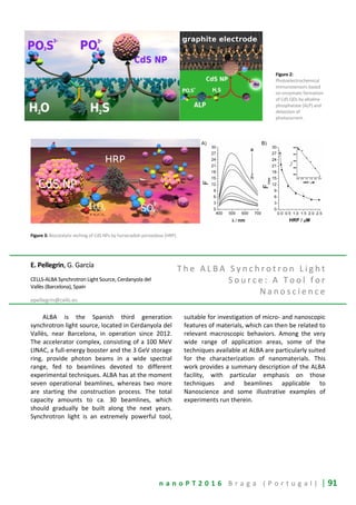
![92 | n a n o P T 2 0 1 6 B r a g a ( P o r t u g a l )
Manuel Pernia Leal1,2
, Carmen Muñoz-
Hernández1
, Sara Rivera-Fernández3
, Jaime M.
Franco
4
, David Pozo
4
, Jesús M. de la Fuente
3,5
,
Catherine C. Berry
2
b and María Luisa García-
Martín1
1
BIONAND, Andalusian Centre for Nanomedicine and
Biotechnology, BIONAND (Junta de Andalucía-Universidad
de Málaga), Málaga, Spain
2
Centre for Cell Engineering, Glasgow University, U.K
3
Institute of Nanoscience of Aragon, University of Zaragoza,
Zaragoza, Spain
4
CABIMER, Andalusian Center for Molecular Biology and
Regenerative Medicine, Seville, Spain
5
Institute of Materials Science of Aragon, CSIC, University of
Zaragoza, Spain
mpernia@bionand.es
O p t i m i z a t i o n o f b l o o d
c i r c u l a t i n g t i m e s o f
m a g n e t i c n a n o p a r t i c l e s
b a s e d o n t h e e f f e c t o f P E G
m o l e c u l a r w e i g h t c o a t i n g
a n d n a n o p a r t i c l e s i z e
f o l l o w e d b y M a g n e t i c
R e s o n a n c e I m a g i n g
Magnetic Resonance based Molecular Imaging
has emerged as a very promising technique for early
detection and treatment monitoring of a wide
variety of diseases, among them, cancer,
neurodegenerative disorders, stroke, etc. The
limited sensitivity and specificity of conventional
MRI are being overcome by the development of
novel contrast agents, most of them based on
nanotechnology approaches, with improved
magnetic and biological properties. In this work, we
report a facile and robust ligand-exchange method
to synthesize magnetic nanoparticles based on iron
oxide and manganese ferrite nanoparticles as MRI
contrast agents with long circulation times. The
selection of the right molecular weight PEG coating
on the nanoparticles and the nanoparticle size are
crucial points in the design that will determine the
fate of the magnetic nanoparticles. Therefore,
PEGylated small magnetic nanoparticles (PEG-
MNPs), using PEG MWs ranging from 600 to 8000,
were synthesized, resulting in highly stable and
water-soluble nanoparticles. Semi-quantitative and
quantitative MRI studies allowed us to track the
pharmacokinetics and biodistribution of
intravenously injected PEG-MNPs (HD < 50 nm) in
vivo up to one week. Results show that high MW
PEGs (6000-8000) lead to nanoparticle aggregation
and low MW PEGs (≤1500) are not able to stabilize
the 6 nm iron oxide nanoparticles in physiological
medium or confer stealth properties, thus leading to
rapid recognition by the RES. In contrast, PEG3000-
MNPs show excellent in vivo behavior, they do not
aggregate and exhibit better stealth properties,
giving rise to slower liver uptake and longer
circulation times. Moreover, we synthesize
manganese ferrite nanoparticles between 6 and 14
nm covered by a 3kDa polyethylene glycol (PEG)
shell that leads to a great stability and confer the
best stealth properties. These PEGylated MNPs have
shown different relaxivities r1 and r2 depending on
their nanoparticle core size, for instance the 6 nm
PEGylated MNP has a r1 value of 13.3 mM
-1
s
-1
and a
r2 value of 65 mM
-1
s
-1
with a low ratio r2/r1 of 4.9,
resulting in a good dual T1 and T2 contrast agent at
clinical magnetic field. On the other hand, the 14 nm
PEGylated MNP is an excellent T2 contrast agent at
high magnetic field, with a r2 value of 335.6 mM
-1
s
-1
.
The polymer core shell of the PEGylated MNPs
minimizes their cytotoxicity, and permits long blood
circulation times (24 h). This combination of cellular
compatibility, excellent T2 and T1 values at low fields,
together with long circulation times and moderate
liver uptake, make these nanomaterials very
promising contrast agents for molecular imaging.
R e f e r e n c e s
[1] Pernia Leal, M., Muñoz-Hernandez, C., Berry,
C. C. and Garcia-Martin, M. L. In Vivo
Pharmacokinetics of T2 Contrast Agents based
on Iron Oxide Nanoparticles: Optimization of
Blood Circulation times. RSC Advances 2015, 5,
76883.
[2] Pernia Leal, M., Rivera-Fernandez, S., Franco,
J. M., Pozo, D., de La Fuente, J. M. and Garcia-
Martin, M. L. Long-circulating PEGylated
manganese ferrite nanoparticles for MRI-
based molecular imaging. Nanoscale 2015, 7,
2050-2059](https://image.slidesharecdn.com/nanopt2016conferencebook-160222125923/85/NanoPt2016-Conference-Book-92-320.jpg)
![94 | n a n o P T 2 0 1 6 B r a g a ( P o r t u g a l )
membrane protein bacteriorhodopsin (BR) in
buffered solution. Further research towards
automated AFM has been put into the feature
“Experiment Control” which gives the opportunity
to control all main parameters of the AFM
remotely conveniently on any device, such as, a
tablet, PC or mobile phone without interfering
with the setup.
Additionally, we show the capability of
combining AFM with super-resolution techniques.
Firstly, we demonstrate the relation of
cytoskeleton distribution and mechanical
properties of HeLa cells. Alexa647 labeled
microtubules are imaged with dSTORM, while the
cell surface and mechanical information are
measured in parallel by AFM. Secondly, we show
AFM QI elastic moduli data of individual living
fibroblast cells and actin super-resolution STED
images of the same cell acquired in one
experiment. For this research, the JPK
NanoWizard® 4 AFM has been integrated into the
Abberior easy3D STED microscope.
In another study, we monitor and modify the
kinetics of collagen type I fibrillogenesis. It can be
shown that fast AFM imaging can be successfully
applied to understand the real-time kinetics of
collagen type I formation. By further modifying the
used buffer compositions, pH value and potassium
ion content, we demonstrate that we can alter the
kinetics of the fibrillar nanomatrix formation and
successfully study it with high spatial and temporal
resolution. In addition, the dynamics of a calcite
crystal surface at the atomic scale will be
demonstrated.
Tânia V. Pinto1
, P. Costa1
, C. M. Sousa2
, C. A. D.
Sousa
1
, A. Monteiro
3
, C. Pereira
1
,1 O. S. G. P.
Soares4
, C. J. S. M. Silva3
, M. F. R. Pereira4
, P. J.
Coelho2
, C. Freire1
1
REQUIMTE/LAQV, Departamento de Química e
Bioquímica, Faculdade de Ciências, Universidade do Porto,
Porto, Portugal
2
Departamento de Química e CQ-VR, Universidade de Trás-
os-Montes e Alto Douro, Vila Real, Portugal
3
CeNTI, Centro de Nanotecnologia e Materiais Técnicos,
Funcionais e Inteligentes, Vila Nova de Famalicão, Portugal
4
Laboratório de Catálise e Materiais (LCM), Laboratório
Associado LSRE-LCM, Departamento de Engenharia
Química, Faculdade de Engenharia, Universidade do Porto,
Porto, Portugal
tania_v_pinto@hotmail.com
P h o t o s w i t c h a b l e s i l i c a
n a n o p a r t i c l e s f o r t h e
p r o d u c t i o n o f l i g h t
r e s p o n s i v e s m a r t t e x t i l e s :
f r o m f a b r i c a t i o n t o c o a t i n g
t e c h n o l o g y
The design of high-performance
multifunctional textiles has been one of the
greatest challenges for Textile Industry, motivated
by consumers and markets demand for fabrics
with enhanced properties such as
(super)hydrophobicity, antimicrobial and fire
retardancy [1]. Photochromic textiles emerged as a
new niche market for the production of smart
clothing due to their switchable sensing properties
and protection against the harmful effects of UV
radiation; furthermore, they confer fancy color
effects to fashion and interior design decoration
[2–4]. Organic (or inorganic) photochromic dyes
are potential scaffolds to produce smart textiles
due to their switchable color
generation/disappearance in response to solar
light. Concerning organic photo-active species, the
most commonly reported are spiro-based
compounds – spiropyrans, spirooxazines and
naphthopyrans – because of their excellent
photoswitching capability and fatigue resistance
[5,6]. However, the incorporation of photochromic
dyes onto textiles has not been translated into
significant commercial success, mainly assigned to
technical limitations (dye degradation with high
temperatures and low dye uptake) and to their
lower performance upon immobilization [2–4,7,8].
Nevertheless, the immobilization of photochromic
dyes onto inorganic matrices constitutes a
promising route for the design of photochromic
textiles with efficient color switching, high comfort
and dye stability [4]. Moreover, SNPs have been](https://image.slidesharecdn.com/nanopt2016conferencebook-160222125923/85/NanoPt2016-Conference-Book-94-320.jpg)
![n a n o P T 2 0 1 6 B r a g a ( P o r t u g a l ) | 95
successfully applied to textiles to provide novel
functionalities, while preserving the pristine textile
properties (appearance, touch and washing
fastness) [2,9]. Although silica embedded with
photochromic dyes has been used to produce
functional hard surfaces (e.g. glass and plastic
films), its use in textiles has been less explored [4].
To the best of our knowledge, no work concerning
the textiles functionalization with photochromic
silica particles with nano dimensions (<100 nm)
has been published.
The purpose of this work was the fabrication
of a novel generation of light responsive textiles
with enhanced photochromic properties. To
achieve that goal, naphthopyrans were firstly
incorporated onto SNPs (~15 nm particle size) and
then the resulting photochromic nanomaterials
were incorporated onto cotton fabrics by
advanced screen-printing processes. All
nanomaterials were characterized in terms of
morphology, structure and chemical composition
by transmission electron microscopy with energy-
dispersive X-ray spectroscopy (TEM-EDS),
elemental analysis (EA), thermogravimetric
analysis (TG), Fourier transform infrared
spectroscopy (FTIR) and solid-state
29
Si and
13
C
nuclear magnetic resonance (NMR). The
characterization techniques confirmed the
successful immobilization of the photo-active
naphthopyran molecules onto the SNPs surface
and the preservation of their structure. The
photochromic properties in the solid-state were
evaluated by UV-Vis spectroscopy and colorimetry
before and after UV exposure (λ = 365 nm). All
hybrid nanomaterials revealed excellent photo-
switching behavior, showing fast
coloration/decoloration kinetics (coloring in 1 min
and bleaching in less than 2 min), good optical
density (∆OD ~ 1) and good color difference values
(∆E ~ 55); moreover, they presented promising
resistance to photodegradation upon prolonged
exposure to UV light (1 h). In the case of the
functional textiles, FTIR-ATR and TG analyses
proved the incorporation of the hybrid
nanomaterials on the screen-printed textiles.
Additionally, the resulting functional fabrics
showed notable photochromic behavior (Figure 1),
with a fast color change upon UV/visible light
irradiation (within seconds) and good reversibility
(a few minutes) for more than 12 UV/Dark cycles
without loss of their photochromic performance;
furthermore, the textiles showed high resistance
to photodegradation upon prolonged exposure to
UV light (1 h).
Acknowledgments: The work was funded by
Fundação para a Ciência e a Tecnologia (FCT)/MEC
under FEDER under Program PT2020 (projects
UID/QUI/50020/2013 and UID/EQU/50020/2013)
and through project ref. PTDC/CTM-
POL/0813/2012 in the framework of Program
COMPETE. T. V. Pinto (SFRH/BD/89076/2012), P.
Costa (grant under PTDC/CTM-POL/0813/2012
project), C. M. Sousa (SFRH/BD/75930/2011), C. A.
D. Sousa (SFRH/BPD/80100/2011) and O. S. G. P.
Soares (SFRH/BPD/97689/2013) thank FCT for
their grants.
R e f e r e n c e s
[1] S.L.P. Tang, G.K. Stylios, Int J Cloth Sci Tech. 18
(2006) 108–128..
[2] T. Lin, X. Wang, Int J Nanotechnol. 6 (2009)
579–598.
[3] M. Aldib, R.M. Christie, Color Technol. 129
(2013) 131–143.
[4] T. Cheng, T. Lin, R. Brady, X. Wang, Fiber
Polym. 9 (2008) 301–306.
[5] R. Pardo, M. Zayat, D. Levy, Chem Soc Rev. 40
(2011) 672−687.
[6] R. Klajn, Chem. Soc. Rev. 43 (2014) 148–84.
[7] N. Malic, J. a. Campbell, A.S. Ali, C.L. Francis, R.
a. Evans, J Polym Sci Pol Chem. 49 (2011) 476–
486.
[8] A.F. Little, R.M. Christie, Color Technol. 127
(2011) 275–281.
[9] T. Cheng, T. Lin, R. Brady, X. Wang, Fiber
Polym. 9 (2008) 521–526.
F i g u r e s
Figure 1: Schematic representation and photographs of photochromic
textiles prepared by screen-printing using SNPs functionalized with
organic compounds.](https://image.slidesharecdn.com/nanopt2016conferencebook-160222125923/85/NanoPt2016-Conference-Book-95-320.jpg)
![96 | n a n o P T 2 0 1 6 B r a g a ( P o r t u g a l )
Filipa Pires and M. Raposo
CEFITEC, Departamento de Física, Faculdade de Ciências e
Tecnologias, FCT, Universidade Nova de Lisboa, Caparica,
Portugal
af.pires@campus.fct.unl.pt mfr@fct.unl.pt
C a t e c h i n s : a p o w e r f u l w e a p o n
a g a i n s t o x i d a t i v e s t r e s s a n d
D N A l e s i o n s
The number of cancer cases has increased at a
terrifying rate worldwide due to exposure to
harmful mutagenic agents such as radiation,
tobacco, among others that causes mutations at a
DNA level. A health policy based on a balanced
diet involving healthy and plant-based foods seems
to ameliorate and be effective against cancer.
Catechins are the main plant-phenolic
component present on one of the most consumed
beverage in the world after water: the tea [1]. The
catechins intake through green tea ingestion can
alleviate or repair the DNA damage via antioxidant
mechanisms or by modulating the intracellular
redox environment. These dietary-derived
antioxidants molecules exert a chemopreventive
role during disease progression, offering a great
potential to be used in new cancer fighting
strategies [2-4].
One of the goals of our study is to reveal all
the physical processes underlying the action mode
of catechins in order to understand how these
compounds interact with DNA and affect the
biological environment and thus develop or
improve the current drug delivery systems. Our
previous work showed that is a pre-requisite have
a stable delivery system which provides sufficient
time to repair the DNA-damage induced by UV,
avoiding in this way the cell collapse [5, 6].
Thin films of catechin molecules encapsulated
in liposomes (DPPG) were prepared and exposed
to ultra-violet radiation in conditions near of cell
medium to assess the radiation-induced changes in
catechins and DNA. Additional radiation studies
will be carried out in order to evaluate the
photosensitizing properties and the efficacy of
these molecules to modulate DNA-damage
mechanisms.
R e f e r e n c e s
[1] Ross, J.A. & Kasum, C.M. Dietary flavonoids:
bioavailability, metabolic effects, and safety.
Annual review of Nutrition 22, 19-34 (2002).
[2] Ershov, D. et al. Investigation of the
radioprotective properties of some tea
polyphenols. Structural Chemistry 22, 475-482
(2011).
[3] Nikjoo, H., O'Neill, P., Terrissol, M. &
Goodhead, D. Modelling of radiation-induced
DNA damage: the early physical and chemical
event. International journal of radiation
biology 66, 453-457 (1994).
[4] Ho, C.K., Choi, S.w., Siu, P.M. & Benzie, I.F.
Effects of single dose and regular intake of
green tea (Camellia sinensis) on DNA damage,
DNA repair, and heme oxygenase-1 expression
in a randomized controlled human
supplementation study. Molecular nutrition &
food research 58, 1379-1383 (2014).
[5] Gomes, P.J. et al. Energy Thresholds of DNA
Damage Induced by UV Radiation: An XPS
Study. The Journal of Physical Chemistry B
119, 5404-5411 (2015).
[6] Gomes, P.J., da Silva, A.M.G., Ribeiro, P.A.,
Oliveira, O.N. & Raposo, M. Radiation damage
on Langmuir monolayers of the anionic 1.2-
dipalmitoyl-sn-glycero-3-[phospho-rac-(1-
glycerol)](sodium salt)(DPPG) phospholipid at
the air–DNA solution interface. Materials
Science and Engineering: C 58, 576-579 (2016).
F i g u r e s
Figure 1: Infrared spectra of DPPG cast films prepared from DPPG
aqueous solutions without and with UV irradiation. To visualize the
damage induced in DPPG by exposure to radiation, the difference
between the spectra was also added. The vertical arrows indicate the
wavenumber of bands that disappear upon irradiation.](https://image.slidesharecdn.com/nanopt2016conferencebook-160222125923/85/NanoPt2016-Conference-Book-96-320.jpg)
![n a n o P T 2 0 1 6 B r a g a ( P o r t u g a l ) | 97
Table 1: Characteristic infrared absorptions in DPPG cast films
Bernardo Pires, A. Moskaltsova, D.C. Leitão
and S. Cardoso
INESC-Microsystems and Nanotechnologies (MN) and IN,
Lisbon, Portugal
Department of Physics, Instituto Superior Técnico,
Universidade Lisboa, Lisbon, Portugal
bernardojpires@tecnico.ulisboa.com
H i g h P r e c i s i o n M e t h o d o l o g y
C o n t r o l f o r N a n o M T J
F a b r i c a t i o n P r o c e s s u p t o
1 5 0 m m W a f e r s
Spintronic devices have received a great
attention in the past decades, and provided
considerable applications in industry and
electronic information. Among them, one
highlights the spin transfer torque magnetic
random access memory (STT-MRAM), pointed as
the next generation of non-volatile memory with
commercialized products entering the market very
soon, and the already mature HDD read heads
already in sub 100 nm range. Still, current
demands concerning stacking density, devices size
and performance, continuously push the limits of
standard nanofabrication techniques.
The basic building blocks of both structures is
the magnetic tunnel junction (MTJ), which is a
highly scalable technology. Scaling-down the MTJ
critical features down to 30 nm and simultaneously
integrate them on larger area wafers with highly
controlled and standardized process, providing a
very high yield of working devices, is a main
challenge for magnetic storage industries
nowadays. INESC-MN has already demonstrated
successful patterning of nanoscale MTJs down to
100 nm based on a lift-off process with yield of
88% [1]. However, this progress was limited to 25
mm substrates. A more promising route towards
sub 100 nm was also explored using chemical
mechanical polishing (CMP). A 30 nm full MTJ
device was demonstrated, although the process
showed significant challenges in controlling the
CMP end point reflecting dramatically in the final
yield of working devices [2].
This work, relies on a distinct method for
fabrication of sub 100 nm MTJ devices, targeting
large yield in 150 mm substrates. Nano-MTJs were
fabricated using combination of optical lithography
(OL), ion milling and electron beam lithography
(EBL). Then, to achieve functional and operational
devices, the critical steps are the nanopillars
definition and the definition (and opening) of
electrical vias to pillars after passivation (300 nm
SiO2 film). The nanopillars (circles of 30 to 100nm
diameter) are defined by EBL using a Raith-150
System and negative resist [3] followed by two-
step ion milling etching, while the vias (500 nm
size) are defined by EBL using a positive resist
(PMMA) followed by Reactive Ion Etching (RIE)
(see Figure 1).
Final yield of working devices depends on a
large number of factors, such as resist and SiO2
thickness and uniformity, exposure resolution and
alignments, or RIE end-point and RIE uniformity. At
this point, we focus on EBL. We have
systematically studied the misalignment between
consecutive write field (WF) exposures, stage
movement drift and misalignment between BE,
nanopillars and vias, critical for the success of the
fabrication process on 150 mm wafers. Customized
test structures were designed for automatic
quantification of local deviations. Analyses were
performed using SEM images, image process and
data analysis programs (see Figure 2). Our results
provided deviations between WF (of 500 µm)
exposures up to 530 nm in the horizontal direction,
and a maximum of 700 nm in the vertical direction.](https://image.slidesharecdn.com/nanopt2016conferencebook-160222125923/85/NanoPt2016-Conference-Book-97-320.jpg)
![98 | n a n o P T 2 0 1 6 B r a g a ( P o r t u g a l )
We also demonstrate highly uniform (better than
+-1%) negative EBL resist coating up to 80 nm thick
(see Figure 3) which provides the minimum
resolution of our EBL system [2].Gathering all
these factors, we can set a maximum value
tolerance (~250 nm) for the misalignment of the
mix & match exposure between BE, nanopillars
and vias using a WF=500 µm for single isolated
devices. This conditions are crucial in expediting a
large yield MTJ nanofabrication process run for
150 mm wafers towards commercialization of
nanodevices.
R e f e r e n c e s
[1] D.Leitão, E. Paz, A. Silva et al., IEEE
Transactions on Magnetics 50, No. 11 (2014)
[2] R. Macedo, J. Borme, R. Ferreira et al., Journal of
nanoscience and nanotechnology 10, 2010, 1-7
[3] D. Leitão, R. Macedo, A. Silva et al.,
Proceedings of the IEEE Conference on
Nanotechnology, 2012
F i g u r e s
Figure 1: Illustration of the nano fabrication process from bottom electrode definition until metallization of top electrode (a)-(d), highlighting the
critical steps of nanopillars definition (b) and definition and opening of electrical vias to pillars after passivation (c) . Top: 3D model of the fabrication
process. Bottom: Section of the MTJ device during fabrication process.
Figure 2: Statistical analysis of horizontal misalignment
between consecutive write-fields exposures in a 20 x 20 mm2
map on 150 mm wafer. The inset shows: a) SEM image of
misaligned exposed structures and b) design of test structures
for automaticquantification of local deviations
Figure 3: Left: 3D negative EBL resist profile thickness along a 150 mm wafer
measured by ellipsometry; Right: Uniformity thickness profile of negative EBL resist
D. M. F. Prazeres, A. M. M. Rosa, J. R. C.
Trabuco, A. R.M. Almeida
iBB - Institute for Bioengineering and Biosciences,
Department of Bioengineering, Instituto Superior Técnico,
Universidade de Lisboa, Lisboa, Portugal
miguelprazeres@tecnico.ulisboa.pt
C a r b o h y d r a t e b i n d i n g
m o d u l e s a s a g e n e r i c t o o l t o
a n c h o r b i o m o l e c u l e s a n d
m e t a l n a n o p a r t i c l e s o n t h e
s u r f a c e o f p a p e r - b a s e d
b i o s e n s o r s
There is a global demand for affordable,
sensitive, selective and rapid analytical platforms
usable in low-tech contexts to perform health
diagnostics, environmental monitoring and food
quality testing. Paper-based analytical devices have
emerged as one of such platforms, with the
additional advantages of being biodegradable, easy-
to-use and portable [1]. Paper can be modified and
adapted to perform biological assays by adding
appropriate biorecognition and reporting agents (e.g.
antibodies, enzymes, oligonucleotides and DNA
aptamers) to the test areas [2]. Additionally, paper](https://image.slidesharecdn.com/nanopt2016conferencebook-160222125923/85/NanoPt2016-Conference-Book-98-320.jpg)
![n a n o P T 2 0 1 6 B r a g a ( P o r t u g a l ) | 99
surfaces can be modified with metal nanoparticles
(e.g. Au, Ag, and Cu, among others) to introduce
optical and electronic properties better suited for
biosensing applications [3,4]. One of the keys for the
success of these paper surfaces is the ability to
master the immobilization of biomolecules and metal
nanoparticles, while adequately preserving
functionality and stability.
Covalent attachment to the cellulose fibers is not
a strict requirement for the incorporation of either
biomolecules or metal nanoparticles into paper. In
fact, dry paper itself is able to sorb aqueous solutions
in such a way that the non-volatile components of
the solutions are left in the paper structure after
drying. However, impregnation without attachment
may not be a robust strategy to immobilize
biomolecules or metal nanoparticles because
subsequent exposure to aqueous solutions (e.g.
washing buffers or biological samples) is likely to
leach these components. Furthermore, it is difficult
to control the orientation of biomolecules in the
paper structure, especially in the case of antibodies,
with recognition sites taking different positions in
space after random immobilization, resulting in
hindered interactions with their binding target [5].
We have developed an immobilization platform
that uses specialized proteins named Carbohydrate
Binding Modules (CBMs) that have a natural affinity
to cellulose, to anchor biomolecules and metal
nanoparticles on paper surfaces as an alternative to
conventional methods like covalent binding and
physical adsorption. The strategy relies on the fusion
of biosensing molecules (e.g. affinity handles,
enzymes, oligonucleotides) with CBMs and on their
subsequent immobilization on paper via affinity
interactions.
In this communication, specific applications are
presented that rely on CBM3-ZZ, a fusion protein that
combines the cellulose-binding properties of CBM3a
from Clostridium thermocellum with the antibody-
binding properties of a double Z-domain from the
staphylococcal protein A [5]. Using these fusion
proteins, properly oriented antibodies could be
anchored on paper surfaces (Fig. 1a). By further
exploring the recognition ability of these antibodies,
we were able to immobilize 40 nm gold nanoparticles
(AuNPs, Fig. 1b) and capture DNA hybrids labeled
with AuNPs (Fig. 1c) on the surface of
chromatographic paper (Whatman N. 1). Our results
have shown that colorimetric signals could be
generated that differed substantially from the ones
presented when AuNPs or DNA hybrids labeled with
AuNPs were simply deposited on paper, without the
assistance of CBM3–ZZ fusions (Fig. 2). A SEM
analysis revealed that the difference in the
colorimetric signals could be attributed to the fact
that AuNP homogenously distribute in the paper
matrix when immobilized via CBM-3-ZZ fusions,
whereas they tend to aggregate when they are
simply deposited over paper. By plasmon resonance
effect these differences in AuNP aggregation then
generated the observed color differences (Fig. 2).
As a proof-of-concept, a strategy for the
detection of nucleic acids from Trypanosoma brucei,
the causative agent of sleeping sickness was
developed. In order to confine fluids to specific
regions of paper, a wax printing methodology was
used to print hydrophobic barriers that delineate
circular reaction areas. We then combined the
CBM3-ZZ–based anchoring of antibodies with DNA
probes specific for T. brucei conjugated with gold
nanoparticles. The methodology involved i) the pre-
conjugation of CBM3-ZZ with an anti-biotin antibody,
ii) the deposition of the CBM3-ZZ:antibiotin antibody
conjugate on paper, iii) the pre-hybridization of biotin
labeled target with AuNP labeled probes off-paper
and application on the bioactive circular region of
paper and iv) the visual detection of colored signals.
Our results showed that colorimetric readouts in the
form of red spots were generated only when DNA
strands complementary to the probe were tested.
In summary, we have developed a bioaffinity
based platform for the immobilization of
biomolecules and metal nanoparticles on paper that
is compatible with biosensing applications.
Furthermore, we demonstrated that the
methodology enhances plasmon resonance effects
induced by AuNPs on paper surfaces, making it
possible to perform simple molecular and
immunological diagnostics tests.
References
[1] D. M. Cate, J. Adkins, J. Mettakoonpitak, C. S.
Henry, Analytical Chemistry, 87 (2015) 19-41.
[2] R. Pelton, Trends Analytical Chemistry 28 (2009)
925–942.
[3] R. J. B. Pinto, M. C. Neves, C. P. Neto, T.
Trindade, in Nano Composites: New Trends and
Developments, F. Ebrahimi (ed.), Intech, Rijeka,
Chap. 4 (2012) pp. 73-96.
[4] Y. H. Ngo, D. Li, G. P. Simon, G. Garnier,
Langmuir, 28 (2012) 8782-8790.
[5] A. M. M. Rosa, F. Louro, S. M. Martins, J. Inácio,
A. M. Azevedo, D. M. F. Prazeres, Analytical
Chemistry 86 (2014) 4340–4347.](https://image.slidesharecdn.com/nanopt2016conferencebook-160222125923/85/NanoPt2016-Conference-Book-99-320.jpg)
![102 | n a n o P T 2 0 1 6 B r a g a ( P o r t u g a l )
pharmacokinetics problems of the encapsulated
drugs and can be guided and localized to the
therapeutic sites of interest by external magnetic
field gradients [1,2]. The use of magneto-sensitive
liposomes as nanocarriers allows a safer use of
powerful anticancer drugs in therapy with lower
drug dosage and a more efficient treatment, not
only in cancer but also in other diseases.
In this work, manganese ferrite (MnFe2O4)
nanoparticles with superparamagnetic behaviour
at room temperature and size distribution of
26 ± 5 nm, were obtained by coprecipitation
method. Structural and magnetic properties of the
nanoparticles (NPs) were evaluated by XRD, HR-
TEM and SQUID. The synthesized NPs were either
entrapped in liposomes, originating aqueous
magnetoliposomes (AMLs), or covered with a lipid
bilayer, forming solid magnetoliposomes (SMLs),
the latter prepared by a new method, recently
developed for magnetoliposomes based on nickel
ferrite NPs [3]. The resulting liposomes exhibit
sizes below 150 nm (Fig. 1), suitable for biomedical
applications.
Membrane fusion between both types of
magnetoliposomes and GUVs (giant unilamellar
vesicles), used as models of cell membranes, was
confirmed by FRET (Förster Resonance Energy
Transfer) assays [3-5]. For that purpose, the
labeled lipid NBD-C12-HPC and the hydrophobic
probe Nile Red (or the labeled lipid Rhodamine B-
DOPE) were both incorporated in the lipid bilayer
of magnetoliposomes, the NBD moiety acting as
the energy donor and the dye Nile Red (or
Rhodamine B) as the energy acceptor (Fig. 2). After
interaction with GUVs, an increase in the NBD
(donor) emission band and a decrease of acceptor
fluorescence is observed (Fig. 2A), confirming
membrane fusion (Fig. 2B) [5].
A new potential antitumor drug, a
thienopyridine derivative (Fig. 3), was successfully
incorporated in the lipid bilayer of both types of
magnetoliposomes. This thienopyridine derivative
presents very low growth inhibitory concentration
values (GI50), between 3.5 and 6.9 µM, when
tested in vitro against several human tumor cell
lines, namely MCF-7 (breast adenocarcinoma),
A375-C5 (melanoma) and NCI-H460 (non-small cell
lung cancer) and was the most active of a series of
analogues [6]. Moreover, this compound has
shown a very low affinity for the multidrug
resistance protein MDR1 [7], being suitable as an
anticancer agent.
These results point to a promising application
of magnetoliposomes in oncological therapy,
simultaneously as hyperthermia agents and as
nanocarriers for antitumor drugs, taking also
advantage of magnetic directioning.
R e f e r e n c e s
[1] A. S. Lubbe, C. Bergemann, J. Brock, D. G.
McClure, J. Magn. Magn. Mat. 194 (1999) 149-
155.
[2] S. Dandamudi, R. B. Campbell, Biomaterials 28
(2007) 4673-4683.
[3] A. R. O. Rodrigues, I. T. Gomes, B. G. Almeida, J. P.
Araújo, E. M. S. Castanheira, P. J. G. Coutinho,
Phys. Chem. Chem. Phys. 17 (2015) 18011-18021.
[4] A. R. O. Rodrigues, I. T. Gomes, B. G. Almeida, J. P.
Araújo, E. M. S. Castanheira, P. J. G. Coutinho,
Mat. Chem. Phys. 148 (2014) 978-987.
[5] A. R. O. Rodrigues, J. M. F. Ramos, I. T. Gomes, B.
G. Almeida, J. P. Araújo, M.-J. R. P. Queiroz, E. M.
S. Castanheira, P. J. G. Coutinho, submitted to
publication.
[6] M.-J. R. P. Queiroz, R. C. Calhelha, L. Vale-Silva, E.
Pinto, M. S.-J. Nascimento, Eur. J. Med. Chem. 45
(2010) 5732-5738.
[7] C. N. C. Costa, A. C. L. Hortelão, J. M. F. Ramos, A.
D. S. Oliveira, R. C. Calhelha, M.-J. R. P. Queiroz, P.
J. G. Coutinho, E. M. S. Castanheira, Photochem.
Photobiol. Sci. 13 (2014) 1730-1740.
F i g u r e s
Figure 1: TEM image
of solid
magnetoliposomes
(SMLs) containing
MnFe2O4 NPs.](https://image.slidesharecdn.com/nanopt2016conferencebook-160222125923/85/NanoPt2016-Conference-Book-102-320.jpg)
![n a n o P T 2 0 1 6 B r a g a ( P o r t u g a l ) | 103
Figure 2: A. Fluorescence spectra (λexc=400 nm) of AMLs containing both NBD-C12-HPC and
Nile Red, before and after interaction with GUVs. Inset: Spectral overlap between the
fluorescence emission of the donor (NBD-C12-HPC) and the absorption of the acceptor (Nile
Red). B. Schematic representation of membrane fusion between AMLs and GUVs.
Figure 3: Structure of the antitumor
thienopyridine derivative.
M.L. Rodriguez-Mendez1
, C. Garcia-
Hernandez1
, C. Medina-Plaza1
, C. Garcia-
Cabezon
2
,D. Paniagua1
, S. Rodriguez1
, F.
Pedrosa2
, J.A. de Saja3
1
Department of Inorganic Chemistry, Engineers School,
Universidad de Valladolid, Valladolid, Spain
2
Department of Materials Science, Engineers School,
Universidad de Valladolid, Valladolid, Spain
3
Department of Condensed Matter Physics, Faculty of
Sciences, Universidad de Valladolid, Valladolid, Spain
mluz@eii.uva.es
A n t i o x i d a n t s d e t e c t i o n w i t h
n a n o s t r u c t u r e d
e l e c t r o c h e m i c a l s e n s o r s
The use of quick, reliable and cheap sensors
for the detection of chemical compounds
represents an important need in the food industry.
Antioxidants are among the analytes that must be
monitored and measured in order to guarantee
the quality of final products. In particular, phenolic
and polyphenolic compounds are interesting
antioxidants because they inhibit or delay the
oxidation processes by blocking the initiation or of
oxidizing chain reactions.
Previous works have demonstrated that
voltammetric electrodes chemically modified with
electrocatalytic materials can be used to detect
such compounds in musts and wines [1].
Electrodes chemically modified can be an
advantage because the electrocatalytic activity of a
variety of modifiers (carbon nanotubes,
nanoparticles, porphyrins, phthalocyanines, etc)
can reduce the oxidation potential while increasing
the intensity of the response. In turn, electrodes
can be prepared using a variety of techniques from
simple Carbon Paste Electrodes (CPE) or Screen
Printed Electrodes (SPE) to extremely sophisticated
nanostructured sensors prepared using the
Langmuir-Blodgett (LB) or the electrostatic Layer-
by-Layer (LbL) techniques.
Nanostructured sensors have the advantage of
the enhanced number of active sites producing an
increase in the intensity. Moreover, the control of
molecular architectures afforded by these
techniques can led to the development of a variety
of devices where synergy is achieved by combining
distinct materials, including organic-inorganic
hybrids [2].
For instance, combinations of phthalocyanines
with nanoparticles or carbon nanotubes in LB films
have been developed and their structures have
been analyzed. The films have been used as
voltammetric sensors for the detection of
compounds of interest in the food industry (i.e.
citric acid) (Figure 1). The combination of
phthalocyanines with carbon nanotubes (CNT)
produced a clear increase in the intensity of the
responses due to the synergy promoted by the pi-
pi stacking between both components.
Similarly, gold nanoparticles in LB films
produced an increase in the sensitivity towards
phenols and detection limits of 10
-7
mol.L
-1
were
attained. Similar detection limits could be obtained](https://image.slidesharecdn.com/nanopt2016conferencebook-160222125923/85/NanoPt2016-Conference-Book-103-320.jpg)
![104 | n a n o P T 2 0 1 6 B r a g a ( P o r t u g a l )
by combining phthalocyanines with silver
nanoparticles in LbL films.
Finally, voltammetric biosensors have also
been prepared using the LB technique (Figure 2).
LB films combining phthalocyanines and
amphiphilic molecules provided biomimetic
environments where enzymes could preserve their
functionality. The role of the molecular
interactions in the electrocatalytic properties in
biomimetic systems has been studied by
combining enzymes with different electron
mediators and the existence of synergistic effects
has been evidenced. Detection limits as low as 10
-8
mol.L
-1
towards phenols can be attained for the
detection of phenols.
It has been demonstrated that the arrays
formed by voltammetric electrodes (voltammetric
electronic tongue) modified with phthalocyanines
are able to discriminate complex liquids. Arrays of
sensors based on MPcs have been successfully
used to discriminate wines of different qualities,
grape variety or wines prepared using different
techniques or aged in different types of oak barrels
[3]. The capability of discrimination is due to the
sensibility of phthalocyanine sensors towards
redox (i.e. polyphenols) and acids present in wines.
Their electrocatalytic properties play also an
important role in the discrimination capabilities of
the array.
Acknowledgements: The authors are grateful
to FEDER and to the Spanish Ministry of Science-
CICYT (Grant AGL2012-33535), Junta de Castilla y
León (VA-032U13) and FPI-UVa for the financial
support.
R e f e r e n c e s
[1] Rodriguez-Mendez, M.L., Medina, C., De Saja,
J.A., Apetrei, C., Muñoz, R. Eds. Lvova, L.,
Kirsanov, D., Di Natale, C., Legin, A. Pan
Stanford Publisheing, Chapter 4 (2012) 70-109.
[2] Pavinatto, F.J., Fernandes, E.G.R., Alessio, P.,
Constantino, C.J.L., De Saja, J.A., Zucolotto, V.,
Apetrei, C., Oliveira O.N.Jr., Rodriguez, M.L. J.
Mat. Chem. 21 (2011) 4995-5003.
[3] Prieto,N., Gay, M., Vidal, S., Aagaard, O., De
Saja, J.A., Rodriguez-Méndez M.L. Food Chem.
129 (2011) 589-594.
F i g u r e s
Figure 1: LB film based sensors exposed to 0.1 M citric acid. (Blue)
Lutetium bisphthalocyanine films, (red) Lutetium bisphthalocyanine +
CNT films.
Figure 2: Voltammetric biosensor formed by an amphiphile, a
phthalocyanine and an enzyme (tyrosinase).
M. H. M. Sá, M. Goreti F. Sales and Lúcia
Brandão
BioMark/CINTESIS, ISEP, Porto, Portugal
mhmsa@isep.ipp.pt
C a r b o n B l a c k m o d i f i c a t i o n
t o w a r d s e l e c t r o c h e m i c a l
b i o s e n s o r s
The development of innovative electrical
biosensors for early detection of cancer powered by
a passive direct methanol fuel cell (DMFC) is the
core of the work presented. In fact, the current state
of the art of electrical detection methodologies
underpin the progressive drive towards
miniaturised, sensitive and portable biomarker
detection protocols [1], which in our case was
synergistically associated with the molecular
imprinting strategy of the biomarkers [2].
Having as target a protein biomarker of the
rectal colon cancer, carcinoembryonic antigen
(CEA) will be recognized by a proper molecularly
imprinted polymer (MIPs) matrix, assembled inside](https://image.slidesharecdn.com/nanopt2016conferencebook-160222125923/85/NanoPt2016-Conference-Book-104-320.jpg)
![n a n o P T 2 0 1 6 B r a g a ( P o r t u g a l ) | 105
the DMFC. The process of molecular imprinting
involves the formation of recognition cavities by
connecting different polymeric building blocks
under the guidance of a molecular template (or
print molecule) (Figure 1).
Besides the optimization of the
polymer/protein matrix, the proper modification
of electrocatalysts within the DMFC is also
considered. For this purpose, Carbon Black is being
modified with pyrenes, namely 1-Pyrenemethyl
methacrylate (PyMMA) or 1-Pyrenebutyric acid
(PyBA) (Figure 2), and the resulting modification
followed by thermogravimetric analysis and
different spectroscopic techniques, like FTIR,
Raman and UV-Vis.
This approach has been applied with success to the
dispersion of carbon nanotubes [3], showing that
non-covalent interactions (π - π stacking) is
attractive in terms of the stability and
homogeneity of the functionalization. This surface
functionalization is expected to anchor the MIP
formation and compared in terms of the
effectiveness of polymer binding and performance
of the DMFC.
In a first approach, the MIP material is
prepared by free radical co-polymerization of vinyl
based monomers and crosslinkers in a buffered
aqueous medium. Morphological observations and
detailed experimental characterization reveals that
CB surface modifications occurred.
Acknowledgments: The authors acknowledge
funding from European Union’s Horizon 2020
research and innovation program through H2020-
FET-Open-Symbiotic, GA 665046.
R e f e r e n c e s
[1] X. Luo and J. Davis, Chem. Soc. Rev., 42 (2013)
5944.
[2] L. Ye and K. Mosbach, Chem. Mater., 20 (2008)
859.
[3] T. Fujigaya and N. Nakashima, Sci. Technol.
Adv. Mater., 16 (2015) 024802.
F i g u r e s
Figure 1: Schematic presentation of the molecular imprinting
approach at the carbon black surface.
Figure 2: Molecular structure of a) 1-Pyrenemethyl methacrylate (PyMMA) and
b) 1-Pyrenebutyric acid (PyBA).
H. Limborço
1,2
, P.M.P. Salomé
1
, D.G. Stroppa
1
,
R. R-Andrade1,2
, N. Nicoara1,3
, K. Abderrafi1,3
,
J.P. Teixeira
4
, J.P. Leitão
4
, J.C. Gonzalez
2
and
S. Sadewasser1
1
International Iberian Nanotechnology Laboratory, Braga,
Portugal
2
Departamento de Física, Universidade Federal de Minas
Gerais, Belo Horizonte, Minas Gerais, Brazil
3
IMM-Instituto de Microelectrónica de Madrid (CNM-CSIC),
Tres Cantos, Madrid, Spain
4
Departamento de Física and I3N, Universidade de Aveiro,
Aveiro, Portugal
sascha.sadewasser@inl.int
G r o w t h o f C u I n S e 2 n a n o w i r e s
b y m o l e c u l a r b e a m e p i t a x y
w i t h o u t e x t e r n a l c a t a l y s t
Chalcopyrite materials of the composition
Cu(In,Ga)Se2 (CIGSe) represent the light absorbing
layer in the thin-film solar cell technology with the
currently highest power conversion efficiency
(21.7% [1]), outperforming multi-crystalline Si solar
cells. The most efficient CIGSe material is grown in
a three-stage coevaporation process [2]. On the
other hand, the use of semiconductor](https://image.slidesharecdn.com/nanopt2016conferencebook-160222125923/85/NanoPt2016-Conference-Book-105-320.jpg)
![106 | n a n o P T 2 0 1 6 B r a g a ( P o r t u g a l )
nanostructures has received significant attention
in the quest to enhance power conversion
efficiencies of solar cells by quantum effects
and/or light management structures [3].
We report the growth of CuInSe2 nanowires
using a molecular beam epitaxy system where the
elemental constituents Cu, In, and Se are
evaporated from elemental sources at low
evaporation rates of ~0.5 nm/min. The growth of
the wires occurs on top of an underlying CuInSe2
polycrystalline layer that initially forms on the
Si(100) substrate, where the native oxide has not
been removed intentionally. Reference samples,
where the native oxide was removed chemically
prior to the CuInSe2 growth and where the same
growth process is performed, do not exhibit the
presence of the nanowires. The structure and
composition of single wires were analyzed by
transmission electron microscopy (TEM) using
selective area electron diffraction (SAED) and
energy dispersive x-ray spectroscopy (EDX). The
structure of the nanowires is identified as
tetragonal, the same structure observed for
polycrystalline thin-film material. High resolution
(HR) TEM analysis indicates a high crystalline
quality of the nanowires. X-ray diffraction (XRD)
identifies the polycrystalline layer as CuInSe2 and
photoluminescence at low temperature revealed
an emission in the range ~0.8-1.0 eV
demonstrating strong optical activity of the
samples. The visible and near-infrared spectral
part of the optical reflectivity of samples with a
high density of nanowires is reduced compared to
reference samples without nanowires, making the
realized nanowire structures interesting for solar
energy harvesting. A series of growth experiments
with a variation of the growth parameters was
carried out to identify a growth model for the
CuInSe2 nanowires. Based on the observed relation
between nanowire density and growth
parameters, we propose the formation of liquid In-
Se droplets on the polycrystalline CuInSe2 base
layer as a seed for the nanowire growth.
Acknowledgements: We acknowledge financial
support from the Fundação CAPES (Brazil) through the
CAPES-INL collaboration project (04/14), from the
Ministerio de Economía y Competitividad (Mineco,
Spain) through the collaboration project IMM-CSIC
with INL (AIC-B-2011-0806), and from the projects
RECI/FIS-NAN/0183/2012 (COMPETE: FCOMP-01-
0124-FEDER-027494) and UID/CTM/50025/2013 from
the Fundação para a Ciência e a Tecnologia (Portugal).
P.M.P.S. acknowledges financial support from EU
through the FP7 Marie Curie IEF 2012 Action No.
327367.
R e f e r e n c e s
[1] Ph. Jackson, D. Hariskos, R. Wuerz, O. Kiowski,
A. Bauer, T. Magorian Friedlmeier, and M.
Powalla, physica status solidi (RRL) 9, 28 (2015).
[2] A. Chirila et al., Nature Mater. 10, 857 (2011).
[3] A. Polman and H.A. Atwater, Nature Mater.
11, 174 (2012).
Adi Salomon
Bar Ilan University, Ramat-Gan, Isarel
Adi.salomon@biu.ac.il
S t r o n g C o u p l i n g i n P l a s m o n i c
s y s t e m s a n d t h e i r I n t e r a c t i o n
w i t h M o l e c u l e s
We study the optical properties of molecules
deposited metallic nanostructures with respect to
the free molecules. We show theoretically and
experimentally that molecular excited states can
be strongly coupled to plasmonic modes. Upon
coupling new hybrid states are form, the lower and
the higher polariton. These modes have the
characteristic of both molecular and plasmonic
states. As the coupling strength grows, a new
mode emerges, which is attributed to long-range
molecular interactions mediated by the plasmonic
field. The new, molecular-like mode repels the
polariton states, and leads to an opening of energy
gaps. By tuning the plasmonic modes to be on/off
resonance with respect to molecular system
excited state, one can shift these hybrid modes
and by that modify the photo-physical and even
the chemical properties of these molecules, and
form a new kind of tunable hybrid materials.
In the same aspect, we study and demonstrate
the strong coupling between plasmonic modes of
metallic nanocavities (holes). The geometric
parameters of the cavity, the distance between
them and density of electrons participating in the
modes are all determine the nature of
hybridization. We study by cathodeluminescence](https://image.slidesharecdn.com/nanopt2016conferencebook-160222125923/85/NanoPt2016-Conference-Book-106-320.jpg)
![n a n o P T 2 0 1 6 B r a g a ( P o r t u g a l ) | 107
together with linear and nonlinear optical
measurements the nature of coupling in
nanocavities milled either in aluminum or in silver
and discuss their application for hybrid materials
and catalysis. We further use such strong coupling
to strongly enhance the nonlinear responses of the
metallic surface and to tune actively the
electromagnetic field at the sub-micron scale.
R e f e r e n c e s
[1] A. Salomon, R. J. Gordon, Y. Prior, T.
Seideman, and M. Sukharev, Phys. Rev. Lett.,
vol. 109, no. 7, p. 73002, 2012.
[2] A. Salomon, S. Wang, J. A. Hutchison, C. Genet,
and T. W. Ebbesen, ChemPhysChem, vol. 14,
no. 9, pp. 1882–1886, 2013.
[3] A. Salomon, M. Zielinski, R. Kolkowski, J. Zyss,
and Y. Prior, J. Phys. Chem. C, vol. 117, no. 43,
pp. 22377–22382, 2013.
[4] A. Salomon, Y. Prior, M. Fedoruk, J. Feldmann,
R. Kolkowski, and J. Zyss, J. Opt., vol. 16, no.
11, p. 114012, 2014.
[5] M. Sukharev, T. Seideman, R. J. Gordon, A.
Salomon, and Y. Prior, ACS Nano., vol. 8, no. 1,
pp. 807–817, 2014.
[6] A. Salomon, C. Genet, and T. W. Ebbesen, vol.
48, no. 46, pp. 8748–8751, 2009.
F i g u r e s
Figure 1: Images of
Cathtodoluminescence of
metallic nano cavities at
400nm±20nm. (a) SEM image
of the studied plasmonic
structure, the triangular hole
side length is about 200nm
and the distance between
them is about 400nm. The CL
revealed the nature of
coupling and the difference
between (b) silver and
aluminum (c).
Figure 2: Simulations of a similar system. (a)
Transmission spectra for the series of Ag slit arrays
covered by a 10 nm thin film of molecular layer with a
density of 3x 10
25
m
3
. An additional mode is clearly seen
at about 2.64 eV. (b) Anti-crossing behavior of the
hybrid system with RS value of 0.15 eV. The peak
position of the additional mode barely changes with
detuning of SPP mode. (ref[5])
Laura M. Salonen, Marisa P. Sárria, Carlos
Rodríguez-Abreu, Begoña Espiña
International Iberian Nanotechnology Laboratory, Braga,
Portugal
laura.salonen@inl.int
C o v a l e n t O r g a n i c F r a m e w o r k s
f o r t h e C a p t u r e o f W a t e r b o r n e
T o x i n s
Nanoporous 2D covalent organic frameworks
(COFs) are crystalline materials formed by the self-
assembly of organic building blocks, driven by
aromatic stacking interactions in the third
dimension. Due to their structural tunability, large
specific surface area, and low density, COFs show
great promise for a wide variety of applications,
such as catalysis, gas storage, adsorption, and
optoelectronics.
The presence of biotoxins in food and water is
a general threat to human health that causes
yearly many diseases and even mortalities](https://image.slidesharecdn.com/nanopt2016conferencebook-160222125923/85/NanoPt2016-Conference-Book-107-320.jpg)
![108 | n a n o P T 2 0 1 6 B r a g a ( P o r t u g a l )
worldwide. Therefore, to prevent and remediate
the negative impact of toxic outbreaks, it is
important to establish efficient strategies and find
new materials for biotoxin separation and water
detoxification. COFs are interesting candidates for
waterborne biotoxin encapsulation due to their
tunable and uniform pore size and shape, which
would allow for a more selective toxin capture in
comparison to the commonly used macroporous
resins.
We have prepared different COFs and
evaluated their ability to adsorb marine toxin
okadaic acid. Absorption capacity, desorption, and
absorption kinetics were studied. A water-stable
COF derivative was found to capture the toxin
efficiently in both ultrapure water as well as
synthetic seawater, showing the potential of these
materials for water monitoring and detoxification
applications.
F i g u r e s
Olivier Sandre, Gauvin Hemery, Elisabeth
Garanger, Sarah R. MacEwan, Annie Brûlet,
Laure Bataille, Ashutosh Chilkoti, Sébastien
Lecommandoux, Andrew D. Wong, Elizabeth
R. Gillies, Boris Pedrono, Thomas Bayle, David
Jacob
LCPO Univ. Bordeaux / CNRS / Bordeaux-INP, ENSCBP 16
Pessac, France
olivier.sandre@enscbp.fr
I r o n o x i d e n a n o p a r t i c l e s
g r a f t e d w i t h t h e r m o s e n s i t i v e
p o l y m e r s a n d d i b l o c k e l a s t i n -
l i k e p e p t i d e s s t u d i e d b y i n
s i t u d y n a m i c l i g h t
b a c k s c a t t e r i n g u n d e r m a g n e t i c
h y p e r t h e r m i a
Magnetic hyperthermia is envisioned to
become in a near future a well-recognized
therapeutic method by oncologists to fight against
certain incurable cancers such as glioblastoma [1].
On the other hand, local thermometry is emerging
as intensive research area fostered by
fundamental questions on how nanoparticles
convert (electro)magnetic radiations into heat at
the nano-scale and dissipate it into their
surrounding medium, potentially in living tissues.
Hyperthermia can involve plasmonic absorption
(visible or near-infrared) by noble metal NPs,
magnetic induction in the MHz or the GHz
bandwidths, focused ultrasound (FUS) and other
approaches. Recently several studies highlighted
the possible high discrepancy between the local
temperature in the direct vicinity of nanoparticles
(within nm) and the macroscopic bulk solvent
temperature. Thermal gradients of several tens of
°C are authorized by the classical Fourier / Kelvin
model of heat transfer as transient states at the
timescale of picoseconds [2]. However, recent
puzzling results also suggest that stationary
gradients could be maintained between the
surface of nanoparticles and the bulk. Chemical
reactions occurring normally at high temperatures
(homolithic bond cleavage [3], retro Diels-Alder
reaction [4], Fischer-Tropsch reaction catalysis [5],
gene expression in vitro [6, 7]…) were observed
even in the absence of a macroscopic temperature
increase. Cellular toxicity under radiofrequency
magnetic field was thus more likely ascribed to
reactive oxygen species production, a
phenomenon sometimes referred to as “cold
hyperthermia” [8].
The grafting of polymer chains at the surface
of the NPs aids in the comprehension of this
phenomenon, by measuring a macroscopic](https://image.slidesharecdn.com/nanopt2016conferencebook-160222125923/85/NanoPt2016-Conference-Book-108-320.jpg)
![n a n o P T 2 0 1 6 B r a g a ( P o r t u g a l ) | 109
property of the NP suspension (e.g. fluorescence)
and comparing it to a calibration curve built up by
macroscopic heating. The nanometer dimensions
of polymers with a thermo-cleavable bond and a
fluorescent probe enables estimating temperature
locally, i.e. in the near vicinity of the surface of the
NPs [3]. Another approach consists in grafting onto
iron oxide NPs polymer chains which are
thermosensitive, i.e. which exhibit a transition
between swollen and dehydrated states, as
already shown with commercial synthetic
polymers called Jeffamine™ [9].
In this presentation, I will present a novel
dynamic backscattered light intensity setup
combined with MH (Figure 1) enabling to follow
the hydrodynamic diameter variation of
thermosensitive magnetic nanoparticles in situ
while applying a radiofrequency magnetic field
[10]. A fiber-based backscattering setup enabled
positioning of the DLS remote-head as close as
possible to the coil of a magnetic heating inductor
to afford probing of the backscattered light
intensity, hydrodynamic diameter, and
temperature. This approach provides a promising
platform for estimating the response of magnetic
NPs to application of a radiofrequency magnetic
field or for understanding the behavior of other
types of thermogenic NPs. Superparamagnetic iron
oxide NPs were prepared by the coprecipitation of
ferrous and ferric salts and functionalized with
aminosilanes, then azides, using a sol-gel route
followed by a dehydrative coupling reaction.
Thermosensitive poly[2-(dimethylamino)ethyl
methacrylate] (PDMAEMA) with an alkyne end-
group was synthesized by controlled radical
polymerization and was grafted using a copper
assisted azide-alkyne cycloaddition reaction.
Measurement of the colloidal properties by
dynamic light scattering (DLS) indicated that the
PDMAEMA-grafted iron oxide NPs exhibited
changes in their Zeta potential and hydrodynamic
diameter as a function of pH and temperature due
to the grafted PDMAEMA chains. These changes
were accompanied by changes in the proton spin
relaxivities of the NPs, suggesting application as
thermosensitive contrast agents for magnetic
resonance imaging (MRI) [9].
With the aim of improving this approach and
applying it in cellular environments, we develop
another biocompatible and biomimicking coating
based on recombinant proteins of the VPGXG
pentapeptide sequence of elastin, a natural
protein of the extracellular matrix that exhibits
thermosensivitity (X being any amino acid but
proline). More precisely we designed diblock ELP
proteins with a thermosensitive block (sketched in
dark blue on Figure 2) that undergoes a swelling-
deswelling transition at a critical temperature, and
a hydrophilic block (light blue) proving steric
repulsion. In a precedent work, we showed that
diblock ELPs form well defined nanoparticles
above their transition temperature, with a
compaction of their core when temperature
increases [11]. Here we report their grafting onto
iron oxide nanoparticles synthesized by a polyol
route, resulting into magnetic thermosensitive
nanoparticles with high magnetic heating
efficiency, significant temperature-size response
and improved colloidal stability in biological
buffers (e.g. phosphate buffer saline).
Although the size variation still correlates with
the variation of macroscopic temperature (Figure
2) rather than at the nanoscale, this experimental
approach improve the understanding of magnetic
heating by iron oxide NPs in more complex
environments like in intra-cellular compartments.
R e f e r e n c e s
[1] E. A. Périgo, G. Hemery, O. Sandre, D. Ortega,
E. Garaio, F. Plazaola, and F. J. Teran, Applied
Physics Reviews, 2015. DOI:
10.1063/1.4935688
[2] J. Soussi, S. Volz, B. Palpant and Y. Chalopin,
Applied Physics Letters 106 (2015), 093113.
[3] A. Riedinger, P. Guardia, A. Curcio, M. A.
Garcia, R. Cingolani, L. Manna and T.
Pellegrino, Nano Letters 13 (2013), 2399-2406.
[4] T. T. T. N'Guyen, H. T. T. Duong, J. Basuki, V.
Montembault, S. Pascual, C. Guibert, J.
Fresnais, C. Boyer, M. R. Whittaker, T. P. Davis
and L. Fontaine, Angewandte Chemie
International Edition 52 (2014), 14152-14156.
[5] A. Meffre, B. Mehdaoui, V. Connord, J. Carrey,
P. F. Fazzini, S. Lachaize, M. Respaud and B.
Chaudret, Nano Letters 15 (2015), 3241-3248.
[6] J. T. Dias, M. Moros, P. del Pino, S. Rivera, V.
Grazú and J. M. de la Fuente, Angewandte
Chemie International Edition 52 (2013),
11526-11529.
[7] M. Moros, A. Ambrosone, G. Stepien, F.
Fabozzi, V. Marchesano, A. Castaldi, A. Tino,
and J. M. de la Fuente and C. Tortiglione,
Nanomedicine, 10 (2015) 2167-2183.](https://image.slidesharecdn.com/nanopt2016conferencebook-160222125923/85/NanoPt2016-Conference-Book-109-320.jpg)
![110 | n a n o P T 2 0 1 6 B r a g a ( P o r t u g a l )
[8] V. Connord , P. Clerc, N. Hallali, D. El Hajj Diab,
Daniel Fourmy, V. Gigoux, and J. Carrey,
Nanoscale 11 (2015), 2437-2445.
[9] A. Hannecart, D. Stanicki, L. Vander Elst, R. N.
Muller, S. Lecommandoux, J. Thevenot, C.
Bonduelle, A. Trotier, P. Massot, S. Miraux, O.
Sandre and S. Laurent, Nanoscale 7 (2015),
3754-3767.
[10] G. Hemery, E. Garanger, S. Lecommandoux, A.
D. Wong, E. R. Gillies, B. Pedrono, T. Bayle, D.
Jacob, and O. Sandre, Journal of Physics D:
Applied Physics, 48 (2015), 494001.
[11] E. Garanger, S. R. MacEwan, O. Sandre, A.
Brûlet, L. Bataille, A. Chilkoti, and S.
Lecommandoux, Macromolecules, 48 (2015),
6617-662750.
F i g u r e s
Figure 1: Setup for simultaneous application of magnetic hyperthermia
(MH) and dynamic light scattering (DLS) measurement.
Figure 2: Sketch of iron oxide nanoparticles coated with
thermosensitive diblock elastin-like peptides (ELP) and corresponding
DLS curve under MFH.
Alok Shukla
Department of Physics, Indian Institute of Technology
Bombay, Mumbai, India
shukla@phy.iitb.ac.in
T h e o r y o f E l e c t r o n i c S t r u c t u r e
a n d O p t i c a l P r o p e r t i e s o f
G r a p h e n e N a n o d i s k s
Graphene is a material with fascinating
transport properties, but with a limited scope for
opto-electronic applications because of its gapless
nature. One way to overcome this hurdle is to
work with nanostructures of graphene such as
graphene nanoribbons or graphene nanodisks
many of which are gapped because of their
reduced dimensions, and resultant quantum
confinement. However, in order to realize the full
potential of graphene nanostructures in opto-
electronic applications, it is essential to obtain a
deep understanding of their electronic structure
and optical properties. In this talk we will discuss
the theory of electronic structure and optical
properties of graphene nanodisks, within a Pariser-
Parr-Pople (PPP) model Hamiltonian based
correlated electron approach, developed recently
in our group. We will present results of theoretical
calculations of the optical absorption spectra of
graphene nanodisks of different shapes and sizes.
In addition to the linear optical absorption spectra,
results on the nonlinear optical process of two-
photon absorption will also be presented. Large-
scale multi-configuration interaction methodology
employed in this work ensures that our
calculations include electron correlation effects to
a high order.
R e f e r e n c e s
[1] P. Sony and A. Shukla, Comp. Phys. Comm.
181, 821 (2010).
[2] G. Kondayya and A. Shukla, Phys. Rev. B 83,
075413 (2011).
[3] G. Kondayya and A. Shukla, Phys. Rev. B 84,
075442 (2011).](https://image.slidesharecdn.com/nanopt2016conferencebook-160222125923/85/NanoPt2016-Conference-Book-110-320.jpg)
![n a n o P T 2 0 1 6 B r a g a ( P o r t u g a l ) | 111
[4] G. Kondayya and A. Shukla, Comp. Phys.
Comm. 183, 677 (2012).
[5] “A Pariser-Parr-Pople Model Hamiltonian
based approach to the electronic structure
and optical properties of graphene
nanostructures," K. Gundra and A. Shukla,
invited chapter, pages 199-227, in Topological
Modeling of Nanostructures and Extended
Systems, A. R. Ashrafi et al. (eds.), Carbon
Materials: Chemistry and Physics Volume 7, F.
Cataldo and P. Milani (Series Eds), Springer
Science (2013)
[6] K. Aryanpour, A. Shukla, and S. Mazumdar, J.
Chem. Phys. 140, 104301 (2014)
[7] T. Basak, H. Chakraborty, and A. Shukla, Phys.
Rev. B 92, 205404 (2015).
[8] T. Basak and A. Shukla, arXiv:1511.03094.
Carla Silva, António Marques*, Joana Fonseca,
André Pinto, Miguel Ribeiro, José Gonçalves,
Nelson Durães, Nelson Cardoso, João Gomes
CeNTI - Centre for Nanotechnology and Smart Materials,
V.F.Famalicão, Portugal
* amarques@centi.pt
D e v e l o p m e n t o f f i b e r s a n d
t e x t i l e s s t r u c t u r e s f o r e n e r g y
h a r v e s t i n g a n d s t o r a g e
Energy harvesting for autonomous energy
generation is one of the main objectives for many
researchers as the need for off grid energy
generation devices increases. Also, the storage of
the generated energy has been addressed in the last
years, with the aim of producing thin, lightweight
and easily integrated battery/supercapacitor.
Solar cells for electricity generation using
photoelectric materials have been a reality for
many years. The more efficient and durable solar
cells are bulky, rigid and present many limitations
regarding their integration in flexible structures.
Other PV technologies are available to produce
light and flexible solar cells but so far the
cost/efficiency/durability is still an issue to be
tackled.
One way to develop structures that collect and
store sun energy is to design and develop these
features directly integrated on a complex flexible
fibrous matrix and yarn. This approach provides
added functionalities in a textile format, with
benefits of reduced weight, an unobtrusive
appearance, flexibility, conformability, easier
storage and transportation than existing systems.
This development provides a wide range of new
application and design opportunities in smart
clothing (e.g. biomedical diagnostics and
monitoring, sensing and display), telecoms (e.g.
power for mobile devices), transport and safety
(e.g. integrated power in inflatable rafts, safety
clothing), disaster relief (e.g. smart energy
generating tents, rescue gear) and leisure wear
(e.g. sports goods incorporating sensors).
Currently the different fibers and structures
are being optimized. The development of these
fibers requires not only the materials development
but also the development of the structure of the
fibers and the coating techniques. Experimental
results regarding the optimization of the fibers
structure, the mechanical and electrical
characterization of the fibers and the performance
of the fibers will be presented.](https://image.slidesharecdn.com/nanopt2016conferencebook-160222125923/85/NanoPt2016-Conference-Book-111-320.jpg)
![112 | n a n o P T 2 0 1 6 B r a g a ( P o r t u g a l )
Cláudia G. Silva1
, Maria J. Sampaio1
, João W.L.
Oliveira2
, Sónia A.C. Carabineiro1
, Daniel L.
Baptista
2
, Adrián M.T. Silva
1
, Joaquim L. Faria
1
1
LCM – Laboratory of Catalysis and Materials, Associate
Laboratory LSRE-LCM, Faculty of Engineering, University of
Porto, Porto, Portugal
2
Instituto de Física, Universidade Federal do Rio Grande do
Sul, Porto Alegre, Brazil
cgsilva@fe.up.pt
A u / Z n O n a n o s t r u c t u r e s f o r
p h o t o c a t a l y t i c a p p l i c a t i o n s
Heterogeneous photocatalytic processes,
based on the use of semiconductor nanoparticles
as photocatalysts, have been gaining increasing
commercial interest worldwide mostly in the fields
of environmental cleanup (water/air
purification/disinfection), construction and
architecture (self-cleaning surfaces), energy
generation (photovoltaics and H2 production) and
synthesis of high added-value compounds such as
fuels and fine chemicals. These processes have
salient advantages, such as the possibility of being
conducted at ambient conditions of pressure and
temperature, with the additional benefit of being
driven by sunlight, an inexhaustible and clean
energy source.
The wide spread use of titanium dioxide (TiO2)
in conjunction with other specialty materials, such
as paints, sunscreens and food colouring, led to
massive consumption of this commodity and
triggered the interest on alternative materials
capable of efficiencies similar, or even higher, than
TiO2 for specific applications.
Zinc oxide (ZnO) with a bandgap similar to that
of TiO2, has been investigated as a potential
alternative in photocatalytic applications specially
because its great morphological versatility and
lower cost [5]. Depending on the synthesis method
and preparation conditions, ZnO materials showing
different shapes at the micro- and nanoscale can
be obtained, such as nanospheres, nanowires,
nanotubes, nanorings and nanotetrapods.
However, its similarity to TiO2 is
simultaneously, its major drawback, namely with
its 3.2 eV bandgap mostly absorbs UV light, which
accounts for only 5% of the total solar spectrum
reaching Earth’s surface. Numerous attempts have
been made to improve the inherently low
efficiency of ZnO (and TiO2) in harvesting sunlight
by shifting the spectral response into the visible
and/or by retarding the recombination of electrons
and holes. The major practices involve catalyst
modification by metal and non-metal doping,
metal loading, dye photosensitization, mixing with
other semiconductors, and addition of inert
supports or carbon materials [1].
One promising strategy to enhance the
photocatalytic activity of semiconductor materials
is the introduction of noble metal nanoparticles
such as Au, Ag or Pt onto their surfaces. The first
report on the positive effect of adding metal
nanoparticles to semiconductor photocatalysts
dates back to the 70s, with the pioneering work of
Fujishima and Honda on the photoelectrochemical
generation of hydrogen by using a Pt/TiO2
electrode [2]. Since then, many studies have
focused on the role of metal nanoparticles as co-
catalysts in semiconductor-based photocatalysts. A
variety of explanations have been advanced for
rationalizing the observed improvement in
photoefficiency, including: i) increased absorption
due to surface plasmons and light-trapping effects;
ii) improved charge separation as a result of
localized electromagnetic field; iii) promotion of
electron transfer to adsorbed species; or iv)
electron storage effects that can drive the Fermi
level to more negative potentials. Moreover,
different effects are observed depending on the
type of metal nanoparticles, their sizes and shapes.
Metal nanoparticles of silver and gold exhibit
surface plasmons in the visible spectral range and
can absorb visible light via surface plasmon
resonance, i.e., through collective oscillations of
the conduction band electrons in the metal
particles driven by the electromagnetic field of
incident light. The plasmonic effect is often
presented as the main contribution for the
enhanced photoactivity of Au-loaded metal oxides
upon visible light excitation. However, it has been
found that photocharging effects, which would
arise from storage of electrons within the metal
core, may also play a role. Moreover, the optical
properties of Au nanoparticles are influenced by](https://image.slidesharecdn.com/nanopt2016conferencebook-160222125923/85/NanoPt2016-Conference-Book-112-320.jpg)
![n a n o P T 2 0 1 6 B r a g a ( P o r t u g a l ) | 113
many factors, namely the dielectric constants of
both the metal and the surrounding material, the
particle size, the particle shape of the metal and
the surrounding environment.
Several authors have reported on the positive
effect of loading Au onto ZnO materials in
photocatalytic applications, mostly dealing with
water decolourization, but also with organic
synthesis and hydrogen production.
In this work, gold was loaded with minute
amounts of Au nanoparticles (< 1 wt.%) by a
double impregnation method on several ZnO
samples with different micro/nanoscale
morphologies (Figure 1): “needle”-like structures
(ZnO-n), rods (ZnO-r), “flower”-like ZnO (ZnO-f)
and spheroidal/needle structures (ZnO-t).
Materials were characterized by spectroscopic,
microscopic and N2 adsorption techniques, and
tested for two distinct applications: solar
photocatalytic oxidation of phenol in aqueous
solutions and photocatalytic production of H2 from
ethanol.
Results indicate that the photoefficiency of
the Au/ZnO materials depend on the ZnO
morphology, gold nanoparticle dimensions and
shapes. Also, Au/ZnO photoexcitation mechanisms
vary depending on the wavelength of irradiation
and also on the intrinsic properties of the catalyst,
namely ZnO radiation absorption and gold
nanoparticle size.
Acknowledgements: This work was co-
financed by FCT and FEDER under Programme
PT2020 (Project UID/EQU/50020/2013). MJS
gratefully acknowledges her Ph.D. scholarship
(SFRH/BD/79878/2011) from FCT. CGS, SACC and
AMTS acknowledge the FCT Investigator
Programme (IF/00514/2014, IF/1381/2013 and
IF/01501/2013, respectively) with financing from
the European Social Fund and the Human Potential
Operational Programme. This work was partially
supported by Brazilian agencies CNPq and CAPES.
DLB and JWLO thank DIMAT/NULAM for the use of
Electron Microscopy facilities at INMETRO, Brazil.
R e f e r e n c e s
[1] C.G. Silva, M.J. Sampaio, R.R.N. Marques, L.A.
Ferreira, P.B. Tavares, A.M.T. Silva, J.L. Faria,
Applied Catalysis B: Environmental, 178 (2015)
82.
[2] A. Fujishima, K. Honda, Nature 238 (1972) 37.
[3] C.G. Silva, M.J. Sampaio, S.A.C. Carabineiro,
J.W.L. Oliveira, D.L. Baptista, R. Bacsa, B.F.
Machado, P. Serp, J.L. Figueiredo, A.M.T. Silva,
J.L. Faria, Journal of Catalysis, 316 (2014) 182.
[4] M.J. Sampaio, J.W.L. Oliveira, C.I.L. Sombrio, D.L.
Baptista, S.R. Teixeira, S.A.C. Carabineiro, C.G.
Silva, J.L. Faria, Applied Catalysis A: General, in
press, doi: 10.1016/j.apcata.2015.10.013.
F i g u r e s
Figure 1: SEM micrographs of ZnO-n (a), ZnO-r (b), ZnO-f (c) and ZnO-t (d); STEM and HRTEM micrographs of Au/ZnO-t (e and f, respectively).](https://image.slidesharecdn.com/nanopt2016conferencebook-160222125923/85/NanoPt2016-Conference-Book-113-320.jpg)
![114 | n a n o P T 2 0 1 6 B r a g a ( P o r t u g a l )
J. P. Silva1
, C. Gonçalves2
, J. Sousa2
, C. Costa1
,
R. Gomes2
, A. G. Castro2
, J. M. Pedrosa2
, R. A.
Appelberg
3
, F. M. Gama
1
1
CEB - Centre for Biological Engineering, University of
Minho, Braga, Portugal
2
Life and Health Sciences Research Institute (ICVS), School
of Health Sciences, University of Minho, Braga, Portugal;
ICVS/3B's-PT Government Associate Laboratory,
Guimarães,Portugal
3
Department of Immunophysiology, University of Porto,
Porto, Portugal
jpsilva@deb.uminho.pt
A n t i m i c r o b i a l p e p t i d e d e l i v e r y
f r o m s e l f - a s s e m b l i n g
H y a l u r o n i c a c i d N a n o p a r t i c l e s
f o r t u b e r c u l o s i s t r e a t m e n t
Tuberculosis (TB), a disease caused by the
highly virulent human pathogen M. tuberculosis,
has recently joined HIV as the deadliest infectious
diseases. In 2014, more than 9 million people
worldwide were diagnosed with TB, 1.5 million of
which died from the disease. Bacille Calmette
Guérin (BCG) vaccine fails to prevent adult TB and
current treatments rely on longlasting, multiple
antibiotic therapies that often result in treatment
failure and in the current emergence of Multi-Drug
Resistant (MDR) strains. Treatment costs can reach
very high amounts (especially for MDR-TB) and the
low patient compliance to the treatment regimen
become crucial drawbacks to the therapy. For
these reasons, new developments in TB therapy
have become imperative.
In this context, AntiMicrobial Peptides (AMPs),
commonly defined as small, cationic and
amphipathic peptides that play a key role in the
innate immune system, arise as promising
candidates for TB treatment. The involvement of
the only known human cathelicidin (a family of
AMPs), LL37, in the intracellular killing of
mycobacteria has been reported. Moreover,
several analogues of LL37, including the more
cationic and hydrophobic 18-mer LLKKK18 have
been engineered to boost the therapeutic
potential of LL37 [1]. Indeed, we recently showed
the ability of this peptide to reduce the
mycobacterial load of the opportunistic strain M.
avium in axenic cultures [2].
We developed a new approach for TB
treatment, based on the intra-tracheal
administration of LLKKK18 loaded into self-
assembling Hyaluronic Acid (HA) nanoparticles
(NPs), previously developed at our lab [3]. These
NPs may facilitate AMP targeting to activated
macrophages since these express the CD44
receptor, which binds HA, thus enhancing its
internalization.
This loaded peptide was internalized by bone
marrow-derived macrophages, as indicated by
labeling the peptide with a fluorescent tag, and it
effectively co-localized with mycobacteria
(demonstrated by confocal microscopy) within
infected macrophages. This resulted in a significant
reduction of the mycobacterial load in
macrophages infected with either the
opportunistic M. avium strain 2447 or the human
pathogen M. tuberculosis H37Rv. More
remarkable, the LLKKK18-loaded HA nanoparticles
significantly reduced the infection levels of both
M. avium and M. tuberculosis (Fig. 1) in infected
mice after just a 5-administration regimen carried
out over a period of 10 days. Nevertheless, further
studies are currently being held to increase the
peptide’s effect.
Overall, we have developed a promising new
approach towards anti-tuberculosis therapy, based
on the high potential of LLKKK18 to fight
mycobacteria. Additionally, the use of an AMP
involves a much lower risk of acquired resistance
by mycobacteria, while being comparatively
cheaper and not requiring long-lasting treatments,
as mandatory for MDR-TB.
R e f e r e n c e s
[1] Ciornei CD, Sigurdardottir T, Schmidtchen A,
Bodelsson M. Antimicrob. Agents Chemother.
49 (2005) 2845-2850.
[2] Santos JC, Silva-Gomes S, Silva JP, Gama FM,
Rosa G, Gallo RL, Appelberg R. Immunity,
Inflammation and Disease 2 (2014) 1-12.
[3] Pedrosa SS, Goncalves C, David L, Gama M.
Macromol Biosci 14 (2014) 1556-68.](https://image.slidesharecdn.com/nanopt2016conferencebook-160222125923/85/NanoPt2016-Conference-Book-114-320.jpg)
![n a n o P T 2 0 1 6 B r a g a ( P o r t u g a l ) | 115
F i g u r e s
Figure 1: In vivo killing of M. tuberculosis induced by LLKKK18 (AMP)-
loaded HA nanoparticles. C57BL/6 mice were infected with M.
tuberculosis via the pulmonary route. After 3 months, five doses of the
treatments were administered intra-tracheally every other day. Data
represents the mean ± SD for at least 6 mice per group. *** p < 0.001,
compared to control. # p < 0.05, compared to HA.
B.M.S. Teixeira1
, A.A. Timopheev2
, M. Seifert3
,
R. Schmidt
3
, M.R. Soares
4
, V. Neu
3
and N.A.
Sobolev1,5
1
Physics Department and I3N, Univ. of Aveiro, Portugal
2
SPINTEC, CEA, Grenoble, France
3
Institute for Metallic Materials, IFW Dresden, Germany
4
Central Analysis Laboratory and CICECO, University of
Aveiro, Portugal
5
National University of Science and Technology “MISiS”,
Moscow, Russia
bmsteixeira@ua.pt
E f f e c t o f s p i n r e o r i e n t a t i o n
t r a n s i t i o n i n N d C o 5 / F e
b i l a y e r s
Exchange-coupled hard / soft magnetic phases
are candidates for permanent magnets with
enhanced energy densities [1]. As a suitable hard
phase, various ferromagnetic rare-earth / transition-
metal alloys like SmCo5 and NdCo5 offer high
magnetocrystalline anisotropy together with decent
saturation magnetization [2]. Besides, NdCo5
(including thin films [3]) exhibits a temperature
driven spin reorientation transition (SRT), in which
the magnetization easy direction rotates from the
hexagonal c-axis (T>T2) to the basal plane (T<T1).
In this work, a NdCo5 (37 nm) / Fe (22 nm)
bilayer has been grown by pulsed laser deposition
on Cr-buffered MgO (110) substrate (Fig. 1) and
investigated by vibrating sample magnetometry
(VSM) and ferromagnetic resonance (FMR,
measured at ~9.4 GHz). At 350 K (Fig. 2a) the c-axis
is the magnetization easy axis. With the
temperature decreasing to 290 K (Fig. 2b), hysteresis
appears both along the a- and c-axes, as a
consequence of the magnetization easy-direction
rotation away from the c- to the a-axis. Below 255 K
(Fig. 2c) the rotation is complete. Remanence values
(Fig. 2d) also indicate T1≅255 K and T2≅350 K. The
Fe layer’s influence is seen by the rounding of
hysteresis loops vertices and by the S-shape of the
hard-axis curves. Fitting a macrospin model to the
results, we estimated a coupling energy of 1.4
erg·cm
-2
at 350 K and effective coupling fields, Hex,
on each layer, of a few hundred Oe.
FMR modes were simulated for uncoupled and
coupled layers (Fig. 3a). The FMR response of two
ferromagnetically coupled FM layers is described by
two normal modes: the acoustic-mode, A-M, of
lower-frequency and in-phase precession of the
moments in each layer; and the optical-mode, O-M,
of a higher frequency and out-of-phase precession.
In case of the strong coupling, A-M gives the
information about averaged magnetic parameters of
the bilayer, while O-M allows one to estimate the
interlayer coupling strength [4]. In our case,
however, the layers are quite thick, which results in
weak effective coupling fields in both layers, and the
resonance response in each layer is not modified
strongly by the existing coupling. Thus, we can
identify the FMR signals in the sample as those
originating mainly from the individual responses of
the Fe and NdCo5 layers. In NdCo5, the high internal
anisotropy dominates over Hex, so that the high-
frequency FMR peak resembles that of a single
NdCo5 layer. Moreover, in most cases the precession
frequency is much higher than the working
frequency of the spectrometer, i.e. the FMR signal is
unobservable. At the same time, the Fe layer is](https://image.slidesharecdn.com/nanopt2016conferencebook-160222125923/85/NanoPt2016-Conference-Book-115-320.jpg)
![116 | n a n o P T 2 0 1 6 B r a g a ( P o r t u g a l )
magnetically soft, its FMR frequency is much lower
and more easily detectable. The Fe moment’s
precession in the exchange field of NdCo5 layer gives
rise to partial transfer of magnetic anisotropy from
the latter, similarly to the exchange bias effect. The
anisotropy transferred from the NdCo5 to the Fe
layer is measurable, allowing one to follow the SRT
in NdCo5 by tracking the Fe FMR peak field variation
with temperature, which constitutes the novelty of
this work.
The Fe FMR peak field variation with
temperature (Fig. 3c,d) depends on the applied field
direction. With decreasing temperature, the FMR
signal undergoes a shift to higher fields for H ∥ c,
while for H ∥ a the peak goes deeper into negative
values below 350 K. This temperature dependence is
qualitatively the same as that we measured and
simulated (Fig. 3b) for a single NdCo5 layer. It also
agrees with the SRT temperature range as
determined by VSM. This is an indirect observation
of SRT through the Fe signal, as a result of the
interaction between the layers. Through the
magnetic coupling between the Fe and NdCo5 layers,
the anisotropic behaviour of the latter is transferred
to the former, thus allowing a control of the
anisotropy direction in Fe, which may find use in
novel magnetic devices.
The work has been supported by FCT of
Portugal through the projects and grants
BI/UI96/7195/2015 and UID/CTM/50025/2013,
RECI/FIS-NAN/0183/2012 (FCOMP-01-0124-FEDER-
027494), as well as by NUST „MISiS” through grant
no. K3-2015-003.
R e f e r e n c e s
[1] E.E. Fullerton, J.S. Jiang, S.D. Bader, IEEE Trans.
Magn., 200 (1999) 392.
[2] A. Ermolenko, IEEE Trans. Magn., MAG-12
(1976) 992.
[3] M. Seifert et al, New J. Phys., 15 (2013) 013019.
[4] A. Layadi, J. Appl. Phys., 83 (1998) 3738-3743.
F i g u r e s
Figure 1: Sketch of the texture relation NdCo5 film - MgO (110)
substrate.
Figure 2: (a-c) Hysteresis measured along NdCo5’s c- (solid line) and a-
axis (dashed line) at different temperatures. The SRT is observed in: (a)
easy c-axis regime; (b) easy-cone (biaxial) regime and (c) easy-plane
(easy a-axis) regime. (d) Remanence values taken from hysteresis loops
were used to estimate the SRT temperatures as 255 K and 350 K.
Figure 3: (a) Simulated FMR modes for uncoupled (solid line) and coupled (dashed line) NdCo5/Fe bilayers. Horizontal dashed line is the microwave
frequency (9.37 GHz). (b) Simulated peak position with varying temperature for a single NdCo5 layer with H ∥ a (open squares) and H ∥ c (solid
circles); (c-d) FMR signal of Fe with H ∥ a (b) and H ∥ c (c), showing the same qualitative temperature dependence as that of a NdCo5 single layer.](https://image.slidesharecdn.com/nanopt2016conferencebook-160222125923/85/NanoPt2016-Conference-Book-116-320.jpg)
![n a n o P T 2 0 1 6 B r a g a ( P o r t u g a l ) | 117
J. P. Teixeira1
, P.M.P Salomé2
, Jan Keller3
,
R-Ribeiro Andrade2,4
, N. Nicoara2
,
D. G. Stroppa
2
, M. Edoff
3
, T. Törndahl
3
,
S. Sadewasser
2
, J.P. Leitão
1
1
I3N and Department of Physics, University of Aveiro,
Aveiro, Portugal
2
International Iberian Nanotechnology Laboratory, Braga,
Portugal
3
Ångström Laboratory, Solid State Electronics, Ångström
Solar Center, Uppsala University, Uppsala, Sweden
4
Departamento de Física, Instituto de Ciências Exatas, Univ.
Federal de Minas Gerais, Belo Horizonte, MG, Brasil
jenniferpassos@ua.pt
E v a l u a t i o n o f C d S a n d Z n x S n y O z
b u f f e r l a y e r s i n C I G S s o l a r
c e l l s
Thin film solar cells based on Cu(In,Ga)Se2
(CIGS) have recently achieved a power conversion
efficiency of 21.7% [1], being this value
comparable with the record of multicrystalline Si
based solar cells [2]. The record cells are fabricated
using a CdS buffer layer, however there are many
advantages in replacing the CdS for other material.
The ideal buffer layer should have the same
electrical properties as CdS, but a higher bandgap
energy, contain only non-toxic elements and allow
the deposition by a vacuum compatible technique
[3]. Buffer layers thinner than the current
thickness of 70 nm of the CdS layer are also
wanted since then, these layers would be
effectively more transparent. In this work we focus
on a 20-30 nm alternative buffer material ZnxSnyOz
(ZnSnO) and the comparison of its properties and
electrical performance with traditional CdS.
The two buffer layers and resulting devices are
analyzed using several techniques: glow discharge
optical emission spectroscopy (GDOES), x-ray
fluorescence (XRF), current-voltage (J-V) under
illumination, Kelvin Probe Force Microscopy
(KPFM), surface photovoltage (SPV), capacitance-
voltage (C-V), transmission electron microscopy
(TEM) and photoluminescence (PL). In this talk, we
will focus on the electronic levels’ structure in both
samples as investigated by PL. Normalized spectra
of CdS and ZnSnO samples measured at 10 K and
with an excitation power of ~3.6 mW, are
presented in Fig. 1. Both samples show a broad
band emission centered at ~1.09 eV, being the one
from the CdS sample slightly blueshifted with
regards to the emission from the ZnSnO sample.
Both emissions reveal some asymmetry, and
higher on the CdS sample. Frequently this
asymmetry is more pronounced as the
compensation ratio increases [5-7]. Thus, the
results showing a higher asymmetry suggest a
higher density of ionized defects for the CdS
sample, than for the ZnSnO sample. In order to
fully understand the differences between the two
emissions, we also performed excitation power
dependence measurements. The results show a
blueshift of 13.5 meV/decade and 10.5
meV/decade for the CdS and the ZnSnO samples,
respectively (Fig. 2). Such high blueshift values are
typical of highly doped and compensated
semiconductors and can be explained by the
electrostatic fluctuating potentials model [5, 8].
The higher blueshift as well as the higher
asymmetry of the emission for the CdS sample,
suggests a stronger influence of the fluctuating
potentials in that sample as a consequence of a
larger density of ionized defects for the CdS
sample. The results from PL suggest a better
surface passivation of defects at the interface
CIGS/buffer in the ZnSnO sample in comparison
with the CdS one. Such interpretation is also
validated by the higher ideality factor and the
higher saturation current (J0) of the CdS device
compared with the ZnSnO one. Additionally, TEM
analysis showed for localized areas of the
interface, a diffusion of Cd into the CIGS layer and
an out-diffusion of Cu into the CdS layer, which
contribute to a higher density of defects near the
interface CIGS/CdS in comparison with the
CIGS/ZnSnO interface.
We confirmed that the alternative buffer
layers ZnSnO can provide devices with
performances very close to CdS, 14.9% and 14.6%,
respectively, and the general trend that Cd-free
buffer layers usually provide solar cells with higher
values of short circuit current (Jsc) and with lower](https://image.slidesharecdn.com/nanopt2016conferencebook-160222125923/85/NanoPt2016-Conference-Book-117-320.jpg)
![118 | n a n o P T 2 0 1 6 B r a g a ( P o r t u g a l )
values of open circuit voltage(Voc) and fill factor
(FF). The overall PL results show a strong influence
of fluctuating potentials in both samples, being
higher for the CdS sample. The conjugation of the
PL results with the other measurements, namely C-
V, J-V, and TEM, suggest a better surface
passivation of defects at the interface CIGS/ZnSnO
in comparison with the CIGS/CdS one. This work
shows that by replacing the CdS layer with the
ZnSnO, we create an interface with better
properties. However, there are limitations to the
Voc and FF of the ZnSnO devices that need to be
further investigated.
R e f e r e n c e s
[1] Manz AG press release, September 23, 2014.
[2] M. A. Green, K. Emery, Y. Hishikawa, W. Warta
and E. D. Dunlop, Prog. Photovoltaics, 23
(2015) 1.
[3] N. Naghavi, D. Abou-Ras, N. Allsop, N. Barreau,
S. Bücheler, A. Ennaoui, C.-H. Fischer, C.
Guillen, D. Hariskos, J. Herrero, R. Klenk, K.
Kushiya, D. Lincot, R. Menner,T. Nakada, C.
Platzer-Björkman, S. Spiering, A.N. Tiwari and
T. Törndahl, Progress in Photovoltaics:
Research and Applications, 18 (2010) 411.
[4] J. Lindahl, U. Zimmermann, P. Szaniawski, T.
Törndahl, A. Hultqvist, P. Salomé, C. Platzer-
Björkman, and M. Edoff, IEEE JOURNAL OF
PHOTOVOLTAICS, 3 (2013.) 3.
[5] P. W. Yu, Journal of Applied Physics, 48 (1977)
5043.
[6] P. W. Yu,Journal of Applied Physics,47 (1976)677.
[7] J. P. Teixeira, R. A. Sousa, M. G. Sousa, A. F. da
Cunha, P. A. Fernandes, P. M. P. Salomé, J. P.
Leitão, Physical Review B, 90 (2014) 235202.
[8] J.P. Teixeira, R.A. Sousa, M.G.Sousa, A.F. da
Cunha, P.A. Fernandes, P. M.P. Salomé, J.C.
González, J.P. Leitão, Applied Physics Letters,
105, (2014) 163901.
F i g u r e s
0.95 1 1.05 1.1 1.15 1.2
0.0
0.2
0.4
0.6
0.8
1.0 CdS
ZnSnO
PLIntensity(arb.units)
Energy (eV)
Figure 1: Normalized PL spectra of CdS and ZnSnO samples measured
at 5 K and with an excitation power of ~3.6 mW.
0 2 4 6 8 10 12 14 16 18 20
1.060
1.065
1.070
1.075
1.080
1.085
1.090
1.095
1.100
CdS
ZnSnO
PeakEnergy(eV)
Power (mW)
Figure 2: Dependence on the excitation power of the peak energy of
the broad and asymmetric bands for CdS and ZnSnO samples.
Liliana A.A.N.A. Truta and M. Goreti F. Sales
BioMark-CINTESIS/ISEP/School of Engineering, Polytechnic
Institute of Porto, Portugal
lilianatruta@gmail.com
T h e p o t e n t i a l o f a r t i f i c i a l
a n t i b o d i e s a s b i o s e n s i n g
d e v i c e s f o r m o n i t o r i n g t h e
I n t e r l e u k i n 2 c a n c e r b i o m a r k e r
Cancer is among the major causes of death
throughout the world. This disease is commonly
known as the transformation from normal cells
into abnormal cells that divide without control and
can invade nearby tissues of the human body.
Tumor markers are biomolecules, usually proteins,
that are produced by the body in response to
cancer growth, and that may be detected in
biological samples, like blood, urine and tissues.
Interleukine 2 (IL-2) is a glycoprotein with
numerous functions, the most important one
being the stimulation of antigen-activated T cell
proliferation [1]. It promotes the growth and
activity of these cells, and consequently, affects
the development of inflammatory processes from](https://image.slidesharecdn.com/nanopt2016conferencebook-160222125923/85/NanoPt2016-Conference-Book-118-320.jpg)
![n a n o P T 2 0 1 6 B r a g a ( P o r t u g a l ) | 119
the immune system. The discovery of novel non-
invasive biomarkers, such as IL-2, and its fast
determination at low cost is presently required, to
enable its use over wide screening programs and
applications in point-of-care context.
As an original approach, the current work
proposes a novel artificial antibody for IL-2
detection based on molecular imprinted polymer
(MIP) technology. The electrical biosensor was
tailored on top of a disposable conductive glass
covered by fluorine doped tin oxide (FTO),
previously modified with the electrodeposition of
platinum particles, using a conventional
electrochemical cell of three electrodes, following
a bottom-up approach. The several stages of this
process included the biochemical modification of
the platinum particles and the assembly of a MIP
or non-imprinted polymer (NIP) layer, which were
characterized by electrochemical impedance
spectroscopy (EIS) and cyclic voltammetry (CV)
(Figure 1). The analytical performance of the
devices provided sensitive readings of IL-2 from
concentrations below 0.0010 up to 10 µg/mL. The
surface morphology of these sensory materials
was characterized by Scanning Electron
Microscopy (SEM) (Figure 2), and compared with
regard to their chemical modifications.
In conclusion, the devices developed are a
promising tool for monitoring the IL-2 in point-of-
care applications, due to their simplicity of
manufacture, low-cost, good sensitivity and
selectivity.
Acknowledgements: European Research
Council is acknowledged for funding this work
through the Starting Grant 3P’s (GA 311086,
MGFS).
R e f e r e n c e s
[1] Owens, O.J., Taggart, C., Wilson, R., Walker,
J.J., McKillop, J.H., Kennedy, J.H., British
Journal of Cancer 68 (1993) 364-367.
F i g u r e s
Figure 1: Schematic design of the sensor synthesis for IL-2 detection: (A) electrodeposition of the platinum particles on top of FTO surface; (B)
incubation of aniline and IL-2; (C) electropolymerization of 4-aminothiophenol; (D) removal of protein with proteinase K; and (E) rebinding of IL-2
biomarker.
Figure 2: SEM characterization: (A) Platinum particles electrodeposited on top of FTO glass; (B) MIP material; and (C) NIP materials.](https://image.slidesharecdn.com/nanopt2016conferencebook-160222125923/85/NanoPt2016-Conference-Book-119-320.jpg)
![120 | n a n o P T 2 0 1 6 B r a g a ( P o r t u g a l )
Xiaoguang Wang, Wei Li, Dehua Xiong and
Lifeng Liu*
International Iberian Nanotechnology Laboratory (INL),
Braga, Portugal
lifeng.liu@inl.int
F a c i l e c o n s t r u c t i o n o f 3 D
i n t e g r a t e d n i c k e l p h o s p h i d e
c o m p o s i t e a s w i d e p H - t o l e r a n t
e l e c t r o d e f o r h y d r o g e n
e v o l u t i o n r e a c t i o n
Hydrogen, H2, has been proposed to be a clean
and carbon-neutral fuel to replace the fossil fuels.
Compared with steam reforming of natural gas,
water electrolysis represents a much cleaner and
more sustainable approach to H2 generation, but is
underdeveloped. Platinum (Pt) has so far been the
most efficient and commonly used electrocatalysts
for hydrogen evolution reaction (HER). But it is not
practical and economically viable to use Pt on a
large scale because of its high cost and scarcity in
the earth crust. To deploy electrolyzers widely and
to make the electrolyzed H2 fuel economically
competitive, it is important to develop
inexpensive, earth-abundant electrocatalysts to
promote the HER. Transition metal carbides,
sulfides, selenides, and nitrides have triggered a
worldwide investigation on their electrocatalytic
performance towards HER, due to their unique
electronic configuration similar to that of Pt near
the Fermi level [1,2]. Very recently, transition
metal phosphides (TMPs), such as Ni2P [3], Ni5P4
[4], CoP [5], FeP [6], Fe2P [7], MoP [8], Cu3P [9],
etc, have emerged as a new class of catalysts
which have shown sufficiently high electrocatalytic
activity and excellent stability toward the HER in
acidic electrolytes.
Here, we report a facile route to construct
integrated 3D nickel phosphide composite
electrodes using gas-solid reaction between
phosphorous vapor and nickel deposit. This
contributes to the architecture of nanostructured
nickel phosphide uniformly supported onto a 3D
conductive network electrode. In acid solution
(pH=0), to afford a cathodic current density of 10,
20, 100 mA cm
-2
overpotentials as small as 98, 116
and 162 mV are needed, respectively, for this
novel 3D nickel phosphide composite electrode. In
alkaline solution (pH=14), to afford a cathodic
current density of 10, 20, 100 mA cm
-2
only
overpotentials of 117, 150 and 250 mV are
required. In addition, the integrated electrode also
exhibits excellent long-term stability and
durability, retaining its microstructure even after
extended electrocatalytic tests. Therefore, this
integrated 3D nickel phosphide composite
electrode will find promising prospects in actual
large scale application for electrochemical
hydrogen production.
R e f e r e n c e s
[1] X. X. Zou, Y. Zhang, Chem. Soc. Rev., 44 (2015)
5148-5180.
[2] C. G. Morales-Guio, L. A. Stern, X. L. Hu, Chem.
Soc. Rev., 43 (2014) 6555-6569.
[3] X. G. Wang, Y. V. Kolen’ko, L. F. Liu, Chem.
Commun., 51 (2015) 6738-6741.
[4] X. G. Wang, Y. V. Kolen’ko, X. Q. Bao, K. Kovnir,
L. F. Liu, Angew. Chem. Int. Ed., 54 (2015)
8188-8192.
[5] E. J. Popczun, C. G. Read, C. W. Roske, N. S.
Lewis, R. E. Schaak, Angew. Chem. Int. Ed., 126
(2014) 5531-5534.
[6] P. Jiang, Q. Liu, Y. H. Liang, J. Q. Tian, A. M.
Asiri, X. P. Sun, Angew. Chem. Int. Ed., 53
(2014) 1-6.
[7] Z. P. Huang, C. C. Lv, Z. Z. Chen, Z. B. Chen, F.
Tian, C. Zhang, Nano Energy, 12 (2015) 666-
674.
[8] P. Xiao, M. A. Sk, L. Thia, X. M. Ge, R. J. Lim, J.
Y. Wang, K. H. Lim, X. Wang, Energy Environ.
Sci., 7 (2014) 2624-2629.
[9] J. Q. Tian, Q. Liu, N. Y. Cheng, A. M. Asiri, X. P.
Sun, Angew. Chem. Int. Ed., 53 (2014) 9577-
9581.](https://image.slidesharecdn.com/nanopt2016conferencebook-160222125923/85/NanoPt2016-Conference-Book-120-320.jpg)
![n a n o P T 2 0 1 6 B r a g a ( P o r t u g a l ) | 121
M. Zukalova, B. Pitna Laskova and L. Kavan
J. Heyrovský Institute of Physical Chemistry, v.v.i., AS CR,
Prague, Czech Republic
marketa.zukalova@jh-inst.cas.cz
L i ( N a ) i n s e r t i o n i n T i O 2
p o l y m o r p h s a n d t h e i r
c o m p o s i t e s w i t h g r a p h e n e f o r
b a t t e r y a p p l i c a t i o n s
Both TiO2 polymorphs (anatase and TiO2 (B))
and TiO2 based ternary oxides are attractive
candidates for anodes in rechargeable Li-ion
batteries, due to their low cost, non-toxicity, cycling
stability at high charging rate and reasonable
capacity [1, 2]. Li4Ti5O12 (spinel, LTO) has attracted
attention as a promising candidate for Li-ion battery
anode material due to its excellent Li-ion
insertion/extraction reversibility with zero structural
change [3]. Due to limited size of reserves and
higher cost to obtain Li, Na-based compounds have
made a comeback and several reports on Na storage
in LTO and TiO2 have been published recently as
well[4]. The reversible reaction, Li4Ti5O12 + 3Li
+
+ 3e
–
= Li7Ti5O12, takes place at a relatively high potential
of 1.55 V vs. Li
+
/Li, hence one can avoid the dendrite
problem, differently from carbon-based materials,
though at the expense of the lower working
potential difference in resultant batteries. This
character of LTO should also prevent the Na-
dendrite deposition, and LTO should be a promising
candidate of a negative electrode material for Na-
ion batteries. However, there are still issues to be
addressed; poor electrical conductivity and sluggish
Li/Na ion diffusion resulting in poor rate capability.
Efforts to improve the rate capability of TiO2 and
LTO include a synthesis of nanosized particles to
shorten the Li+/Na+ diffusion path and coating with
conductive species. The maximal Li-insertion
coefficient x (in LixTiO2) is usually close to 0.5 for
anatase, but larger reversible capacities, x = 0.8
were also reported in certain anatase
nanostructures[2]. Similar or even larger insertion
coefficients were obtained for TiO2 (B). An opened
channel structure of this metastable monoclinic
modification of titanium dioxide is particularly
suitable for Na storage.
Graphene has superior electronic conductivity
and is an ideal conductive additive for hybrid
nanostructured electrodes. Electrochemical tests
reveal that the presence of reduced graphene oxide
can increase the capacity and cycling stability of LTO
anodes, especially at higher C rate[5].
In our work we carried out systematical
screening of morphology-dependent and particle
size-dependent electrochemical performance of
different TiO2 polymorphs, LTO and their composites
with graphene prepared by both dry and wet
coating with graphene oxide during cyclic
voltammetry of Li insertion and
chronopotentiometry. The LTO-graphene composite
containing 5% of graphene made by wet coating
exhibited improved specific capacity of 169mAh/g as
compared to that of pure LTO (143 mAh/g). Li
insertion in TiO2(B) was considerably facilitated by
reduced graphene oxide coating (Figure 1). The
specific capacity calculated from cathodic branch of
cyclic voltammogram increased from 105 mAh/g
(TiO2(B)) to 188mAh/g for TiO2(B) composite with
graphene. In addition to this an influence of coating
procedure on properties of final composite was
studied as well. Electrochemical behavior of
composites made by wet coating was superior to dry
coated or non-coated TiO2 and LTO.
This work was supported by the Grant Agency
of the Czech Republic (contract No. 15-06511S).
R e f e r e n c e s
[1] B. Laskova, O. Frank, M. Zukalova, M. Bousa, M.
Dracinsky, L. Kavan, Chemistry of Materials, 25
(2013) 3710-3717.
[2] B. Laskova, M. Zukalova, A. Zukal, M. Bousa, L. Kavan,
Journal of Power Sources, 246 (2014) 103-109.
[3] L. Kavan, J. Prochazka, T.M. Spitler, M. Kalbac, M.T.
Zukalova, T. Drezen, M. Gratzel, Journal of the
Electrochemical Society, 150 (2003) A1000-A1007.
[4] M. Kitta, K. Kuratani, M. Tabuchi, N. Takeichi, T.
Akita, T. Kiyobayashi, M. Kohyama, Electrochimica
Acta, 148 (2014) 175-179.
[5] J. Zhang, Y. Cai, J. Wu, J. Yao, Electrochimica Acta,
165 (2015) 422-429.
F i g u r e s
Figure 1: Cyclic voltmmograms of Li insertion into TiO2(B) and TiO2(B)-
graphene composite. Scan rate: 01mV/s, electrolyte: M LiPF6 in
EC/DMC (1:1).](https://image.slidesharecdn.com/nanopt2016conferencebook-160222125923/85/NanoPt2016-Conference-Book-121-320.jpg)


![Highly efficient nanoplasmonic SERS on cardboard packaging substrates
Andreia Araújo1*
, Carlos Caro2
, Manuel J Mendes1
, Daniela Nunes1
, Elvira Fortunato1
, Ricardo
Franco2
, Hugo Águas1*
and Rodrigo Martins1*
1 i3N/CENIMAT, Department of Materials Science, Faculty of Science and Technology, Universidade
NOVA de Lisboa and CEMOP/UNINOVA, Campus de Caparica, 2829-516 Caparica, Portugal
2 REQUIMTE/CQFB, Departamento de Química, Faculdade de Ciências e Tecnologia, Universidade
NOVA de Lisboa, 2829-516 Caparica, Portugal
*andreiajoiaraujo@hotmail.com, hma@fct.unl.pt, rm@uninova.pt
Abstract This work reports on highly efficient surface enhanced Raman spectroscopy (SERS)
constructed on low-cost, fully recyclable and highly reproducible cardboard plates, which are commonly
used as disposable packaging material. The active optical component is based on plasmonic silver
nanoparticle structures separated from the metal surface of the cardboard by a nanoscale dielectric
gap. The SERS response of the silver (Ag) nanoparticles of various shapes and sizes were
systematically investigated, and a Raman enhancement factor higher than 106 (Figure 1) for rhodamine
6G detection was achieved [1]. The spectral matching of the plasmonic resonance for maximum Raman
enhancement (Figure 2) with the optimal local electric field enhancement produced by 60 nm-sized Ag
NPs predicted by the electromagnetic simulations (Figure 3) reinforces the outstanding results
achieved. Furthermore, the nanoplasmonic SERS substrate exhibited high reproducibility and stability.
The SERS signals showed that the intensity variation was less than 5%, and the SERS performance
could be maintained for up to at least 6 months.
References
[1] Andreia Araújo, Carlos Caro, Manuel J Mendes, Daniela Nunes, Elvira Fortunato, Ricardo Franco,
Hugo Águas and Rodrigo Martins, Highly efficient nanoplasmonic SERS on cardboard packaging
substrates, Nanotechnology 25 (2014) 415202.
Figure 1 Scheme array of nanoplasmonic carton SERS substrate in the presence of R6G. (A) UV-Vis-NIR
adsorption spectra of laminated carton substrates with increases of NPs sizes, together with the real images of the
substrates. (B) SEM image showing the uniformly dense surface of the carton substrate with Ag NPs that
correspond to ones at the 6 nm Ag film structure, in which the majority of the nanoparticles have sizes around 60
nm. (C) SERS spectra of the carton substrates as a function of mass thicknesses, 2 nm (b), 4 nm (c), 6nm (d) and
8nm (e). Reference in (a).](https://image.slidesharecdn.com/nanopt2016conferencebook-160222125923/85/NanoPt2016-Conference-Book-130-320.jpg)
![Optimization of the functionalization process of silica
nanocontainers used as anti-corrosion coating pigments
D. Almeida1, J. Carneiro1, I. Sousa 1, J. Tedim1, M.G.S. Ferreira1
1
Departamento de Engenharia de Materiais e Cerâmica, CICECO, Universidade de Aveiro,
3810-193 Aveiro, Portugal
The application of protective coatings loaded with corrosion inhibitors confers active
corrosion protection to metallic substrates in addition to coating barrier properties.
However, the direct addition of inhibitors can create some problems associated with the
loss of corrosion inhibitor efficiency and decrease of coating barrier properties due to
detrimental interactions between the coating matrix and the inhibitor. [1]
A possible solution to overcome this limitation is through encapsulation of corrosion
inhibitors in nanocontainers, which limits the coating/inhibitor interaction. Nevertheless,
the addition of nanocontainers may affect the coating barrier properties due to
agglomeration issues thereby limiting the coating performance. The improvement of
coating/particle interaction can be achieved by surface modification, tuning the
nanocontainer surface chemistry, to improve compatibility ultimately resulting in no
change of the coating barrier properties. [2]
The aim of this work is to optimize the surface functionalization process of inhibitor (2-
mercaptobenzatiazole)-loaded silica nanocontainers with -aminopropyltriethoxy silane
(APS). To tailor the functionalization process, temperature, reaction time and silica/silane
ratio, will be adjusted to achieve minimal inhibitor loss during functionalization and
compatibility with the selected coating.
The functionalized nanocontainers were characterized by physicochemical and
spectroscopic techniques. Then, they were included in a coating formulation that was
applied on AA2024 and the corrosion performance tested by electrochemical techniques.
References
[1]
Nanoscale, vol. 4, no. 4, pp. 1287 98, Feb. 2012.
[2] -silane modification of silica
J. Coatings Technol. Res., vol. 11, no. 4, pp. 651 660, 2014.](https://image.slidesharecdn.com/nanopt2016conferencebook-160222125923/85/NanoPt2016-Conference-Book-133-320.jpg)
![Stimuli-Responsive Nanohydrogels for Drug Delivery in anti-cancer therapies
A. Álvarez-Bautista
a,b
, M.E. Blanco
c
, I. Katime
c
, C.M.M Duarte
a,b
.
a
Instituto de Tecnologia Química e Biológica (ITQB), Universidade Nova de Lisboa, Avenida da
Republica, 2780-157 Oeiras, Portugal
b
Instituto de Biologia Experimental e Tecnológica (iBET), Avenida da República, Quinta-do-Marquês,
Estação Agronómica Nacional, Apartado 12, 2781-901 Oeiras, Portugal
c
Science and Technology Faculty, University of the Basque Country (UPV/EHU), Bilbao, Basque
Country, Spain
arturo.alvarez@ibet.pt
Abstract
The studied drug nanocarriers were synthesized by inverse microemulsion polymerization [1,2]. These
nanohydrogels were developed to respond to certain external stimuli such as pH and temperature,
using N isopropylacrilamide as a base monomer and 1 vinyl imidazole as ionizable co monomer. The
pH sensitivity was measured by following the increase or decrease of swelling in nanoparticles by
changing the pH of the medium. Nanoparticles were properly characterized by Fourier Transform
Infrared Spectroscopy (FTIR), differential scanning calorimetry (DSC), Nuclear Magnetic Resonance
(NMR) and Transmission electron microscopy (TEM). Glass transition temperature increased with vinyl
imidazole content. Nanoparticles with average diameter of 68 nm were obtained. Particle size
decreases with increasing pH due to the presence of ionizable groups in the structure. After
characterization, nanohydrogels were functionalized with folic acid taking advantage that the folate
receptor is overexpressed in different types of cancer cells. [2,3]. The nanoparticles were loaded with
different antineoplasic drugs. The amount of loaded and released drugs thorough the nanoparticles was
measured by UV Vis spectroscopy and UHPLC. Finally cellular viability and internationalization studies
were done obtaining promising results.
References
[1] Álvarez Bautista A., Katime I., Mendizábal E., Guerrero Ramírez L. G., Ochoa Gómez J. R, Adv.
Mat. Lett., 4(2) (2013) 115 120.
[2] Álvarez Bautista A, Mendizábal E, Duarte CMM, Katime I. 9:4 (2015)
[2] Aronov, O.; Horowitz, A.T.; Gabizon, A.; Gibson, D.. Bioconjug. Chem. 14 (2003), 563 574
[3] Shmeeda, H.; Mak, L.; Tzemach, D.; Astrahan, P.; Tarshish, M.; Gabizon, A. Mol. Cancer Ther., 5
(2006) 818 824.](https://image.slidesharecdn.com/nanopt2016conferencebook-160222125923/85/NanoPt2016-Conference-Book-134-320.jpg)
![DIFFERENT NANOSTRUCTURED PLATFORMS FOR THE ELECTROCHEMICAL GENOSENSORS
DEVELOPMENT: TRANSGENIC DETECTION
M. Fátima Barroso1
, N. de-los-Santos-Álvarez2, Cristina Delerue-Matos1
1REQUIMTE/LAQV, Instituto Superior de Engenharia do Porto, Instituto Politécnico do Porto, Porto,
Portugal
2Dpto. Química Física y Analítica, Universidad de Oviedo, Av. Julián Clavería 8, 33006 Oviedo, Spain
mfsba@isep.ipp.pt
The use of nanostructured materials has been intensely increased since these nanomaterials constitute
new platforms for biomolecular sensing that provide improved sensitivity and amenability to
miniaturization [1].
Genosensors are DNA biosensors in which the recognized event consists of the hybridization reaction
between complementary DNA strands. The biological recognition elements in genosensors are DNA
sequences acting as capture probes, complementary to the DNA sequence of interest (target). DNA is
especially suited to get selective devices because of the high specificity of the base-pairing interaction
between complementary sequences, even in the presence of mismatches [2].
The design of electrochemical genosensors involves several stages: i) immobilization of a DNA probe
onto a platform; ii) hybridization with a complementary DNA target; iii) labelling and electrochemical
measurement.
In what concerns the first stage, the DNA probe immobilization plays a major importance on the
performance of the electrochemical genosensors.. Gold and carbon surfaces have been used for these
purposeS. However, in order to increase the sensitivity and the surface area, some nanomaterials or
nanostructured electrodes can also be used.
In this case, the use of nanostructured platforms [1] will be compared with the conventional electrodes
(gold and carbon electrodes) regarding the usability, price, limits of detection, precision and dynamic
range.
References
[1] M. F. Barroso, M. Freitas, M. B. P. P. Oliveira, N. de-los-Santos-Álvarez, M. J. Lobo-Castañón, C.
Delerue-Matos, Talanta, 134 (2015) 158-164.
[2] C. L. Manzanares-Palenzuela, B. Martín-Fernández, M. Sánchez-Paniagua López, B. López-Ruiz,
Trends in Analytical Chemistry 66 (2015) 19-31.
Acknowledgements
This work was financial supported by the Marie Curie Actions, International Research Staff Exchange
Scheme FP7-PEOPLE-2013-IRSES (612545), and by the European Union (FEDER funds through
COMPETE) and National Funds (FCT-Fundação para a Ciência e a Tecnologia) through
UID/QUI/50006/2013 and through grants no. PEst-C/EQB/LA0006/2013 and FCOMP-01-0124-FEDER-
037285. The authors also acknowledge Operation NORTE-07-0124-FEDER-000067
NANOCHEMISTRY. Fátima Barroso is grateful to FCT by the grant SFRH/BPD/78845/2011,
respectively financed by POPH QREN Tipologia 4.1 Formação Avançada, subsidized by Fundo Social
Europeu and Ministério da Ciência, Tecnologia e Ensino Superior.](https://image.slidesharecdn.com/nanopt2016conferencebook-160222125923/85/NanoPt2016-Conference-Book-135-320.jpg)
![Functionalized gold nanoparticles for plasmonic biosensing of nucleic acids
David Botequim
1
, Agnieszka Jóskowiak
1
, Sofia Martins
2
, Duarte M. F. Prazeres
2
, Sílvia M. B. Costa
1
,
and Pedro M. R. Paulo
1
1
Centro de Química Estrutural, Instituto Superior Técnico, Universidade de Lisboa, Av. Rovisco Pais 1,
1049-001 Lisboa, Portugal
2
iBB - Institute for Bioengineering and Biosciences, Department of Bioengineering, Instituto Superior
Técnico, Universidade de Lisboa, Av. Rovisco Pais, 1049-001 Lisboa, Portugal
pedro.m.paulo@tecnico.ulisboa.pt
Biosensor devices are essential for application in clinical diagnosis. Among the several possible targets
for biosensing, there is a particular interest in proteins and nucleic acids because some diseases may
be diagnosed from the presence of these biomarkers in blood, urine or body tissues [1].
Nanotechnology plays an increasingly important role in the development of biosensors. For instance,
plasmonic metal nanomaterials are interesting platforms for label-free detection, multiplexing and
miniaturization.
Here, we report the preparation of dimers of spherical gold nanoparticles with high purity using DNA
hybridization for particle assembly. Through this approach, it is shown that it is possible to control the
interparticle gaps for distances below 20 nm [2, 3]. Such narrow gaps are on the resolution limit of
lithography techniques, but are accessible to self-assembly bottom-up approaches, as shown here. The
narrow gap widths allow for large nearfields in the interparticle region, which provide hot-spots with
enhanced plasmonic biosensing.
Furthermore, the use of metal nanostructures as optical antennas to couple more efficiently light in and
from fluorescent dyes [4, 5] provides a way to increase the response signal from biosensors based on
fluorescence signaling. In this sense, plasmonic gold nanorods functionalized with molecular beacons
will be developed for sensing of specific disease markers, e.g. nucleic acids. The enhanced
fluorescence signaling effect is sought here to improve biosensor responses toward its application as a
diagnostic tool.
Acknowledgements: Authors gratefully acknowledge financial support from Fundação para a Ciência e
a Tecnologia, FCT (Pest-OE/QUI/UI0100/2013/2014 and PTDC/CTM-NAN/2700/2012). David Botequim
thanks the BIOTECnico PhD Program and FCT for PD/BD/113630/2015 grant.
References
[1] H. K. Hunta, A. M. Armani, Label-free biological and chemical sensors. Nanoscale 2010, 2, 1544-
1559.
[2] M. P. Busson, B. Rolly, B. Stout, N. Bonod, E. Larquet, A. Polman, S. Bidault, Optical and topological
characterization of gold nanoparticle dimers linked by a single DNA double strand. Nano Lett. 2011, 11,
5060–5065.
[3] X. Lan , Z. Chen , B.-J. Liu , B. Ren , J. Henzie , Q. Wang, DNA-directed gold nanodimers with
tunable sizes and interparticle distances and their surface plasmonic properties. Small 2013, 9, 2308–
2315.
[4] V. Giannini, A. I. Fernández-domínguez, S. C. Heck, S. A. Maier, Plasmonic nanoantennas:
fundamentals and their use in controlling the radiative properties of nanoemitters. Chem. Rev. 2011,
111, 3888–3912.
[5] l. Novotny, N. van Hulst, Antennas for light. Nature Photon. 2011, 8, 83-90.](https://image.slidesharecdn.com/nanopt2016conferencebook-160222125923/85/NanoPt2016-Conference-Book-141-320.jpg)

![Maximizing biomolecules signal detection for study of single protein-ligand interaction
events
Patrícia M. Carvalho, Gabriela Guerra, Ana S. Martins, Sónia Gonçalves, Tiago F. Outeiro,
Hugo Vicente Miranda, Nuno C. Santos, Ivo C. Martins
Instituto de Medicina Molecular, Faculdade de Medicina, Universidade de Lisboa
Av. Prof. Egas Moniz 1649-028, Lisbon, Portugal
pcarvalho@medicina.ulisboa.pt
Abstract
Amyloid fibrils are formed via the amyloidogenesis process, by which peptides or proteins
monomers spontaneously self-associate into highly ordered aggregates with quasi-crystalline
structures [1-7]. Mature amyloid fibrils, often associated with human neurodegenerative
pathologies such as , are in most cases relatively
innocuous, as shown by us [1-5]. In fact, amyloid fibrils even have physiological roles, including
in humans (reviewed in [5-7]). Toxicity is mostly due to precursor aggregates, oligomers and
protofibrils [1-3]. Importantly, the likelihood of amyloidogenesis can be predicted from the amino
acid sequence [4]. The fibrils rich in -sheet architecture provides them high stability and
mechanical strength, allowing chemical reactions to occur in their vicinity without affecting them
[5-7]. Given this low toxicity, ordered and stable structure, which can be predicted and
manipulated to produce diverse topographies, amyloid fibrils have been suggested as potential
novel biomaterials for nanotechnology and nanomedicine, namely as bioactive gels and in
biosensing [5-7].
References
[1] International Patent Office, Patent Nr WO/2008/028939A1
[2] Martins IC et al. EMBO J, 27 (2008) 224-233
[3] Kuperstein I et al., EMBO J, 29 (2010) 3408-3420
[4] Maurer-Stroh et al., Nat Methods, 7 (2010) 237-242
[5] Hauser CAE et al., Chem Soc Rev, 43 (2014) 5326-5345
[6] Cherny I et al., Angew Chem Int Ed Engl, 47 (2008) 4062-4092
[7] Gazit E, Nanomed (Lond), 9 (2014) 2433-2436](https://image.slidesharecdn.com/nanopt2016conferencebook-160222125923/85/NanoPt2016-Conference-Book-143-320.jpg)
![Flow cytometer with magnetic detection for automated cancer cell quantification
Alexandre Chícharo
1,2
, Marco Martins
1
, Susana Cardoso
2,3
,Lorena Diéguez
1
, Begoña Espiña
1
, Paulo
Freitas
1,2,3
1
International Iberian Nanotechnology Laboratory, Braga, Portugal
2
Instituto Superior Técnico, University of Lisbon, Lisbon, Portugal
3
INESC-MN, Lisbon, Portugal
alexandre.chicharo@inl.int
Abstract
Flow cytometry is a compelling single-cell characterization tool used in a wide spread of applications,
from biomedical research to clinical diagnostics. Flow cytometers analyze a continuous narrow stream
of cells based of the labeling with fluorescent dyes. In specific cases, the sample of cells requires
extensive purification steps in order to deliver improved detection of fluorescent labelled cells in a
complex sample background. Recently, these bulk equipment are being miniaturized offering a point-of-
care diagnostic devices [1] able to reduce size, costs and offering cell detection in different types of
samples, such as whole blood.
Aligned with this goal, we present a miniaturized cytometer that integrates magnetoresistive sensors
(MRs), a microfluidic channel for sample delivery over the sensors, and, magnetic labels that present a
stable and specific signal, unaffected by different sample background. The system is being optimized for
the quantification of circulating tumour cells (CTCs) of advanced cancer patient blood samples.
Although particularly rare, CTCs are suspected to play a crucial role in metastatic carcinomas.
The Lab-on-a-chip system (Fig.1) is able to detect and distinguish small events such as of single
magnetic beads [2] and magnetically labeled single cells [3]. Colorectal adenocarcinoma cells (SW480)
were chosen as cell model, presenting a similar morphology to CTCs and expression of EpCAM
receptors. First, adequate functionalization tests of 1µm-superparamagnetic beads were performed
using control antibodies and tested for adequate amount of antibodies on their surface. Then, we label
SW480 cells with the anti-EpCAM functionalized magnetic beads (Fig.2.e) and determine the best
, ensuring a
high signal. Finally, we demonstrate the system capability for the quantification of cancer cells, which
indicates that it can be suitable to perform detection of CTCs in a patient sample.
References
[1] Costa C, Abal M, López-López R, Muinelo-Romay L., Sensors (Basel). MDPI 14 (2014) 4856 75.
[2] Vila A, Martins VC, Chicharo A, et al., IEEE Transactions on Magnetics 50 (2014) 1-4.
[3] Loureiro J, Andrade PZ, Cardoso S, da Silva CL, Cabral JM, Freitas PP., Lab on a Chip 11 (2011)
2255-61.
Figures
Figure 1: a) Acquisition and amplifier setup. b) Microchip biosensor. c) Hydrodynamic focusing of cell
sample by two parallel sheaths flow. d) Sample focused over SV sensors spaced by 150µm. e)
Characteristic bipolar peak of the detected signal by a magnetic labeled cell. Inset: Magnetic labeled
SW480 cell.](https://image.slidesharecdn.com/nanopt2016conferencebook-160222125923/85/NanoPt2016-Conference-Book-144-320.jpg)
![In vitro drug release study of a novel hexagonal liquid crystalline nanosystem
Marlus Chorilli, Francesca Victorelli, Giovana Calixto
School of Pharmaceutical Sciences, UNESP Sao Paulo State University, Campus Araraquara,
Department of Drugs and Medicines, Rodovia Araraquara-Jaú Km 01, Araraquara, Sao Paulo, Brazil,
chorilli@fcfar.unesp.br
Abstract
Liquid crystalline systems can be considered ordered micelles with the molecular arrangement
structurally similar to a solid crystal, but with the fluidity of a liquid. Thus, these structures make SLC
useful as drug delivery systems because they can control release of drugs. Furthermore, these
structures exhibit a broad potential for solubilization of hydrophilic and/or lipophilic compounds, because
they are alternated by hydrophobic and hydrophilic regions. Hence, it becomes possible to incorporate
the cationic polymers such as chitosan (CS) and polyethyleneimine (PEI) to try to increase the affinity of
SLC by the biological surface [1]. CS is a cationic polymer with high hydrophilicity which gives excellent
bioadhesive properties [2]. PEI is also a cationic polymer with low toxicity and promotes cellular uptake
of drugs, since these polymers have the ability to interact by attractive forces, with the extracellular
membrane proteins, which have a negative charge [3]. Therefore, the aim was to study the drug release
profile from a liquid crystalline system consisting of oleic acid (OA) as the oil phase, polyoxpropylene-
(5) -polyoxyethylene- (20) -cetyl alcohol (PRO) as surfactant and QS and PEI dispersion as aqueous
phase, using metronidazole as a model drug. Firstly, a ternary phase diagram was developed at 25.5 °
C mixing manually fifty-four different proportions (0 to 100% (w/w)) of each phase. After, a small amount
of all the formulations was placed on the glass slide covered with cover slip to verify isotropy and
anisotropy by polarized light microscopy (PLM). Following the analysis of the phase diagram of Figure
1, the formulation F was selected for in vitro drug release study because F showed a hexagonal liquid
crystalline structure with 20% oil phase, 40% surfactant and 40% aqueous phase. In vitro release of
MTZ from F was determined using paratus (Hanson Research Corporation, Chatsworth,
CA). Synthetic cellulose acetate membrane (molar mass cut-off 12 14 kDa) with an area of 1.77 cm
2
was previously treated with Milli-Q water (Milliporeˇ , Bedford, MA) for 5 min. The samples with about
300 mg of F containing 0.5% MTZ were placed on the membrane surface at the donor compartment.
The latter compartment was filled with 7mL of receptor solution phosphate buffer (pH 7.4). The receptor
solution was constantly stirred at 300 rpm and maintained at 32.0±0.5 ºC in sink conditions. The release
samples (2 mL) were collected automatically after 5, 30, 60, 120, 240, 480, 720 minutes using a
Micropipette system (Hanson 0700-1251) and replaced by same amount of fresh dissolution medium. At
the end of the experiment, the amount of MTZ released from F at each time was analyzed by
spectrophotometer at 320 nm (HP 8453 Agilent, London, UK). The results are expressed as average of
six measurements and the error is reported as standard deviation (SD). Observing the diagram, it is
noted that, at concentrations below 40% surfactant, there was a phase separation region with a small
region emulsion from 10 to 30% surfactant and from 10 to 60% oil phase. From 40% to 50% surfactant,
from 30% to 50% water and less than 40% oil, was obtained a CL region. Data were analyzed and it
was found that from 40% concentration of surfactant, with the addition of water in the system, there is a
change in the type of aggregate formed, from an emulsion region to a liquid-crystalline region. In
microscopic analysis it was found that the formulations showed hexagonal liquid crystalline phases in
the structure, as evidenced by the presence of striae, shown in Figure 2. It was observed that the
system starts to be structured in the range of 40% of surfactant, where the formulations have become
more structured by the presence of stretch marks. Therefore, it is concluded that it is possible to obtain
different LC mesophases using dispersions of chitosan and polyethyleneimine as the aqueous phase to
be used as drug delivery system. Release data illustrated in Figure 3 show that aqueous solution of
0.5% metronidazole was increased until 12 h and then reached a plateau. Already the formulation F
with 0.5% released 31% of metronidazole suggesting that the developed SLC can control the release of
metronidazole. Therefpre, this SLC can be exploited as a platform to drug delivery systems.
References
[1] SALMAZI, Rafael et al. A curcumin-loaded liquid crystal precursor mucoadhesive system for the
treatment of vaginal candidiasis. International journal of nanomedicine, v. 10, p. 4815, 2015.
[2] JERE, D. et al. Chitosan-graft-polyethylenimine for Akt1 siRNA delivery to lung cancer cells.
Pharmaceutical Nanotechnology, v. 378, p. 194-200, 2009.](https://image.slidesharecdn.com/nanopt2016conferencebook-160222125923/85/NanoPt2016-Conference-Book-145-320.jpg)
![[3] GÜNTHER, M. et al. Polyethylenimines for RNAi-mediates gene targeting in vivo and siRNA delivery
to the lung. Eur J Pharm Sci, v.77, p. 438-449, 2011
Figures
OLEIC ACID
0 10 20 30 40 50 60 70 80 90 100
PRO
0
10
20
30
40
50
60
70
80
90
100
PEIDISPERSION
+CSDISPERSION
0
10
20
30
40
50
60
70
80
90
100
Phase separation
Emulsion
Dark field + striae
Striae
Dark field
Figure 1. Ternary phase diagram of PRO, oleic acid, and chitosan and PEI dispersion F is the circulated
point.
Figure 2. Photomicrographs of the F.
Figure 3. The in vitro release profiles of MTZ from formulation (F)and aqueous solution. The values
represent the mean±SD of six replicates](https://image.slidesharecdn.com/nanopt2016conferencebook-160222125923/85/NanoPt2016-Conference-Book-146-320.jpg)
![Pathway towards high power and low critical current density spin-transfer
oscillators using MgO barriers with intermediate thicknesses
J.D. Costa1,2
, S. Serrano-Guisan1, B. Lacoste1, T. Böhnert1, M. Tarequzzaman1, E. Paz1, J. Borme1, J.
Ventura2, R. Ferreira1 & P.P. Freitas1
1) International Iberian Nanotechnology Laboratory (INL), Av. Mestre José Veiga s/n, 4715-330 Braga, Portugal
2) IN-IFIMUP, Rua do Campo Alegre, 687 4169-007 Porto, Portugal
diogo.costa@visitor.inl.int
The term spintronics refers to phenomena in which it is the spin and not the charge of the electron that
plays the most significant role in electronic components. In particular, magnetic tunnel junctions (MTJs)
are spintronic structures constituted by two ferromagnetic layers separated by a nanometric insulating
barrier. Fully crystalline CoFeB(001)/MgO/CoFeB(001) MTJs depict giant tunnel magnetoresistance
(TMR) of up to 600%. Such giant TMR effect arises from the conservation of the coherence of the
electron wave function during tunneling across crystalline MgO and from the smaller decay rate of the
spin up states in the barrier when compared to that of spin down states (spin filtering effect).
Current research is focusing on the recently discovered possibility to effectively and selectively
manipulate the magnetization of nano-magnets using local spin polarized electrical currents (spin
transfer torque; STT). The nanofabrication of MTJs in conjugation with high TMR and low resistance ×
area (RA) product allowed the development of novel devices that explore the STT mechanism. STT
controlled magnetic random access memories and Spin Transfer Torque Nano-Oscillators (STNOs) are
the best positioned technologies to reach real world commercialization. STNOs take advantage of the
STT effect to achieve RF emission from persistent magnetic precession driven by DC currents.
However, requirements such as large output powers (Pout ~ 1 µW) and narrow linewidths (
were not achieved so far. Several types of STNOs have been proposed (vortex, homogeneous, point
contact, spin hall) and among these homogeneous nano-oscillators are interesting due to the
simultaneous combination of large power and high frequency. However, the largest measured Pout (280
nW) in homogeneous oscillators was obtained in structures taking profit of perpendicular magnetic
anisotropy [1] which results in a decrease of frequency (<1 GHz). In this work we present an alternate
route to obtain large output power in homogeneous oscillators without the constraints introduced by the
perpendicular magnetic anisotropy.
Here, an MTJ stack incorporating an MgO wedge (RA ranging from below 1 to ~ 4 2 over a 200
mm wafer) was deposited with the purpose of clarifying the tradeoffs between endurance to large
currents and reliability in ultra-thin and thin MgO barriers. Circular devices with diameters of 200 nm
were then fabricated. Upon nanofabrication the static electrical properties of the nano-pillars were
measured and TMR ratios up to 100% achieved. The dynamic properties of representative devices were
then studied by measuring the RF emission and extracting key figures of merit (output power, line width,
frequency, etc.) as a function of biasing conditions (field and voltage/current) for MTJs in different
positions along the MgO wedge covering different ranges of RA values.
Besides oscillations in the low RxA region (~ 2), which is usually the target in MTJ based
STNO applications, large and good quality oscillations were also obtained in devices with intermediate
MgO. A output power of the order of 200 nW for 11 2 with a reasonable linewidth (~100 MHz) could
be achieved. Furthermore, it was verified that there is an optimal region (between 7.5 12.5 2)
where the highest Pout can be obtained. These results were corroborated using macrospin simulations.
The existence of this optimal intermediate RA region that maximizes Pout is a valuable asset to the
development of STNOs. The maximization of the output power in conjugation with a low emission
linewidth might soon launch STNOs into the market.
[1] Z. Zeng, P. K. Amiri, I. N. Krivorotov, H. Zhao, G. Finocchio, J.-P. Wang, J. a Katine, Y. Huai, J.
-power coherent microwave emission from
magnetic tunnel junction nano-oscillators with perpendicular ani ACS Nano, vol. 6, no.
7, pp. 6115 21, Jul. 2012.](https://image.slidesharecdn.com/nanopt2016conferencebook-160222125923/85/NanoPt2016-Conference-Book-147-320.jpg)
![Micromachining of AFM Cantilevers for Scanning Magnetoresistance Microscopy Applications
M. Costa
a,b
, J. Gaspar
a
, R. Ferreira
a
, M. Martins
a
, S. Cardoso
b, c
, and P. P. Freitas
a, c
a
International Iberian Nanotechnology Laboratory, Braga, 4715-330, Portugal
b
Physics Department, Instituto Superior Técnico (IST), Lisbon, 1049-001, Portugal
c
INESC-MN/Institute for Nanosciences and Nanotechnologies, Lisbon, 1000-029, Portugal
margaret.costa@inl.int
Various techniques of scanning magnetoresistance microscopy (SMRM) have been previously
developed to enable the simultaneous imaging of surface topography and stray magnetic field
distributions in order to overcome limitations of magnetic force microscopy (MFM) technique. GMR
read-heads [1], micro-hall devices [2] and TMR sensors integrated on piezoeletric stage [3] have been
used but lack of acceptable spatial resolution for imaging. To overcome this, magnetoresistive sensors
are here integrated into standard atomic force microscopy (AFM) cantilevers and used to
simultaneously map both topography and magnetic fields.
This novel device consists of a 400-µm-long, 60-µm-wide, 25-µm-thick tipless cantilever with 2 spin
valve (SV) sensors at its end that can be used individually or in differential mode, as illustrated in Fig. 1.
The cantilever chip is mounted with the sensor pads wirebonded to a support PCB for connection to
electronic instrumentation and readout. The fabrication process depicted in Fig. 2 consists of defining
SV sensors by optical lithography and reactive ion etching followed by a lift-off technique to pattern
metal contacts on top of a silicon-on-insulator substrate. The sensors are 25-µm-long and 2.5-µm-wide
and are passivated by physical vapor deposition of 250 nm of aluminum oxide (Al2O3). The cantilevers
are micromachined by deep reactive ion etching and the handle is machined. The process is concluded
by an HF vapor release step. Figure 3 shows an SEM graph of a fabricated cantilever with a close-up of
the magnetic sensors.
The finished set of devices has been characterized electrically and mechanically. Measured values for
the cantilevers stiffness, k, and resonance frequency, fres, are 620 N/m and 250 kHz, respectively. In
terms of magnetic response, the SV sensors with a resistance, R
magnetoresistance ratio, MR, and sensitivity, dV/dH, of 3.8 % and 61.33 µV/Oe, respectively, for a bias
current, ibias, of 1 mA. For magnetic imaging measurements, the SV devices are connected in a quarter-
bridge configuration, whose offset-free output is then amplified and measured. The quarter-bridge
incorporating the SV sensor is then calibrated under a known uniform magnetic field being therefore
possible to accurately quantify the magnitude of the stray fields of a given sample averaged over the
sensor area.
The enhanced capability of the fabricated devices is illustrated in Figure 4, with a 100x100 µm
2
scan
over a region with 1x20µm
2
patterned CoFe structures. The cantilever deflection map containing the
topographic information is show in Fig. 4.c and the magnetic information, synchronously obtained from
the integrated sensor output, is given in Fig. 4.d. By comparing the two, one can notice the match
between the magnetic and the topographic information, apart from a shift in the patterns fingerprint as a
result of the offset between contact point and position of the sensor in the cantilever.
This work demonstrates the capability of the fabricated cantilever to be used for SMRM purposes.
Besides topographic data, the SV sensor detects magnetic fields with a sensitivity of 61.33 µV/Oe and
spatial resolution better than 1 µm.
[1] L. Chang et al., IEEE TRANSACTIONS ON MAGNETICS, VOL. 47, p2548 2011.
[2] M. Chan, et al., IEEE TRANSACTIONS ON MAGNETICS, VOL. 45, p4816 2009.
[3] G. Boero et al., SENSORS and ACTUATORS:A VOL 106, p314 2003.](https://image.slidesharecdn.com/nanopt2016conferencebook-160222125923/85/NanoPt2016-Conference-Book-148-320.jpg)
![The challenge of using 3D Computational Simulation to develop 3D Drug Delivery Systems
made from nano Polymeric Porous Materials
Joana M.R. Curto
1
*, N.V.D.F. Martins
1
, J. S. Ferreira
1
, P.E.M. Videira
1
, E.L.T. Conceição
2,3
, A.T.G.
Portugal
3
, R. M.S. Simões
1
, M. J. Santos Silva
1
1
FibEnTec, Fiber Materials and Environmental Technologies Research Unit, University of Beira Interior
2
SABIC Technology Center, Riyadh, Saudi Arabia,
3
Research Center for Chemical Process Engineering and Forest Products, Chemistry Engineering,
Department, University of Coimbra, Portugal.
*
Dep. Química, Ávila e Bolâma n.º 54, 6200-001, Covilhã, Portugal.
jmrc@ubi.pt
Abstract The development of effective drug delivery systems (DDS) is a long process where every
stage of the development is important. We propose the use of a 3D computational simulator to develop
3D nano structures where porosity and thickness are characterized in three dimensions
[1]
.The use of
3D is increasingly important to the development of new nano systems
[2]
. This is particularly important for
the development of properties that are only fully accessed with the 3D structure of the material, like for
example to study the interaction of the porous structures with liquid droplets
[3]
, for applications were the
therapeutic molecule is a liquid. In the development of DDS, the 3D porosity is also relevant to optimize
the therapeutic molecule retention, transport and release. The 3D characterization
the scientific community since scanning electron microscope (SEM) images are two dimensional. To
solve this difficulty we present an innovative methodology that uses our own computational simulator to
produce 3D structures departing from 2D SEM data. The 3D computational simulator, that has been
programed using Matlab
®
, has been validated for nano and micro structures
[4]
.
Several porous structures of Polyvinyl Alcohol (PVA) and Polyamide have been produced by
electrospinning and characterized using SEM. The structures were analysed using 2D images of the xy
cut, and 2D images of the thickness, in the z or out of plane direction . The 2D pore dimensions were
quantified using SEM images both manually, with the vector placement method, and using an image
analysis software, Esprit 1.9 from Bruker
®
. Using the 2D SEM data from both cuts as Inputs, the 3D
computational simulator was used to obtain the 3D structure, and 3D porosity was calculates and saved
for each voxel in a Matlab
®
matrix file. To optimize the DDS porosity and thickness, one thousand
structures have been simulated changing input parameters. This design of computer experiments was
done using a space filling design, the Latin hypercube sampling design
[5]
. The computational simulation
data has been organized using regression/decision trees with one thousand simulated structures where
input parameters, like fiber width and fiber flexibility, were changed according to the computational plan
of experiments. The regression/decision trees obtained proved that the fiber flexibility is the property
that most influences both the porosity and the thickness of the 3D structures.The 3D computational
simulator proved to be a very useful tool to predict 3D structures and relevant properties, saving time
and resources in the development of improved drug delivery systems.
Ackowledgements we thank FCT for financial support of FibEnTec, Fiber Materials and Environmental
Technologies Research Unit (Refª UID/Multi/00195/2013).
References
[1] CURTO, J.M.R., VIDEIRA, P.E.M., CONCEIÇÃO, E.L.T., PORTUGAL, A.T.G., SIMÕES,
R.M.S. and SILVA, M.J.S., 2015, Optimization of polymeric nanomaterials for biomedicine
applications using computational simulation. 3
rd
Imaginenano: Nanospain BioMed, Bilbao,
Spain. 2015. P. 6.
[2] FITZGERALD, K.,
2015a, Life in 3D is Never Flat: 3D Models to Optimise Drug Delivery. Journal of controlled
release . 2015. Vol. 215, p. 39 54.
DOI 10.1016/j.jconrel.2015.07.020.
[3] CURTO, J.M.R., MENDES, A.O., CONCEIÇÃO, E.L.T., PORTUGAL, A.T.G., FIADEIRO, P.T.,
RAMOS, A.M.M., SIMÕES, R.M.S. and SILVA, M.J.S., 2015, Development of an Innovative 3D
Simulator for Structured Polymeric Fibrous Materials and Liquid Droplets. Mechanical and
Materials Engineering of Modern Structure and Component Design. 1. Springer International
Publishing. p. 301 321. ISBN 978-3-319-19442-4. .](https://image.slidesharecdn.com/nanopt2016conferencebook-160222125923/85/NanoPt2016-Conference-Book-150-320.jpg)
![[4] CURTO, J.M.R., CONCEIÇÃO, E.L.T., PORTUGAL, A.T.G. and SIMÕES, R.M.S., 2011, Three
dimensional modelling of fibrous materials and experimental validation. Materialwissenschaft
und Werkstofftechnik. 2011. Vol. 42, no. 5, p. 370 374. DOI 10.1002/mawe.201100790.
[5] SANTNER, T.J., WILLIAMS, B.J., NOTZ, W.I., The design and analysis of computer
experiments, Springer series in statistics, Springer-Verlag, New York, USA, 2003.
Figures:
MATERIAL POLYAMIDE-6
POLYMER
POLYVINYL ALCOHOL
POLYMER
MONOMER
CHEMISTRY
structure
Fiber segment
DIMENSIONS
XY-CUT
Porous structure
DIMENSIONS
XY CUT
Thickness
Z CUT
Figure 1. SEM images of Polyamida-6 and Polyvinyl alcohol polymeric porous structures obtained using electrospinning.
Figure 2. 3D Computational simulation of the porous structure obtained using the developed
Matlab
®
Simulator. A) XY cut of the porous material created using the computational simulator.
B) Z cut with a fiber segment being positioned in the structure during its formation. The fiber
segment is bending in the Z direction and its final position depends on the fiber flexibility and
on the position of other fibers already in the 3D volume; C) Fiber segment position in the 3D
porous material volume.](https://image.slidesharecdn.com/nanopt2016conferencebook-160222125923/85/NanoPt2016-Conference-Book-151-320.jpg)
![Nanocrystalline Diamond for Ultralow Friction in the presence of
H/OH-containing molecules
*M.I. De Barros Bouchet, C. Matta, B. Vacher and J.M. Martin
Laboratory of Tribology and System Dynamics, Ecole Centrale de Lyon, 69134 Ecully, France.
maria-isabel.de-barros@ec-lyon.fr
Abstract
Superhard and ultra-smooth carbon films, like NanoCrystalline Diamond (NCD) and tetrahedral
amorphous carbon (ta-C), are among the most promising coating materials due to their excellent
resistance to abrasion associated with ultralow friction in various environments. When they are
lubricated with environmental friendly molecules, they provide more sustainable solutions compared to
today’s existing coatings and traditional lubricants. Indeed, previous studies have demonstrated that
superlubricity could be reached with OH-containing organic compounds as lubricants
1
such as polyols,
fatty acids, esters and water. This amazing friction behaviour is commonly associated with changes of
the hybridization of carbon atoms from sp
3
to sp
2
states
2
and with the saturation of dangling bonds
generated during sliding at the exposed surface by hydrogen and oxygen from the environment.
3
Recently, based on combined gas phase lubrication (GPL) and first-principles analyses, friction results
with NCD coatings lubricated by H2 or H2O have confirmed the major role of dangling-bond passivation
by H and/or OH species to generate low and ultralow friction in the presence of water and H2.
4
Nevertheless, strong structural transformations of the bulk of the diamond coating under friction cannot
be excluded. Previous studies using Energy Filtered TEM (EFTEM) coupled with electron energy loss
spectroscopy (EELS) have evidenced major structural changes for hard amorphous ta-C films,
consisting in a clear carbon hybridization change from sp
3
to sp
2
, after ultralow friction in the presence
of glycerol. The low-density sp
2
-carbon rich amorphous phase is a manifestation of a tribochemically
transformed phase of matter that is found at the sliding interface in many triboystems.
Ultralow friction of NCD coatings using GPL
For this study, a fairly good quality NCD coating was selected combining high sp3 content of about 94%
and very low surface roughness of 35 nm in RMS. Indeed, the 33 eV maximum energy of the plasmon
peak recorded on this NCD coating indicates a high sp
3
/sp
2
ratio close to pure diamond [Fig. 1]. The
presence of a 5-15 nm thick sp2 rich layer at the top surface of the NCD pristine coating was clearly
revealed by energy-filtered imaging recorded at 6 eV energy-loss. This energy corresponds to the
transition π/π! in graphitic carbon [see inset in Fig. 1].
Fig. 1: Comparison between the EELS spectra of the NCD coating, graphite HOPG and pure diamond. An energy-
filtered TEM image at 6 eV is shown in the figure.
Friction experiments were performed in gas phase environment using a dedicated environmental
controlled analytical tribometer. The diamond-on-diamond friction experiments were carried out with a](https://image.slidesharecdn.com/nanopt2016conferencebook-160222125923/85/NanoPt2016-Conference-Book-152-320.jpg)
![reciprocating pin-on-flat tribometer located in an UHV chamber in the presence of 1 mbar of pure
glycerol or water vapors. The contact pressure was about 300 MPa and the sliding speed was fixed at
0.001 m/s. The friction coefficient of NCD/NCD friction pair under UHV suddenly increases to high
value, of about 0.7, after a transient period corresponding to a few sliding cycles. This low friction
regime is thought to correspond to the removal of hydrogen from the carbon surface [Fig. 2]. The high
friction regime is qualitatively explained by the breaking and formation of C-C bonds between the two
surfaces containing dangling bonds. In comparison, the introduction of 1 mbar of glycerol or water
drastically decreases friction from 0.25 in the first 100 cycles until a low 0.05 level.
0 100 200 300 400 500
Glycerol
H2O
UHV
0
0.1
0.2
0.3
0.4
0.5
0.6
0.7
0.8
Frictioncoefficient
Number of cycles
Fig. 2: Friction coefficient vs. the number of passes for NCD/NCD friction pairs at 25°C in UHV, in 1 mbar of
glycerol vapor and 1 mbar of H2O vapor.
Experimental evidence of tribo-induced rehybridization in NCD by FIB/EFTEM analysis
The structural modification at the surface and in the bulk of NCD coating after low friction experiment
was investigated by EFTEM. The image recorded at 6 eV energy-loss presented in figure 3 can be
compared with the image performed on the pristine NCD coating [Fig. 1]. It can be seen that the
thickness of the sp
2
-rich carbon layer at the top surface has increased from 10-15 nm to about 40 nm
after the test. To go further in the characterization of the modified top layer, HRTEM images were
performed. They showed some residual grains with a size below 5 nm embedded inside the amorphous
sp
2
-rich matrix suggesting an attrition mechanism on the NCD during loading by pressure and shear.
Fig. 3: EFTEM image recorded at 6 eV on the rubbed NCD coating inside the friction track.
Conclusion
The GPL experiments combined with EFTEM characterizations provide valuable insights into the
mechanisms underlying the tribofilm formation between two lubricated NCD coatings. According to
EFTEM analysis that followed the ultralow friction experiments, the tribofilm on NCD consists of an a-C
layer containing embedded diamonds nanograins with less than 5 nm in diameter.
5
References
[1] Kano M. et al.,.Tribol Lett, 18 (2005) 245-51.
[2] Gardos M. et al., J Mater Res, 5 (1990) 2599-2609.
[3] Kim HI. et al., Tribol. Lett. 21 (2006) 51-56.
[4] De Barros Bouchet MI et al., J Phys Chem C, 116 (2012) 6966-72.
[5] De Barros Bouchet MI et al., Carbon Journal, 87 (2015) 317-329.](https://image.slidesharecdn.com/nanopt2016conferencebook-160222125923/85/NanoPt2016-Conference-Book-153-320.jpg)
![AlN Layers for Bistable Energy Harvesting Microdevices
R. A. Dias a, H. Fonseca a, M. Costa a, L. A. Rocha b and J. Gaspar a
a INL International Iberian Nanotechnology Laboratory, Braga, 4715-330, Portugal
b CMEMS, University of Minho, Guimaraes, 4800-058, Portugal
Rosana.Dias@inl.int
Abstract
This work focuses on two points: 1- presenting a novel concept for piezoelectric energy harvesting using
AlN-based buckled microdevices and 2- characterizing these sputtered AlN layers, the selected material
for this application due to process integration reasons.
Bistable piezoelectric energy harvester principle
The proposed energy harvester approach relies on a buckled microstructure comprising a proof-mass
suspended on axially prestressed beams. The level of prestress necessary to induce buckling is achieved
by suitable stacks of compressive/tensile layers deposited on the suspensions/springs. A film of charge-
generating AlN is incorporated in the stack, between metal electrode layers, being axially compressed or
stretched when the structure moves from one stable buckled position to the opposite one. For harvesting
energy from such a device, the AlN layer must be located off the neutral axis of the membrane. This bi-
stability feature can generate high displacement amplitudes even for small applied forces. Macro-scale
implementations of buckling for energy harvesting have been reported [1], but not at micro-scale. The use
of buckled structures not only yields high displacements but also enables such displacements in a much
broader frequency range than the near-resonance operating devices. Prior to fabricating the bi-stable
microstructures, the chosen piezoelectrically active material, AlN, must be comprehensively characterized
(electrical, mechanical, thin-film and piezoelectrically).
AlN material characterization
Structure - The AlN c-axis orientation and grain of the AlN-film are visible on SEM pictures, Fig.1, although
some voids and misaligned crystallites are also observable. Grain and orientation have also been
confirmed by AFM and x-ray diffraction analysis (AlN(002) peak at 36º), cf. Fig. 2. Electrical - Impedance
and IV curves measurements have been performed on several sized devices, with leak resistance values
-nm-thick-AlN devices of different areas indicate a
dielectric relative permittivity of 15 above the bulk reported values of 10.5-11 [2], which can be explained
by migration of the top metal into the AlN layer. The dissipation factor found, 2% at 10 kHz, is below the
values reported in literature (0.1-0.5% at 1kHz [2]), indicating a large equivalent series resistance
(possibly due to the use of doped Si as bottom electrode). A dielectric breakdown field around 0.4MV/cm
has been observed, for current densities between 0.01 and 1A/cm2. The I-V curves fit well to Poole-
Frenkel type conduction. Mechanical - A mean residual stress of +436MPa, Fig.3, has been retrieved
resonance frequency fres measurements of AlN microbridges, Fig. 4, (344GPa reported for bulk AlN [3]).
Piezoelectric - Different size cantilevers have been fabricated from 25-µm-device SOI wafers using a 3-
mask process: one for patterning of the metal/AlN/metal stack on the frontside, another for the DRIE of
the device layer, and another for the backside trench DRIE, Figs. 5 and 6. Selected S1 and S2 structures
(capacitances of 0.7nF and 0.3nF) have been piezoelectrically characterized. Frequency sweeps from
1kHz to 10kHz with 50mV actuation reveal fres-values of 7.69kHz for S1 and 7.52kHz for S2. In the direct
mode (voltage generated by mechanically actuating the beam), frequencies up to 1 kHz have been tested.
The voltage output has shown to be linear with displacement. S1 presents a sensitivity of 0.25µV/µm/Hz
in the direct mode, Fig.7, and 0.28µm/V@fres in the inverse piezoelectric mode, whereas 0.28µV/µm/Hz
and 16nm/V@fres have been obtained for S2.
References
[1] F. Cottone Cottone, L. Gammaitoni, H. Vocca, M Ferrari and V. Ferrari, Smart Materials and Structures,
vol. 21 (2012) p. 035021.
[2] S. Marauska, V. Hrkac, T. Dankwort, R. Jahns, H.J. Quenzer, R. Knöchel, L. Kienle and B. Wagner,
Microsystem Technologies., vol. 18, no. 6 (2012) pp. 787 795.
[3] James F. Shackelford and W. Alexander, CRC Materials Science and Engineering Handbook, Third
Edition. 2001.](https://image.slidesharecdn.com/nanopt2016conferencebook-160222125923/85/NanoPt2016-Conference-Book-155-320.jpg)
![Fig.1. Cross section SEM of the AlN layer. Fig.2. X-ray diffraction of the AlN film.
Fig.3. Residual stress distribution on wafer. Fig.4. Resonance frequency vs. length measurements
(inset: SEM of microbridges).
Fig. 5.Microfabrication process. Fig. 6. Picture of fabricated structures.
Fig.7. Direct piezoelectric mode characterization results of S1: voltage vs. displacement and sensitivity.
0
5000
10000
15000
20000
25000
30000
30 40 50 60
Intensity
Angle (deg)
10
1
10
2
10
3
10
2
10
3
10
4
R2
=0.99788
Beam length [ m]
f
res
[kHz]
Measured data
Best stress/modulus fit
0
50
100
150
200
0 2000 4000
Outputvoltage[V]
Displacement [nm]
35 Hz
75 Hz
100 Hz
300 Hz
530 Hz
750 Hz
1 kHz
0
0.1
0.2
0.3
0.4
0 500 1000 1500
Outputvoltagesensitivity
[V/nm]
Frequency [Hz]](https://image.slidesharecdn.com/nanopt2016conferencebook-160222125923/85/NanoPt2016-Conference-Book-156-320.jpg)
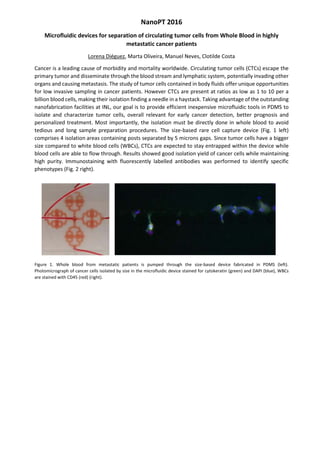
![An Experimental Comparison of Common Methods to Measure Dimensions of Synthetic
Nanoparticles
Peter Eaton
1
, Pedro Quaresma
1
, Cristina Soares
1
, Cristina S. Neves
1
, Miguel Peixoto de Almeida
1
,
Eulália Pereira
1
, Paul West
2
1 UCIBIO, REQUIMTE, Departamento de Química e Bioquímica, Faculdade de Ciências, Universidade
do Porto, 4169-007 Porto, Portugal
2 AFMWorkshop Inc, 1434 E 33rd St., Signal Hill, CA 90755, USA
peter.eaton@fc.up.pt
Abstract Synthetic nanoparticles are being studied for potential use in many applications in diverse
fields, such as medical diagnostics, therapy, structural materials, labeling, etc. Certainly, the main
impetus for this research is the fact that nanoparticles exhibit properties that are very different to the
constituent bulk material. The dimensions of synthetic nanoparticles must be determined with sub-
nanometer accuracy in order to understand their preparation and the structure-property relationship.
A variety of methods have been applied to the determination of such dimensions during the past few
decades of nanotechnology research [1, 2]. These include microscopic techniques, which include direct
imaging of the dry particles, and more indirect methods such as light scattering, which measures the
speed at which the particles move in solution, methods based on sedimentation rate, chromatographic
methods, Coulter counting, which is based on movement of a particle in an electric field, and
crystallographic methods [3].
Nevertheless a direct experimental comparison of the applicability of various methods to a variety of
commonly studied nanoparticle types and mixtures thereof is lacking.
In the work described in this paper, we sought to assess the suitability of a number of very commonly
applied methods to the characterization of the dimensions of several common types of nanoparticle.
Concretely, the characterization methods we tested were dynamic light scattering (DLS), and the
microscopic techniques transmission electron microscopy, atomic force microscopy, and scanning
electron microscopy. While all of the microscopic techniques are capable of imaging and measuring
dried samples of nanoparticles of a variety of materials, they work in different ways, have different
methods of contrast formation, and cannot all achieve the sample maximum resolution. For more details
of these techniques and their applicability in nanoscience, the following references are recommended
[4, 5, 6].
These methods are perhaps the most commonly used methods for characterization in use at this time.
We tested them with only spherical particles, but for each nanoparticle material tested, we produced
and tested two different sizes, and we tested if the techniques could distinguish these two size
populations in mixed samples. The nominal sizes ranged from around 15 nanometers to almost 100 nm.
We tested three different materials, gold, polystyrene, and silica, and compare the results from each
method on each sample.
Overall, we saw that all three microscopic methods were able to characterize all the samples studied,
although with varying degrees of accuracy. The mixed samples, in particular, presented challenges for
some methods. The information available from DLS is useful in assessing the in-solution behavior of the
nanoparticle samples. On the other hand, this technique gives accurate dimensions only for samples
with low polydispersity. We were able to draw conclusions regarding the most appropriate method to
use based on the type of information required, and the type of sample studied.
References
[1] Hassellov, M., et al., Ecotoxicology, 17 (2008).
[2] Tiede, K., et al., Food Addit Contam Part A Chem Anal Control Expo Risk Assess, 25 (2008).
[3] Brar, S.K. and M. Verma, TrAC Trends in Analytical Chemistry, 30 (2011).
[4] Smith, D.J., Characterisation of Nanomaterials Using Transmission Electron Microscopy, in
Nanocharacterisation, A.I. Kirkland and J.L. Hutchison, Editors. 2007, The Royal Society of Chemistry.
[5] Eaton, P. and West, P., Atomic Force Microscopy, OUP (2010) 256 pp.
[6] Zhou, W., et al., Fundamentals of Scanning Electron Microscopy (SEM), in Scanning Microscopy for
Nanotechnology: Techniques and Applications, W. Zhou and Z.L. Wang, Editors. 2006, Springer.](https://image.slidesharecdn.com/nanopt2016conferencebook-160222125923/85/NanoPt2016-Conference-Book-158-320.jpg)
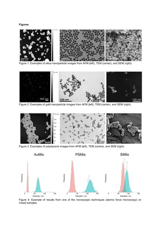
![Synthesis, characterization, biodistribution and toxicological evaluation of star-shaped gold
nanoparticles. Influence of size, shape, and capping agent.
Maria Enea, Joana Costa, Diana Dias da Silva, Eulália Pereira, Helena Carmo and Maria de Lourdes
Bastos.
UCIBIO/REQUIMTE, Laboratory of Toxicology, Department of Biological Sciences,
Faculty of Pharmacy of the University of Porto, Rua de Jorge Viterbo Ferreira 228, 4050-313 Porto,
Portugal.
eneavmaria@gmail.com
The unique properties of gold nanoparticles (AuNPs), such as their multifunctionality potential (ranging
from clinical diagnostics to therapeutics), make them highly attractive [1].
The current work aimed at i) synthesizing and characterizing AuNPs of different shape (stars vs
spheres), size and surface characteristics, and further ii) assessing the in vivo distribution of the novel
synthesized agents, and iii) assessing the influence of size, shape and coating agent on the in vitro
toxicological effects.
By using different methodologies based upon seed-mediated growth synthesis, spherical and star-
shaped AuNPs were synthesized and coated with 11-mercaptoundecanoic acid (MUA) or with sodium
citrate. Transmission electron microscopy (TEM), dynamic light scattering (DLS), and UV-Vis
spectrophotometry were employed for the characterization of the AuNPs. The gold concentration of the
samples was obtained by graphite furnace atomic absorption spectrometry (GFAAS) and the
concentration of nanospheres was determined using the UV-Vis spectrum, based on the mathematical
equation of Haiss et al. [2]. The effect of the shape on the AuNPs biodistribution was evaluated on
Wistar rats. A dose of 0.6 mg Au/ kg of MUA-coated gold nanostars (54 nm of diameter) or of citrate-
coated gold nanospheres (58 nm of diameter) was given per os and the quantification of gold was
determined on different organs and biological fluids was assessed 24h later. Also, the toxicity of the
AuNPs was evaluated in vitro using HepaRG cells (non-differentiated and differentiated for 15 days with
2% dimethyl sulfoxide), Caco-2 cells and primary rat hepatocytes. The performance of two distinct
viability assays, namely (3-(4,5-dimethylthiazol-2-yl)-2,5-diphenyltetrazolium bromide) tetrazolium (MTT)
reduction and neutral red (NR) incorporation assays, was assessed after 4 and 24 hour incubations. In
the case of Caco-2 cells, one additional assay, i.e. the lactate dehydrogenase (LDH) release assay, was
also performed. Six concentrations of each treatment (1 M, 5 M, 10 M, 20 M, 40 M, and 60 M)
were tested. Solvent (2.2 mM sodium citrate and 33 M MUA), negative (cell culture media) and positive
(1% triton-X100) controls were also included in each experiment.
Three batches of MUA-capped gold nanostars ranging from 54 nm to 72 nm of diameter and three
batches of citrate-capped gold nanospheres (diameter from 15 nm to 67 nm) were produced. For the
sake of comparison MUA-capped gold nanospheres of 15 nm were also synthesized. Preliminary in vivo
data demonstrated that for both types of gold nanoparticles, low levels of AuNPs were detected in the
biological samples (in the majority of organs analyzed the data was below the limit of the quantification
of the method). In what concerns the toxicity of the synthesized AuNPs, our results indicated detrimental
effects on all the tested cellular models, at the highest concentrations. The toxicity profiles of the tested
AuNPs were not the same for all cell lines but in all cases a concentration-dependent relationship was
established. Caco-2 enterocytes proved to be the most resistant model, while the rat primary and
HepaRG hepatocytes were the most sensitive, suggesting that metabolism is not involved in the
observed toxicity. Regarding the shape, nanospheres showed higher toxicity, when compared with the
stars. The star-shapped NPs with higher diameter displayed greater injuriousness than smaller NPs in
HepaRG non-differentiated and in primary rat hepatocytes. In what concerns the coating agent, neither
MUA or sodium citrate seem to affect the toxicological profile of gold NPs.
In respect to their toxicity, these preliminary results suggest that our novel gold nanomaterials have high
potential to be considered promising candidates for industry, but further investigations are required,](https://image.slidesharecdn.com/nanopt2016conferencebook-160222125923/85/NanoPt2016-Conference-Book-161-320.jpg)
![particularly aiming at elucidating the oral biodistribution profile, when different doses of the AuNPs are
administered.
References
[1] Ashraf S, Pelaz B, Del Pino P, Carril M, Escudero A, Parak WJ, Soliman MG, Zhang Q, Carrillo-
Carrion C. Top Curr Chem, 370 (2016) 169-202.
[2] Haiss W, Thanh NTK, Aveyard J, Fernig DG. Anal Chem, 79 (2007) 4215–21.
Figures
Centrifugation UV/Vis
Concentration determined by
the method of Haiss et al.,
2007
!
+ 400 L 50mM HAuCl4
+ 563.48 L Seed solution
+ 336.52 L Milli-Q® H2O
+ 450 L 0.1 M ascorbic acid
+ 900 L 0.002M silver nitrate
Ajust pH to ≈ 7
+300 L 10mM MUA
Centrifugation
Nucleation
Milli-Q® H2O
15 mL 1% citrato de sódio
+ 69.2 L 1.44 M HAuCl4
Growth
Gold nanostars – Method of nucleation and growth
• Seed-mediated growth method
Milli-Q® H2O
Seed solution
Gold nanospheres – Method of sodium citrate reduction
• Seed-mediated growth method adapted from Turkevich method
2.2 mM sodium
citrate
1 mL 25mM HAuCl4
1 mL 25mM
HAuCl4
DILUTION
55 mL previous
suspension
+ 53 mL Milli-Q® H2O
2 mL 60 mM sodium
Citrate + 1 mL 25 mM HAuCl4
≈ 70 nm
≈ 60 nm
≈ 15 nm
346 L HAuCl4
Milli-Q® H2O
10 mL sodium citrate
DILUTION
55 mL previous
suspension
+ 53 mL miliQ H2O
1 mL 25mM HAuCl4 1 mL 25mM HAuCl4
1 mL 25mM
HAuCl4
2 mL 60 mM sodium
Citrate + 1 mL 25 mM HAuCl4
Figure 1. Synthesis of gold nanoparticles. Figure 2. Synthesis of gold nanoparticles.
• Treatment of cells with 0.5 mg/mL MTT
• 30 minutes of incubation at 37 ºC
• Solubilisation of the formazan crystals formed with DMSO
• Measurement of absorbance at 550 nm
• Treatment of cells with 50 g/mL Neutral Red (NR) solution
• 30 minutes of incubation at 37 ºC
• Solubilisation of the incorporated dye with lysis solution
• Measurement of absorbance at 540 nm
• 50 L of supernatant (5x diluted) of each well
• Addition of 40 L 0.05 M KH2PO4 buffer solution
• Addition of 200 L 0.15 mg/mL β-NADH
• Addition of 25 L 22.7 mM sodium pyruvate
• Measurement of NADH oxidation to NAD+ at 540 nm (kinetic reaction)
MTT reduction
assay
Neutral Red
Incorporation
assay
LDH leakage
assay !
Viability of Caco-2 cells
MTT
NR
4 hour incubation 24 hour incubation
LDH
AuPs biodistribution profile
Per os administration
Figure 3. In vitro assessment of cytotoxicity. Figure 4. Experimental results of toxicological and biodistribution assays.](https://image.slidesharecdn.com/nanopt2016conferencebook-160222125923/85/NanoPt2016-Conference-Book-162-320.jpg)
![Development of a multiplexed system for ischemic stroke using a magnetoresistive (MR) biochip
platform
E. Fernandes*1, V. Martins2, D.Y. Petrovykh 1, T. Dias2, J. Germano2, T. Sobrino3, J. Castillo3, J. Rivas1,
S. Cardoso2, P.P. Freitas1,2
1INL International Iberian Nanotechnology Laboratory, Portugal;
2INESC-MN Instituto de Engenharia de Sistemas e Computadores-Microsistemas e Nanotecnologias
and IN-Institute of Nanoscience and Nanotechnology, Portugal;
3Clinical Neurosciences Research Laboratory, Neurovascular Area, Department of Neurology, Hospital
Clínico Universitario, IDIS, University of Santiago de Compostela, Spain
elisabete.fernandes@inl.int
Abstract
Modern development of diagnostic/prognostic devices for complex diseases is centered on systems
based on the detection of multiple biomarkers to provide actionable information for stratification of
patients and personalized medicine. For example, for ischemic stroke patients, the elevated levels of
several biomarkers in blood have been shown to accurately predict a high risk of developing negative
side effects associated with thrombolytic therapy [1,2]. Detecting these biomarkers, however, is a
challenge that requires addressing assay parameters such as dynamic range, cross-reactivity, and
effects of the complex biological matrix. In this work, we developed a detection system based on
multiple immunoassays by combining a recognition/labelling step (in solution) with a detection step
using a magnetoresistive (MR) biochip platform (on chip) [3,4]. The work started with two predominant
stroke biomarkers present in serum, cellular fibronectin (cFN) and matrix metallopeptidase 9 (MMP9),
and will be extended eventually to six targets. Calibration experiments for cFN and MMP9 in sequential
and multiplex formats were performed to address the parameters described above.
The experiments involved assays using single or multiple biomarkers. Here, targets were initially
captured/concentrated in solution onto 250-nm magnetic nanoparticles (MNPs) functionalized with the
respective biotinylated polyclonal antibodies. The sample was then passed using a microfluidic system
over an array of 30 spin-valve sensors that are functionalized with monoclonal antibodies: this
arrangement of a sandwich assay allows us to decrease false positives by performing the most
selective capture on the chip. Moreover, the efficiency of this capture step was enhanced by bringing
the MNPs in contact with the sensor sites using the magnetic particle attraction (magnetic focusing)
functionality of this platform. Our biochip showed sensitivity for clinically relevant low concentrations of
MMP9 and cFN down to 1 ng/mL and 4 µg/mL, respectively. In the multiplex assay format, and using
higher analyte concentrations, we were able to differentiate each target and to compare those
concentrations to the respective values in single-target measurements (calibration curves).
References
[1] M. Rodríguez-Yáñez, T. Sobrino, S. Arias, F. Vázquez-Herrero, D. Brea, M. Blanco, R. Leira, M.
Castellanos, J. Serena, J. Vivancos, A. Dávalos, and J. Castillo. Stroke, 42 (2011) 2813 2818.
2011.
[2] M. Castellanos, T. Sobrino, M. Millán, M. García, J. Arenillas, F. Nombela, D. Brea, N. Perez De
La Ossa, J. Serena, J. Vivancos, J. Castillo, and A. Dávalos. Stroke, 38 (2007) 1855 1859.
[3] J. Germano, V. C. Martins, F. a. Cardoso, T. M. Almeida, L. Sousa, P. P. Freitas, and M. S.
Piedade. Sensors, 9 (2009) 4119 4137.
[4] V. C. Martins, F. a. Cardoso, J. Germano, S. Cardoso, L. Sousa, M. Piedade, P. P. Freitas, and
L. P. Fonseca. Biosensors and Bioelectronics, 24 (2009) 2690 2695.](https://image.slidesharecdn.com/nanopt2016conferencebook-160222125923/85/NanoPt2016-Conference-Book-164-320.jpg)
![Electrochemical Determination of Vitamin B-12 in Food and
Pharmaceutical Samples by Poly (PBHQ)/MWCNTs/GCE
Hayati Filik, Asiye Aslıhan Avan, Sevda Aydar
Istanbul University, Faculty of Engineering, Department of Chemistry, 34320 Avcılar, Istanbul, Turkey
E-mail: filik@istanbul.edu.tr
Water-soluble vitamin B-12 (or vitamin B12 and vitamin B12) exists in several patterns, called
cobalamins; cyanocobalamin (CN-CbA[Co(III)]) is the main one used in vitamin supplements and
pharmaceuticals. VB-12 is inherently found in animal foods. In the cobalamin molecules, cobalt
normally exists in the Co (III) state. However, under different pH values, the cobalt center is reduced
to Co(II) or even Co(I) state, which are usually denoted as reduced (VB-12r) and super-reduced (VB-
12s), respectively. One of the most effective antioxidants in food and medicine is VB-12, the only
naturally gifted biomolecule with a carbon–metal bond. The daily intake of VB-12 is as low as 1 to 2 µg
when compared to supplements. Hence, the deficiency may be at the nanogram to picogram level,
which is a challenging task to analyze.
This study focused on the development of poly(2,2
´
-(1,4-phenylenedivinylene) bis-8-
hydroxyquinaldine)/multi-walled carbon nanotube hybrid film modified GCE for the electrochemical
monitoring of VB-12. The obtained poly (PBHQ) /MWCNTs hybrid film has significantly altered film
morphologies and improved electrochemical properties. Estimation of VB-12 in pharmaceutical
supplements was assessed sensitively by using poly (PBHQ) /MWCNTs modified glassy carbon
electrode.
Poly(2,2
´
-(1,4-phenylenedivinylene) bis-8-hydroxyquinaldine)/multi-walled carbon nanotubes modified
glassy carbon electrode (poly (PBHQ)/MWCNTs/GCE) was developed and applied for the
electrochemical estimation of vitamin B-12 (VB-12). Compared to multi-walled carbon nanotubes
modified glassy carbon electrode, well-defined redox peaks were observed in phosphate buffer
solution at pH 2.5. In contrast with the ill-defined redox peaks observed with unmodified glassy carbon
electrode surfaces. The poly(2,2
´
-(1,4-phenylenedivinylene) bis-8-hydroxyquinaldine based electrode
displayed a good linear range of 0.1 µM to 10 µM VB-12 with a low detection limit of 0.03 µM. To
further study the practical applicability of the proposed sensing procedure, the estimation of real
samples was employed with satisfactory consequences. In addition, multiwalled carbon nanotubes
(MWCNTs) were used as sorbent for solid phase extraction (SPE) of vitamin B-12 from cereal food
samples. Solid phase extraction parameters, such as the amount of MWCNTs, sample volume, pH,
and type and amount of the eluent were optimized.](https://image.slidesharecdn.com/nanopt2016conferencebook-160222125923/85/NanoPt2016-Conference-Book-165-320.jpg)
![Flexible Magnetoresistive Devices with High-Performance Sensors
H. Fonseca
a
, E. Paz
a
, R. Ferreira
a
, S. Cardoso
b
, J. Gaspar
a
, and P. P. Freitas
a,b
a
International Iberian Nanotechnology Laboratory, Braga, 4715-330, Portugal
b
INESC-MN/Institute for Nanosciences and Nanotechnologies, Lisbon, 1000-029, Portugal
helder.fonseca@inl.int
This work reports on the integration for the first time of magnetic tunnel junction (MTJ) sensing devices
with magnetoresistance responses above 150% on flexible substrates, as opposed to previous attempts
in which figures below 53% have been obtained [1]-[3]. These are able to bend and conform to non-
planar geometries, non-conformal and hard-to-reach regions of space for magnetic sensors processed
in conventional rigid substrates, paving the way for new spintronic applications. Their fabrication
process is based on polyimide (PI) materials due to their flexibility, thermal stability, chemical
resistance, high mechanical modulus, and biocompatibility. Magnetoresistive performance is
characterized in terms of controlled mechanical load conditions.
The fabrication summarized in Fig. 1 begins with the definition of the MTJ sensors on a PI layer atop
SiO2/Si. The MTJ stack is patterned by photolithography/ion milling and annealed to obtain magnetic
sensors as detailed elsewhere for rigid substrates [4]. The subsequent step is another PI coating acting
as encapsulation. The PI layers are patterned to define the shape of the flexible device and probes are
finally detached from the rigid substrate by means of HF vapor that selectively removes the underlying
sacrificial layer. The overall flexible probe thickness is slightly larger than 20 µm. Layouts of devices
fabricated using such technology are shown in Figs. (2) and (3), corresponding to long magnetic
sensing stripes and neural insertion probes, respectively. The stripes consist of ca. 50-mm-long, 4.5-
mm-wide structures with MTJ arrays located at their centers, each MTJ connected in a 4-wire
configuration, and are used to analyze magnetoresistive performance as a function of mechanical
loading. As for the neural insertion/magnetic recording probes, Fig. (3), they consist of ca. 30-mm-long
devices with an opening of 90 µm at one end, compatible with surgery tools used for brain insertion.
Devices comprising square-shaped impedance electrodes with 30 µm and MTJ sensors with pillars
ranging from 4 to 20 µm have been processed, Figs. (4) and (5).
The effect of mechanical load through bending on MTJ sensors processed on the long stripes has been
characterized using the double 4 point bending bridge (4PBB) setup depicted in Figs. (6) and (7). It
consists of a system of automated stages that displaces inner pins with respect to outer ones in which
the structure is accommodated and therefore bends with a well-known, controlled radius of curvature
[5]. This configuration allows for inducing compressive/tensile stresses by simply moving the inner pins
to the left/right by a given displacement d. A Cu-wire coil, whose axis is centered and solidary to the
inner pins, is placed above the bended structure for generating fields, Hz, parallel to the sensing
direction of the loaded sensors. Sensors are located within the inner pins region and deviate from the
coil axis by less than 1 mm for the range of displacements used in this study, -5 mm < d < 5 mm. The
measured field / current calibration curves of the coil shown in Figs. (8) and (9) show that Hz at the
sensors position, z = 0, is uniform within the 1-mm-deviation from the coil axis. Transfer curves of
sensors resistance are recorded as a function of magnetic field for sequences of radius of curvature
imposed to the sensor.
Figure (10) shows the transfer curve of a sensor with area (pillar dimension), A, of 8x8 µm
2
in a
released, unloaded probe with resistance, Rmin, magnetoresistance ratio, MR, and sensitivity, dV/dH, of
145 W, 171% and 250 µV/Oe, respectively, in agreement with literature for rigid substrates [4]. The
resistance of sensors with different dimensions follows the expected 1/A dependence, Fig. (11.a), and
an average MR of 166 % is obtained for MTJ devices with areas of up to almost 300 µm
2
. The
sequence of optical graphs in Fig. (12) shows the mechanical test evolution for displacement values
from -5 mm to 5 mm corresponding to curvature radii, , between ca. 5 mm and (non-deformed state)
for compression and tension. As it can be observed from Fig. (13.a), there is a relative variation in MR
as bending increases, i.e. for smaller , by about 1% for radii down to 5 mm. A similar but more
pronounced effect is observed in terms of sensitivity, Fig. (13.b), which relatively increases/decreases
by about 7.5% as the radius of curvature decreases from the non-deflected state down to 5 mm for
positive/negative displacements.](https://image.slidesharecdn.com/nanopt2016conferencebook-160222125923/85/NanoPt2016-Conference-Book-166-320.jpg)
![[1] Melzer et al., Nano Lett., vol. 11, no. 6, p. 2522 (2011).
[2] Y. Chen et al., Advanced Materials, vol. 20, no. 17, p. 3224 (2008).
[3] C. Barraud et al., Appl. Phys. Lett., vol. 96, no. 7, p. 072502 (2010).
[4] R. Ferreira et al., IEEE Trans. Magn., vol 48, no. 11, p. 3719 (2012).
[5] P. Alpuim et al., J. Appl. Phys., vol. 109, no. 12, p. 123717 (2011).
Figure (1) Fabrication process flow of MTJ sensors encapsulated by PI layers. (2)-(3) Layouts of
flexible bending test stripes and neural insertion probes with magnetic sensors, respectively,
and (4)-(5) optical graphs of fabricated devices. (6)-(7) 4PBB setup used to characterize
sensors magnetoresistance response as a function of bending and (8)-(9) field generated by
coil used in apparatus. (10) Representative output vs. applied magnetic field of a 8x8 µm2
pillar
with no applied mechanical stress and (11) sensors resistance and magnetoresistance
distribution as a function of device area. (12) Optical graphs of mechanical loading sequence
and (13) relative variations of magnetoresistance and sensitivity dependence on radius of
curvature imposed to devices.](https://image.slidesharecdn.com/nanopt2016conferencebook-160222125923/85/NanoPt2016-Conference-Book-167-320.jpg)
![Paper-based Nanostructured Plasmonic Surfaces for ultra-sensitive detection of trace analytes
by Surface Enhanced Raman Spectroscopy
Ricardo Franco
1
, Maria João Oliveira
1,2
, Pedro Quaresma
3
, Eulália Pereira
3
, Elvira Fortunato
2
, Rodrigo
Martins
2
, Hugo Águas
2
1 - REQUIMTE-UCIBIO, Departamento de Química, Faculdade de Ciências e Tecnologia, Universidade
Nova de Lisboa, 2829-516 Caparica, Portugal; 2 - CENIMAT-I3N, Departamento de Ciência dos
Materiais, Faculdade de Ciências e Tecnologia, Universidade Nova de Lisboa, 2829-516 Caparica,
Portugal; 3 - REQUIMTE-UCIBIO, Departamento de Química e Bioquímica, Faculdade de Ciências,
Universidade do Porto, 4169-007 Porto, Portugal ricardo.franco@fct.unl.pt
Abstract
Surface Enhanced Raman Spectroscopy (SERS) is a highly sensitive analytical technique, based on
light dispersion by analytes in the vicinity of plasmonic nanostructures. In fact, the Raman signal can be
amplified by several orders of magnitude, when molecules are adsorbed at the surface of metal
nanoparticles (NPs), with coinage metals (gold or silver) providing the highest enhancements. Hot-
spots, where the enhancement factor (EF) is higher, occur mainly between NPs with a distance < 10 nm
or at sharp edges of non-spherical NPs such as star shaped NPs [1]. For analytical applications in
portable sensors, substrates that are easy to produce and disposable are highly desirable, such as
paper-based materials [2]. In this work, SERS substrates were produced by simple deposition of
solutions containing spherical silver nanoparticles (AgNPs) or star-shaped silver nanoparticles (AgNSs),
on two different types of paper. Both nanoparticles were synthesized by well-known and highly
reproducible chemical methods [3]. Paper used was either with high porosity (Whatman filter paper no.
1) or with low porosity (office paper). The porosity of the paper was a determinant factor for the type of
distribution observed for the NPs along the structure of the paper (Figure 1). The paper substrates were
tested for SERS activity using rhodamine-6G - a model dye for SERS – and a 633 nm laser. Both
papers treated with NPs showed no paper-derived fluorescence, a general problem reported for Raman
measurements, especially in office paper-based substrates, and that we were able to eliminate using
deposition of known amounts of NPs from solution. With these easy to produce plasmonic surfaces, a
limit of detection for R6G as low as 10
-9
M could be achieved for the office paper substrate with
deposition of AgNS. This same substrate allowed an analytical EF of 10
6
, a result in the same order of
magnitude as the EF obtained for paper surfaces screen printed with AgNP, a much more laborious and
expensive production process [4]. The paper-based plasmonic surfaces revealed to be stable for at
least 5 weeks, always with good reproducibility, and also when different AgNSs synthesis batches were
tested. Studies are underway with these promising paper-based substrates to further improve the
obtained EF as well as expanding the work to relevant analytical molecules such as pesticides and food
toxins.
References
[1] Schlucker, S. Angew Chem Int Ed Engl, 53(19) (2014) 4756.
[2] Araújo, A. et al. Nanotechnology, 25(41) (2014) 415202.
[3] Garcia-Leis A. et al. J. Phys. Chem. C, 117(15) (2013) 7791.
[4] Lu-Lu, Q. et al. Anal. Chem. Acta, 792 (2013) 86.](https://image.slidesharecdn.com/nanopt2016conferencebook-160222125923/85/NanoPt2016-Conference-Book-168-320.jpg)
![Poly(N-isopropylacrylamide)-grafted membranes as bacteriophage smart-delivery systems for
food-packaging applications
Pablo Fuciños
1
, Carla Carvalho
1,2
, Lorena Diéguez
1
, Lorenzo Pastrana
1
, Joana Azeredo
2
1
INL - International Iberian Nanotechnology Laboratory, Braga, Portugal
2
Centre of Biological Engineering, University of Minho, Braga, Portugal
pablo.fucinos@inl.int
Abstract
Concern about microbial food-borne diseases is growing worldwide. Each year, in the European Union
alone, Campylobacter bacteria, the most frequent food-borne pathogen, causes more than 236,000
human cases [1]. Bacteriophages (phages), viruses that specifically infect and kill bacteria, may provide
a natural, specific, effective, and non-toxic tool to reduce food-borne bacteria [2]. Phages that predate
food-borne Campylobacter strains were isolated, and preliminary results indicate that phages may be a
suitable alternative to classic antimicrobials [3]. However, inconsistencies on the effectiveness of phage
treatments were also reported, mainly related to the phage resistance in the environment, and the
appropriate dosage and correct moment of administration [3,4]. Environmentally-sensitive controlled
release systems may be used in smart packaging applications to increase the effectiveness of
bacteriophage treatments, protecting the phages until the release is required (e.g. under environmental
temperature promoting microbial growth). In this work, smart thermoresponsive membranes were tested
for the controlled release of Campylobacter bacteriophages. Poly(N-isopropylacrylamide) (PNIPAM)
gates were prepared onto 0.2 µm MWCO polycarbonate membranes using a plasma-graft pore-filling
polymerization method [5]. The transfer of Campylobacter bacteriophages across the PNIPAM-grafted
membranes was assayed at two different temperatures (4ºC and 37ºC), below and above the PNIPAM
lower critical solution temperature (LCST). The obtained results showed that bacteriophage diffusion
trough the PNIPAM gates was strongly dependent on the environmental temperature, allowing the use
of these membranes for bacteriophage smart delivery applications.
Acknowledgements
This work was supported by a Marie Curie COFUND Action (Project No: 600375. NanoTRAINforGrowth
- INL Fellowship programme in nanotechnologies for biomedical, environment and food applications)
References
[1]. EFSA (European Food Safety Authority) and ECDC (European Centre for Disease Prevention and
Control), The European Union summary report on trends and sources of zoonoses, zoonotic
agents and food-borne outbreaks in 2014, EFSA Journal, 13 (2015) 4329.
[2]. J. Mahony, O. McAuliffe, R.P. Ross, D. van Sinderen, Bacteriophages as biocontrol agents of food
pathogens, Curr. Opin. Biotechnol., 22 (2011) 157 163.
[3]. C.M. Carvalho, B.W. Gannon, D.E. Halfhide, S.B. Santos, C.M. Hayes, J.M. Roe, et al., The in vivo
efficacy of two administration routes of a phage cocktail to reduce numbers of Campylobacter
coli and Campylobacter jejuni in chickens, BMC Microbiol., 10 (2010) 232.
[4]. Scientific Opinion of the Panel on Biological Hazards on a request from European Commission,
The use and mode of action of bacteriophages in food production, EFSA Journal, 1076 (2009),
1-26.
[5]. Chu, L.-Y., Niitsuma, T., Yamaguchi, T. and Nakao, S.-i., Thermoresponsive transport through
porous membranes with grafted PNIPAM gates. AIChE J., 49 (2003) 896-909.](https://image.slidesharecdn.com/nanopt2016conferencebook-160222125923/85/NanoPt2016-Conference-Book-172-320.jpg)
![CARBON-BASED NANOMATERIALS FOR GOLD (III) RECOVERY: KINETICS AND LOADING
INVESTIGATIONS
I. García-Díaz, F.A. López, O. Rodríguez, F.J. Alguacil
National Center for Metallurgical Research, Avda. Gregorio del Amo, 8, 28040, Madrid, Spain
irenegd@cenim.csic.es
Abstract
Currently, the development of different smart technology to recover or eliminate strategic or toxic metals
from liquid effluent is constant. Among the different process developed to the treatment of liquid
effluents bearing these types of metallic elements, include chemical or electrochemical precipitation,
membrane based technology, ion exchange and adsorption [1,2].
Adsorption is a high efficiency, cost-effectiveness and easily handling method to recover pollutants or
strategic metals [3,4]. Nowadays a research challenge is the development of new adsorbents. Among
nanomaterials, carbon nanomaterials have adequate properties to be used as metal adsorbent [5,6].
On the other hand gold is one of the most precious elements in the world. The price in 2015 of this
strategic metal is about 1.376 $/kg [7]. Besides its uses in jewellery it is highly used in different
industries, so it is important the gold recovery from liquid effluents generated from these various
industries. The adsorption method is a way to treat these types of effluents, characterized for their low
gold concentration.
The aim of this research was to optimize various operational parameters, and thus obtain efficient
carbon nanotubes processing for gold (III)-bearing effluents.
The adsorption of gold (III) by carbon nanofibers (CNF), carbon multiwalled (MWCN) and carbon
multiwalled with carboxyl group (MWCN_ox) systems were investigated. The experimental parameters
which may influence gold adsorption were investigated, i.e. stirring speed of the aqueous solutions,
adsorbent dosage, acid concentration, temperature etc.
Figure 1 shows the effect of the acid concentration on the Au(III) adsorption to the three adsorbent. It
can be seen that the adsorption Au(III) decreases with the increase of HCl concentration. Probably it is
due the existence of other gold species, such as HAuCl4, at the higher hydrochloric solution against the
presence of the predominant AuCl4
-
species in the more dilute HCl solutions which are more adsorbable
than the gold-acid form.
SEM studies of gold loaded carbon nanomaterials show dark particles on the surface, Figure 2. The
EDS analysis of the dark particles show
metallic gold. Probably this reduction occurs on the carbon surface, related with the metal reduction [8].
The isotherm and kinetic studies of the carbon nanomaterial-Au(III) system show a different behavior in
function of the carbon nanomaterial used as adsorbent. The experimental data obtained using the
MWCNT and CNF fit better to a pseudo second order equation and an isotherm Freundlich model.
The three carbon nanomaterials, appeared to be a promising material for the recovery of Au(III) from
this type of acid solutions in the optimal experimental for each one of them condition, Table1.
Table 1.- Adsorption of gold on the optimal conditions by carbon nanomaterials (mmol Au/g
nanomaterials)
Optimal condition CNF MWCNT MWCNT_ox
0.005 Au g/l; 0.1 M HCl; 20ºC; 2000
rpm, 0.006 g solid
0.34
0.005 Au g/l; 0.1 M HCl; 20ºC; 1000
rpm; 0.005 g solid
0.15
0.005 Au g/l; 0.1 M HCl; 20ºC; 1500
rpm; 0.006 g solid
0.23](https://image.slidesharecdn.com/nanopt2016conferencebook-160222125923/85/NanoPt2016-Conference-Book-173-320.jpg)
![0 10 20 30 40 50 60
0
1
2
3
4
5
qt
(mg/g)
t (min)
0.1 M HCl
1.0 M HCl
10.0 M HCl
0 10 20 30 40 50 60
0
1
2
3
4
5
qt
(mg/g)
t (min)
0.1 M HCl
1.0 M HCl
10.0 M HCl
0 10 20 30 40 50 60
0
5
10
15
20
qt
(mg/g)
t (min)
0.1 M HCl
1.0 M HCl
10.0 M HCl
References
[1] Alguacil F.J., López F.A., García-Díaz I., Rodríguez O., Chemical Engineering and Processing:
Process intensification, (2015) in press.
[2] Alguacil F.J., García-Díaz I., López F.A., Journal of Industrial and Engineering Chemistry, 19[4]
(2015) 1086-1091.
[3] Cho D.-W, Jun W., Sigdel A., Kwan O-H., Lee S.-H., Kabra A.-N., Jeon B.-H., Geosystem
Engineering 16[3] (2013) 200-208.
[4] Alguacil F.J., López F.A., García-Díaz I., Desalination and Water treatment (2015) 1-13
[5] Alguacil, F.J., Cerpa, A., Lado, I., López, F.A. Rev. Metal. 50(3), (2014) e025. doi:
http://dx.doi.org/10.3989/revmetalm.025.
[6] N.M. Mubarak, J.N., Sahu, E.C. Agdullah, N.S. Jayakumar, Separation and Purification Reviewer, 43
(2014) 311-338.
[7] http://www.metalprices.com
[8] Pang S.-K., Yung K.-C. Chemical Engineering Science, 107 (2014) 58-65
Acknowledgements
To the CSIC Agency (Spain) for support. Thanks to the Grupo Antolín Carbon for supplying carbon nanofibers. Dra. I. García-Díaz
expresses her gratitude to the Ministry of Economy and Competitiveness for their Postdoctoral Junior Grants (Ref. FPDI-2013-
16391) contracts co-financed by the European Social Fund.
Figure 1.- Influence of the HCl concentration in the adsorption of Au(III).
a) CNF: 0.025 g, aqueous phase- 0.005 g/l Au(III), T=20ºC, stirring speed: 2000 rpm. b) MWCNT: 0.1 g aqueous
phase- 0.005 g/L Au(III), T= 20ºC, stirring speed: 1000 rpm. c) MWCNT_ox: 0.1 g, aqueous phase- 0.005 g/L
Au(III), T= 20ºC, stirring speed 1000 rpm.
Figure 2.- TEM micrographs CNF loaded with Au and the elemental composition analysis of Au loaded
in the CNF, dark particles.
a) b)
c)](https://image.slidesharecdn.com/nanopt2016conferencebook-160222125923/85/NanoPt2016-Conference-Book-174-320.jpg)
![Polypyrrol/AuNP composites deposited by different electrochemical methods. Sensing
properties towards catechol
C. Garcia-Hernandez
1
, C. Garcia-Cabezon
2
, C. Medina-Plaza
1
, F. Martin-Pedrosa
2
, Y. Blanco
2
, J.A. de
Saja
3
, M.L. Rodriguez-Mendez
1
.
1
Department of Inorganic Chemistry, Engineers School, Universidad de Valladolid, 47011 Valladolid,
Spain. E-mail: mluz@eii.uva.es.
2
Department of Materials Science, Engineers School, Universidad de Valladolid, 47011 Valladolid,
Spain.
3
Department of Condensed Matter Physics, Faculty of Sciences, Universidad de Valladolid, 47011
Valladolid, Spain.
Abstract
Polypyrrole (Ppy) is one of the most extensively studied conducting polymers due to its good electrical
conductivity and redox properties [1]. Ppy films can be easily generated by electropolymerization as a
strong adherent layer using different electrochemical techniques [2]. Electrodes chemically modified
with Ppy have a good electrocatalytic activity. The structure and sensing properties of the Ppy films are
considerably influenced by the electrochemical method used for the polymerization (potentiostatic,
galvanostatic or potentiodynamic), by the electrochemical conditions (voltage, intensity, scan rate, etc.),
and by the other experimental conditions such as the nature and concentration of the doping agent or
the nature of the substrate [3].
Recently, composite nanomaterials based on conducting polymers and metal nanoparticles (NPs) have
been developed. Gold nanoparticles (AuNPs) have attracted considerable interest because of their
unique optical, electronic and catalytic properties [4]. Conducting PPy/AuNP composites exhibit
improved physical and chemical properties over their single-component counterparts and are the focus
of intensive research. Ppy/AuNP composites can be prepared by chemical and electrochemical
polymerization. Electrochemical methods provide a better control of the structure and properties of the
composite by controlling the electrochemical conditions during film generation [5]. It could be expected
that the electrocatalytic and the sensing properties of the Ppy/AuNPs films directly depend on the
polymerization conditions.
One of the fields where electrochemical sensors are having an important success is in the detection of
phenolic compounds, which are strong antioxidant reagents present in foods, with beneficial effects on
human health [6]. As phenols are electroactive compounds, they can be detected by amperometric or
voltammetric techniques using a great variety of electrodes. Ppy/AuNPs composites could be good
candidates as electrocatalytic materials for the detection of phenols.
The objective of this work was to develop new voltammetric sensors based on electrodeposited
Ppy/AuNPs for the detection of catechol (an antioxidant of interest in the food industry) and to evaluate
the influence of the electrodeposition method in their performance. For this purpose Ppy/AuNP films
doped with 1-decanesulfonic acid (DSA) were deposited using different methods. The first approach
consisted on the electrodeposition of the Ppy/AuNPs films from a solution containing the monomer and
the trichloroauric acid (cogeneration method). The second approach consisted of the electrodeposition
of the Ppy/AuNPs composited from a solution containing the monomer and gold nanoparticles
previously formed (trapping method). In both methods, electrodeposition was carried out by
chronoamperometry (CA) and by chronopotentiometry (CP). Particular attention was paid to the study of
the influence of the substrate used for the electrodeposition that was carried out onto classical platinum
electrodes and on stainless steel substrates. This aspect could play a crucial role not only in the
structure, properties and performance of the sensor but also in the final price.
Using CA, the polymerization charge was strongly dependent on the presence of AuNPs and the mass
deposited in the absence of AuNPs was higher than the mass deposited in the presence of gold. The
charge calculated for films obtained by cogeneration was superior than by trapping. That is, the amount
of polymer deposited followed the same trend whatever CP or CA were used. This result also points to
the role of AuNPs in the nucleation of Ppy, difficulting the oxidation of the monomers.
Scanning electron microscopy (SEM) demonstrated that in all cases gold nanoparticles of similar size
(30-40 nm) were uniformly dispersed in the Ppy matrix. The amount of AuNPs incorporated in the Ppy
films was higher when electropolymerization was carried out by CP. Besides, cogeneration method
allowed for the incorporation of a higher number of AuNPs than trapping (Figure 1).
Electrochemical Impedance Spectroscopy (EIS) experiments demonstrated that the insertion of AuNPs
modified the electrical behavior and increased the conductivity. The cogeneration method combined
with chronopotentiometry seemed to be the most suitable electrodeposition technique to prepare
electrochemical sensors.experiments demonstrated that the insertion of AuNPs increased the
conductivity. The electrocatalytic and sensing properties towards catechol of Ppy/AuNP electrodes were](https://image.slidesharecdn.com/nanopt2016conferencebook-160222125923/85/NanoPt2016-Conference-Book-175-320.jpg)
![analyzed. Catechol produced the expected well-shaped redox pair generated by the two-electron
oxidation/reduction of the orto-dihydroquinone to benzoquinone. The reversibility of the peaks was
improved with the incorporation of the AuNPs and the intensity of the peaks increased with the
concentration of AuNPs. These effects were stronger in films deposited by CP than in films deposited by
CA, due to the higher concentration of nanoparticles. In contrast, the method to insert the nanoparticles
(trapping or cogeneration) only produced small changes in the intensities and positions of the peaks,
probably due to the minimal differences in the AuNPs concentration. The electrocatalytic effect was
stronger in films deposited on platinum than in SS. The limits of detection (LOD) were in the range from
10 to 10 mol/L (Figure 2). LODs attained using films deposited on platinum were lower due to a
synergy between AuNPs and platinum that facilitates the electron transfer, improving the electrocatalytic
properties. Such synergistic effects are not so pronounced on stainless steel, but acceptable LODs are
attained with lower price sensors.
References
[1] Ramanavicius, A.; Ramanaviciene, A; Malinauskas, A. Electrochim. Acta, 51 (2006) 6025.
[2] Li, C.M.; Sun, C.Q.; Chen, W.; Pan, L. Surf. Coat. Tech. 198 (2005) 474.
[3] Chillawar, R.R.; Tadi, K.K.; Motghare, R.V. J. Anal. Chem. 70 (2015) 399.
[4] Yoon, H. Nanomaterials 3 (2013) 524.
[5] Rapecki, T.; Donten, M.; Stojek, Z. Electrochem. Commun. 12 (2010) 624.
[6] Hurtado E.; Gomez, M.; Carrasco, A.; Fernandez, A. J. Pharm. Biomed. Anal. 53 (2010) 1130.
Acknowledgements
The authors are grateful to FEDER and to the Spanish Ministry of Science-CICYT (Grant AGL2012-
33535), Junta de Castilla y León (VA-032U13) and FPI-UVa for the financial support.
Figures
Figure 1. SEM images of Ppy/AuNP films deposited on stainless steel by (left) Cogeneration-CP
and (right) Cogeneration-CA.
Figure 2. Voltammograms registered using electrodes deposited by CP on stainless steel
immersed in 1·10
-5
to 1·10
-3
mol/L solutions of catechol: (left) Ppy-CP and (right) Ppy/AuNP-
Trapping-CP.](https://image.slidesharecdn.com/nanopt2016conferencebook-160222125923/85/NanoPt2016-Conference-Book-176-320.jpg)
![Nanofabrication of silicon nitride photonic crystals membranes
Valentim, P. T.,1, 2, 3 Vasco, J. P.,2, 3 Fonseca, H.,1 Borme, J.,1 Assis, P.-L.,2, 3 Rodrigues, W. N.,2,
3 Quivy, A. A.,3, 4 Guimarães, P. S. S.,2, 3 Gaspar, J.1
1
INL- International Iberian Nanotechnology Laboratory, Braga, Portugal
2
Departamento de Física, Universidade Federal de Minas Gerais, Belo Horizonte, MG, Brazil
3
DISSE-INCT de Nanodispositivos Semicondutores, Brazil 4Instituto de Física da Universidade de São
Paulo, CP 66318, 05314-970 São Paulo, SP, Brazil
joao.gaspar@inl.int
We report on the nanofabrication of silicon nitride (SiNx) L3 photonic crystals nanocavities with high
geometrical quality. Lately, these kind of devices have attracted much attention due to their capability
for confining, guiding and modifying the light transportation within the matter. These can also interact
with novel materials such as transition metal dichalcogenides (TMDC) and antibodies within the visible
range of the electromagnetic spectrum [1]. The aim of this work is to develop an efficient fabrication
process and study the emission properties of such cavities both with photoluminescence and reflectivity
experiments at room temperature. Theoretical calculations were carried out using guided mode
expansion approach to help us establish the optimal geometrical parameters of our structures, such as
lattice parameter (a), radius (r) and thickness (t), which in our case, were chosen to be a = 270 nm, r =
83.7 nm and t = 270 nm, respectively. Taking into account the refractive index for SiNx (n = 2.01), the
theoretical fundamental L3 photonic mode is expected to be around 672 nm and has a theoretical
quality factor (Q) of 4300. Figure 1 bellow shows the schematics of our structure.
It is known from literature that fabrication imperfections are the major causes for cavities low quality
factors [2]. To overcome these challenges, we have developed a method for producing high quality
factor cavities using MEMS/NEMS fabrication based technologies. Firstly, using a plasma enhanced
chemical vapor deposition (PECVD) system, we deposit a 270-nm-thick layer of SiNx on the front side
of a 725 µm-thick double side polished (DSP) silicon wafer. A 3500-nm-thick layer of silicon dioxide
(SiO2) is then deposited on the backside. The photonic crystal cavity (PHC) pattern is produced on the
front side of the wafer by the means of a negative tone resist E-beam lithography, development and
deposition 25 nm-thick layer of Al followed by lift-off in a Microstrip solution at 60°C under ultrasonic
agitation. By the end of this step we have fabricated a metallic aluminum hard mask that will be used to
transfer the PHC pattern into the SiNx layer. After that, the sample is etched in a fluorine based reactive
ion etch (RIE) process to remove only the areas on the SiNx layer that are not protected by the Al mask.
Then, on the back side of the wafer, a conventional optical lithography is combined with a RIE plasma to
make small apertures on the SiO2 layer that will serve as a hard mask for deep reactive ion etch (DRIE)
of silicon. During this process most of Si is removed, leaving just a 100 µm-thick layer left. The last step
is an anisotropic Tetramethylammonium hydroxide (TMAH) wet etch. Along this part, the last 100 µm of
Si are slowly etched, in a rate of 45 µm/h, enabling the gentle releasing of the patterned SiNx
suspended membranes. The outcome are free-standing silicon nitride layers exhibiting very good holes
circularity and very straight side walls, both desirable features of high quality structures necessary to
study cavity quantum electrodynamic (cQED) phenomena.
We are currently implementing a cross-polarization measurement system that will allow us to perform
microphotoluminescence and reflectivity (transmission) experiments at room temperature on the
samples. The first objective is to study how the quality factor of these cavities changes with respect to
the lattice parameter, hole size and membrane thickness. Afterwards, we intend to investigate the
coupling behavior between the cavity mode and external light sources, as well as, the coupling between
two photonic cavities containing external light emitters.](https://image.slidesharecdn.com/nanopt2016conferencebook-160222125923/85/NanoPt2016-Conference-Book-177-320.jpg)
![[1] Gan, X. et al., App. Phys. Letters 103, 181119 (2013);
[2] Lim, K-m., et al. Microelectronic Engineering 88, 994-998 (2011).](https://image.slidesharecdn.com/nanopt2016conferencebook-160222125923/85/NanoPt2016-Conference-Book-178-320.jpg)
![Large-Stroke MEMS Electrostatic Comb Drive Actuators for Magnetic Field Modulators
I. R. B. Ribeiro
a b d
, R. A. Dias
d
, L. A. Rocha
c d
, H. Fonseca
d
, J. Gaspar
d
a
Universidade Federal de Viçosa, Viçosa,36570-900, Brazil
b
Instituto Federal de Educação, Ciência e Tecnologia do Espírito Santo, Alegre, 29500-000, Brazil
c
Universidade do Minho, Braga, 4710-057, Portugal
d
International Iberian Nanotechnology Laboratory, Braga, 4715-330, Portugal
joao.gaspar@inl.int
MEMS electrostatic comb drive actuators represent a very useful class of MEMS devices that include
gyroscopes, accelerometers, micro-positioners and oscillators [1]. An actuator capable of static
displacements greater than 245 µm has been recently reported, known as the clamped paired double
parallelogram (C-DP-DP) [2]. The purpose of this work is to fabricate a device capable of inducing such
large displacements in AC at a convenient location where a magnetic flux guide can be integrated, for a
magnetic field modulation application, coupled to a magnetic sensor that in turn senses a modulated,
amplified field. For this, we take advantage of the principle described in [2], where the geometry is
rearranged for accommodating further magnetic components and not only its DC but also dynamic
behaviours are both characterized for the first time.
Based on the geometry and dimensions featured in [2], the layout has been changed by removing the
fingers of the central part of combs and introducing a long piston with a horizontal support (Figs. 1 and
2). The fabrication process consists of micromachining the actuator on SOI (silicon on insulator) wafers
with 25 µm device layer thickness, buried oxide of 2 µm, and silicon handle wafer of 625 µm. A metal
layer is first deposited by sputtering and then patterned for defining contacts/pads. It follows the
lithography and deep reactive ion etching (DRIE) for micromachining anchors, springs, suspended
structures and proof mass. The structure is finally released by HF (hydrofluoric acid) vapor etch, which
selectively removes the underneath oxide layer.
Microstructures have been fabricated with several geometric variations (namely number of comb drive
fingers and dimensions, suspended springs/flexures and piston size). The results presented refer to the
structure shown in figure 1, given that the behavior of other structures is similar. A DC potential
difference is first applied between the combs using probes and the displacement is monitored from
image acquisition and analysis using a microsystem analyzer MSA-500 from Polytec, as shown in the
Figs. 3 and 4. From these figures, one can observe the elastic deformation of the spring, as well as the
large displacement of the “hammer”-shaped piston. Figure 5 presents a plot of the measured
displacement versus actuation voltage, where ca. 175 µm are obtained for an actuation DC voltage of
160 V.
The dynamic behavior of the devices has been studied both theoretically and experimentally. In the
theoretical analysis, one has used the MEMS module of the commercial FEM software COMSOL, from
which only the first and second mode shapes correspond to translational vibrations. The experimental
measurements have been performed by stroboscopic video microscopy using the MSA 500 to obtain
accurate amplitude and phase information of in-plane resonances. Figure 6 shows the results of one
measurement with VDC = 75 V and VAC = 5 V. This first mode shape appears at approximately 420 Hz,
with a vibration amplitude of ca. 60 µm. The difference between the simulated and experimental values
has been found to be below 1.35%.
Modifications to the reference geometry and its manufacturing process have been accomplished
successfully. Current work is on integrating this geometry with magnetic flux guides and sensors to
obtain on-chip amplification and modulation the low-DC magnetic fields.
[1] M. Imboden et al., J. Microelectromech. Syst. 23 (2014) 1063-1072.
[2] M. Olftania et.al, J. of Microelectromech. Syst. 22 (2013) 483-494.](https://image.slidesharecdn.com/nanopt2016conferencebook-160222125923/85/NanoPt2016-Conference-Book-179-320.jpg)
![Figure 1 Synthesis of plastic antibody and calibration in real sample.
Acknowledgement: The European Research Council is acknowledged, through ERC-StG-
2012- GA 311086 (given MGF Sales).
polymerization
extraction
glutamic acid
acrylamide
bis-acrylamide
water
APS
TEMED
heating
rebinding
nitrogenUS
-6.5 -5.5 -4.5 -3.5 -2.5
Log [GLU,mol/L]
Calibration in Urine
MIP pTop
NIP
Ctr pTop](https://image.slidesharecdn.com/nanopt2016conferencebook-160222125923/85/NanoPt2016-Conference-Book-182-320.jpg)
![Nitric Oxide Reductase stabilization using carbon nanotubes
F. Gomes
a,b,*
, C. M. Cordas
b
, L. Maia
b
, I. Moura
b
, C. Delerue-Matos
a
, J. J. G. Moura
b
, S. Morais
a
a
LAQV, REQUIMTE, Instituto Superior de Engenharia do Instituto Politécnico do Porto, Rua Dr. António
Bernardino de Almeida, 4200-072 Porto, Portugal.
b
UCIBIO, REQUIMTE, Departamento de Química, Faculdade de Ciências e Tecnologia, Universidade
Nova de Lisboa, Campus de Caparica, 2829-516 Caparica, Portugal.
* filog@isep.ipp.pt
Abstract
Nitric Oxide Reductase (NOR) is a membrane enzyme containing a heme c (NorC) and two b-type
hemes plus a non-heme iron (NorB) isolated from the denitrifying organism Pseudomonas nautica. This
enzyme is involved in the denitrification mechanism, where it catalyzes the nitric oxide radical (•
NO)
reduction to nitrous oxide (N2O): 2•
NO + 2e− + 2H
+
→ N2O + H2O [1].
In addition, as expected from its structural similarity to cytochrome oxidases, NOR is also able to reduce
O2 to H2O: O2 + 4H
+
+ 4e
–
→ 2H2O [1].
An important condition to develop enzymatic sensors is to succeed in the stabilization of the enzymes
on solid supports. There are many approaches for enzyme immobilization onto electrode surfaces such
as covalent attachment, entrapment in polymers, simple physical adsorption and cross-linking. The
main objectives are to optimize the immobilization procedure, however the efficiency of the enzyme and
its stability must be preserved. Moreover, many anchorage sites are required to obtain high enzymatic
catalytic currents, due to the higher size of enzymes when compared to chemical catalysts. To reach
these goals, 3D structures are preferred, and carbon nanotubes (CNT)-based electrodes are very
popular [2].
CNTs are unique structures with remarkable applications in several domains, being interesting in the
stabilization of enzymes on graphite electrodes [3-4]. A promising application of CNTs is their use in
electronic nanodevices and chemical sensors. Such potential applications are due to the ability of CNTs
to promote the electron-transfer reactions of several biomolecules, its excellent biocompatibility and
high reactivity [5]. However, an important limitation for developing such CNTs-based sensors is its very
poor solubility (or insolubility) in several solvents. So, the challenge of solubilizing CNTs has been
addressed through their covalent modification or non-covalent functionalization [6]. Biopolymers,
surfactants and polyelectrolytes have been successfully used as dispersing agents to increase the
solubility of CNTs through non-covalent interactions. A great number of organic solvents have been
described for the best dispersion of CNTs like N,N-dimethylformamide (DMF) [6]. Additionally, chitosan
that has been widely used to improve CNTs dispersion in electrochemical electrodes, is a cheap
renewable resource and presents good biocompatibility [7]. Nafion can also be used as solubilizing
agent for CNTs [8]. In this work we report attempts to develop a NOR-based electrochemical biosensor
device with the incorporation of CNTs-DMF, CNTs-Nafion and CNTs-Chitosan-Glutaraldehyde on the
surface of pyrolytic graphite to improve the electrochemical response and enzyme stability. The
construction of the electrode was characterized by cyclic voltammetry and electrochemical impedance
spectroscopy.
References
[1] AG Duarte, CM Cordas, JJG Moura and I Moura, Biochimica et Biophysica Acta, 1837 (2014) 375–384.
[2] JT Cang-Rong and G Pastorin, Nanotechnology, 25 (2009) 1-20.
[3] VN Popov, Materials Science and Engineering R, 43 (2004) 61–102.
[4] KE Geckeler and T Premkumar, Nanoscale Research Letters, 6 (2011) 136.
[5] PJ Britto, KSV Santhanam and PM Ajayan, Bioelectrochemistry and Bioenergetics, 41 (1996) 121–125.
[6] MM Rahman, H Younes, N Subramanian and AA Ghaferi, Journal of Nanomaterials, 2014 (2014) 1-11.
[7] T Rungrotmongkol, U Arsawang, C Iamsamai, A Vongachariya, ST Dubas, U Ruktanonchai, A Soottitantawat, S
Hannongbua, Chemical Physics Letters, 507 (2011) 134–137.
[8] J Wang, M Musameh and Y Lin, Journal of the American Chemical Society, 125 (2003) 2408–2409.
!
Acknowledgements!
F. Gomes is supported by Fundação para Ciência e Tecnologia (SFRH/BD/52502/2014).](https://image.slidesharecdn.com/nanopt2016conferencebook-160222125923/85/NanoPt2016-Conference-Book-183-320.jpg)
![Natural materials modified and applied to the detection of drugs in the aquatic environment:
quantification of oxytetracycline
Helena I.A.S. Gomes, M. Goreti F. Sales
BioMark-CINTESIS/ISEP, School of Engineering, Polytechnic Institute of Porto, Porto, Portugal
helenaasgomes@gmail.com
Abstract
Water is a renewable but finite resource. By 2030, global demands of water will exceed more than 40%
of the existing resources and more than a third of the world's population will have to deal with water
shortages [1]. Efforts are currently being made throughout Europe towards a reduced and efficient water
use and prevention of any further deterioration of the quality of water [1], [2], [3]. The wide use of
antibiotics in aquaculture has led to the emergence of resistant microbial species [4]. It should be
avoided or minimized. This minimization requires a rigorous control of the amount of drug applied, which
can only be done by means of a simple, inexpensive and on-site analytical process.
For this purpose, the present work describes a novel sensor system made with new chemically modified
supporting materials: paper and eggshell. The modification of such materials aimed to develop an
analytical procedure that is similar to pH monitoring by universal pH paper. It consisted in binding a
metal that could lead to colour development in the presence of a target antibiotic. The cellulose paper
was modified by self-assembling monolayer technique and the eggshell modified by improving the
porosity of the eggshell and subsequent metal adsorption. In both cases, the materials gained a typical
colour after contacting with the antibiotic, being such colour intensity correlated with the antibiotic
concentration. As proof of concept, these approaches were applied to oxytetracycline (OXY), one of the
antibiotics commonly used in aquaculture.
All chemical modifications made on paper and eggshell supports were followed and evaluated by
Raman spectroscopy and FTIR, and further optimized to provide an intense colour change against the
concentration of antibiotic (optimization of complexing reactions between the metal and the antibiotic).
The resulting colour changes were evaluated by visual comparison and/or mathematical manipulation of
the colour coordinates of the pictures collected by a digital camera. For example, in Cu paper material,
the colour gradient was more intense, and observed for lower concentrations of OXY, enabling the
production of quantitative data for OXY concentrations higher 5.0×10⁻⁷ mol/L (equivalent to 30.3ng/mL).
The linear trends found for the paper sensors corresponded to the concentration ranges 5.0×10⁻⁷ to
5.0×10⁻³ for Cu sensor and 5.0×10⁻⁴ to 1.0×10⁻² mol/L for Fe sensor (Figure 1).
In general, this work provided a simple method for screening and discriminating tetracycline drugs, in
aquaculture. This is a promising tool for local, quick and cheap monitoring of antibiotic drugs. The
sensory materials prepared were also characterized with regard to their analytical features, namely the
identification of the colour coordinates providing a linear correlation to concentration, the linear
concentration range and the cross-response against other antibiotics. These materials were also
applied to the analysis of spiked environmental water.
In addition, this work provided a simple way for modifying natural supports and sensitizing these to
tetracycline drugs, more specifically OXY. This was a promising process for local, quick and cheap
monitoring of antibiotic drugs. The results pointed out that this novel device may turn out an alternative
approach to current techniques described in the literature. Overall, the sensory material proposed are
inexpensive; allow quick, low-cost, simple, equipment-free and environmental friendly determinations;
thereby being suitable for field applications.
References
[1] EEA, 2010. The European Environment State and Outlook 2010: Synthesis. European
Environment Agency, Copenhagen
[2] European Commission, 2010. Directive 2000/60/CE of October 23th. Off. J. Eur. Comm., L327/1.
[3] Directive 2006/7/EC of the European Parliament and of the Council of 15 February 2006, Concerning
the management of bathing water quality and repealing Directive 76/160/EEC, Official Journal of the
European Communities, L64, 2006, pp. 37 51.](https://image.slidesharecdn.com/nanopt2016conferencebook-160222125923/85/NanoPt2016-Conference-Book-184-320.jpg)
![y = -0.0734x + 2.1944
R² = 0.9656
2.2
2.3
2.4
2.5
2.6
2.7
-9.0 -7.0 -5.0 -3.0 -1.0Log(2×Hue+Lightness)
Log(OXY, mol/L)
y = -0.1586x + 1.7506
R² = 0.9986
2.0
2.1
2.2
2.3
2.4
-9.0 -7.0 -5.0 -3.0 -1.0
Log(2×Hue+Lightness)
Log(OXY, mol/L)
[4] Helena I.A.S. Gomes, M. Goreti F. Sales, Biosensors and Bioelectronics, 65, (2015), page 1.
Figures
Figure 1 Linear regression to sensor paper: Iron (A) and Copper (B).
A B](https://image.slidesharecdn.com/nanopt2016conferencebook-160222125923/85/NanoPt2016-Conference-Book-185-320.jpg)
![Fabrication of Structural Color with Hierarchical ZnO Structure
Geon Hwee Kim1
, Taechang An2
*, and Geunbae Lim1
*
1. Department of Mechanical Engineering, Pohang University of Science and Technology,
77 Cheongam-Ro, Nam-Gu, Pohang, Gyungbuk 790-784, Republic of Korea
2. Department of Mechanical Design Engineering, Andong National University,
Andong, Gyungbuk, 760-749, Republic of Korea
*Author to whom correspondence should be addressed;
limmems@postech.ac.kr, tcmerias@andong.ac.kr
Abstract
Structural colored surface exhibits various colors due to diffraction of light and interference effects. One
of the structural colored surface in the nature is Morpho r caused by chitin structure
[1]. We fabricated structural colored surface with hierarchical structure of ZnO. Our method can control
structural color by controlling oxidation time. By using masking method, we controlled oxidation time in
one wafer and achieved gradation pattern. This means that we can fabricate the structural colored
surface of any color we purpose.
References
[1] Osamu Sato et al, Accounts of chemical research, 42 (2009) 1-10.
Figures
Figure 1. SEM image of ZnO hierarchical structure.
Figure 2. Gradation patterns of ZnO structural color.](https://image.slidesharecdn.com/nanopt2016conferencebook-160222125923/85/NanoPt2016-Conference-Book-186-320.jpg)
![Graphene-based Nanocomposites for High Rate Electrochemical Energy Storage Devices
Kwang-Bum Kim, Hyun Kyung Kim, and Myoung Seong Kim
Department of Material Science and Engineering, Yonsei University,
134 Shinchon-dong, Seodaemoon-gu, 120-749, Seoul, Republic of Korea
* kbkim@yonsei.ac.kr
Abstract
Graphene, a one-atom-thick, two-dimensional (2D) sp2 carbon structure, has attracted considerable
interest as a next-generation electrode material. This can be attributed to a number of interesting
properties of graphene, such as its good mechanical/chemical stability, high electrical/thermal
conductivity, and a large surface area due to its high surface-to-volume ratio. The combination of these
unique physical and chemical properties means that graphene has significant potential to act as either
an electrochemically active material in itself or as a conductive carbon template suitable for use in
electrochemical capacitor applications.[1-3] At the same time, metal oxide/graphene nanocomposites
are also of considerable interest for electrochemical energy storage applications owing to their
outstanding properties. These excellent properties of metal oxide/graphene nanocomposites are due
synergistic combination of graphene with metal oxide on the nanometer scale.[4-7] In this study, we
report on the synthesis and electrochemical characterization of graphene-based electrode materials for
energy storage applications.
References
[1] S.H Park, S.B Yoon, H.K Kim, J.T Han, H.W Park, J Han, S.M Yun, K.C Roh, and K.B Kim, Sci. Rep.,
2014, 4, 6118
[2] S.H. Park, H.K. Kim, S.B. Yoon, C.W. Lee, D.J. Ahn, S.I. Lee, K.C. Roh and K.B. Kim, Chem. Mater.,
2015, 27 (2), pp 457–465
[3] H.C Youn, S.M. Bak, M.S. Kim, C. Jaye, D.A. Fischer, C.W. Lee, X.Q. Yang, K.C Roh, and K.B. Kim,
ChemSusChem 2015 8(11) 1875 DOI: 10.1002/cssc.201500122
[4] H.C Youn, S.H Park, H.K Kim, H.S Park, K.C Roh, and K.B. Kim, ACS Nano 2014, 8, 2279
[5] C.W. Lee, S.B. Yoon, H.K. Kim, H.C. Youn, J. Han, K.C. Roh and and K.B. Kim, J. Mater. Chem. A,
2015, 3, 2314-2322
[6] H.K .Kim, S.H. Park, S.B. Yoon, C.W. Lee, J.H. Jeong, K.C. Roh, and K.B. Kim, Chem. Mater., 2014,
26 (16), pp 4838–4843
[7] M.S. Kim, K.C. Roh and K.B. Kim, J. Mater. Chem. A, 2014, 2, 10607-10613](https://image.slidesharecdn.com/nanopt2016conferencebook-160222125923/85/NanoPt2016-Conference-Book-187-320.jpg)
![Promoting and Directing Outgrowth of Primary Neurons with Au-SiO2 Nanohybrid
Paromita Kundu, 1,2 Andreea Nae, 1,2 Elmar Neumann1,2Dirk Mayer1,2 and Andreas
Offenhaeusser1,2
1Institute of Bioelectronics (PGI-8), Forschungszentrum Jülich, D 52425 Jülich, Germany
2JARA Fundamentals of Future Information Technology, Germany, Jülich, Germany
Contact: paro.124@gmail.com
Gold nanoparticles finds application in catalysis, sensors and biotechnology. Moreover, Au
nanohybrids, particularly based on SiO2 support, with a wide variety of composition and morphology, have
been studied extensively. Their biocompatibility makes them more appealing in applications like
biosensing, bioimaging, drug delivery, therapeutics and cell engineering. Recently, protein modified silica
particles were studied and a positive influence of the > 200 nm size silica spheres on neuron vitality was
reported in literature [1]. An additional interest in SiO2 spheres is to use them as a three dimensional (3D)
platform to understand the neural networking in 3D [2]. In both cases a surface modification with suitable
ligands or proteins/biomolecules are necessary to promote cell adhesion and growth or cellular uptake to
bring them in application. Other than the chemical nature, neuronal cell attachment, neurites growth and
directionality are strongly affected and influenced by the topography of the (nano)substrates as this can
also control the development of focal adhesions
leading edge (i.e. filopodia, lamellipodia). Here, we demonstrate that the ~ 500 nm SiO2 spheres
decorated with 5-10 nm sized Au nanoparticles [3] facilitates suitable ligands attachment as well as
induces nanotopography with increased surface area than flat SiO2 substrate. This promotes neuronal
adhesion, viability and directionality. We also explored the substrates with Au-SiO2 nanospheres pattern
which are fabricated by dip-coating, enabling axonal guidance which forms the basis of underlying neural
networking mechanism in brain. Fluorescence microscopy (with live-dead staining) and electron
microscopy (with low beam energy) form the primary tools of characterization for materials
microsctructure and to understand the cell growth mechanism and directional influence of the
nanostructures on the neurites. Results shows that cell attachment depends on the surface nature and
non-specific to the nanostructures, however, the neurite growth and directionality can be dictated by
topography and the chemical nature/surface modification of the nanostructures. We also present an
understanding of the nature of interaction of the neurons with the nanospheres developed by studying the
interface using SEM-FIB. It shows a strong interaction between the cellular matrix and the hybrid particles
with engulfment of the spheres in several instances. This was interesting to observe and provides insight
on the Au-SiO2 hybrid spheres as potential candidates for other biomedical applications like drug delivery.
References
[1] Kyungtae Kang, Sung-Eun Choi, Hee Su Jang, Woo Kyung Cho, Yoonkey Nam,
Insung S. Choi and Jin Seok Lee, Angew. Chem. Int. Ed., 51 (2012), 2855](https://image.slidesharecdn.com/nanopt2016conferencebook-160222125923/85/NanoPt2016-Conference-Book-188-320.jpg)
![[2] Sophie Pautot, Claire Wyart and Ehud Y Isacoffi, Nature Methods, 5 (2008), 735
[3] Paromita Kundu, Hamed Heidari, Sara Bals, N. Ravishankar, and Gustaaf Van Tendeloo, Angew.
Chem. Int. Ed., 53 (2014), 3970](https://image.slidesharecdn.com/nanopt2016conferencebook-160222125923/85/NanoPt2016-Conference-Book-189-320.jpg)
![Fe3O4@SiO2 core shell nanoparticles and Fe3O4/CNTs nanocomposites preparation and
morphology control
Changyong Lu
1
, Susagna Ricart
2
, Gerard Tobias
2
, Josep Ros
1
1
Departament de Química Universitat Autònoma de Barcelona, Edifici C Facultat de Ciències
08193 - Cerdanyola del Vallès, Barcelona, Spain
2
Institute of Materials Science of Barcelona (ICMAB), Campus de la UAB, Bellaterra, Barcelona, Spain
E-mail: lu.changyong@e-campus.uab.cat
Abstract
In the biotechnology research, the nontoxic silica provides Fe3O4 nanoparticles, which has potential
application in bio-imaging and drug delivery, water solubility as well as good biocompatibility
[1]
. The
Fe3O4@SiO2 core/shell nanoparticles are promising candidates in the application in biotechnology,
magnetic resonance imaging and separation.
With a novel combination of the easily controlled reverse microemulsion process and fast microwave
synthesis, Fe3O4@SiO2 nanoparticles with very well defined core-shell structures and very thin SiO2
layer were obtain in a quite short time (Figure. 1 a). The nanoparticles were well dispersed in ethanol
without aggregation. In this way the reaction time decreased from 24h (traditional microemulsion
method) to 5min. Changing reaction parameters the morphology of nanoparticles can be controlled.
Functional CNTs with magnetic nanoparticles could combine the features of magnetic nanoparticles and
CNTs, which results in novel physical and chemical properties and therefor promising applications for
example in microelectronic devices and biomedical
[2]
. CNTs were first impregnated by the iron
precursor, and then a controlled microwave irradiation process or normal heating method was applied to
synthesize magnetite nanoparticles inside the CNTs. TEM analyses of CNTs/Fe3O4 nanocomposites,
indicate that the CNTs were fully loaded by Fe3O4 nanoparticles (Figure. 1 b) and the nanocomposites
could be able to be separated by magnet (Figure. 1 b insert).
The synthesized composites could be further decorated by silica forming a protection layer on the
surface. After the acid treatment, the magnetic nanoparticles still remain inside the carbon nanotubes
(Figure. 2) indicating a potential application of this kind of materials in a corrosive environment. These
loaded nanotubes can be further functionalized with the appropriate bioactive molecules to be used in
drug delivery, bio-imaging and targeted therapy etc.
References
[1] Stutz Christian, Bilecka Idalia, Thunemann Andreas F., Niederberger Markus, Borner Hans G.,
Chemical Communications, 48(57) ( 2012), 7176-7178.
[2] Liu X. J., Marangon I., Melinte G., Wilhelm C., Menard-Moyon C., Pichon, B. P., Ersen O., Aubertin
K., Baaziz W., Pham-Huu C., Begin-Colin S., Bianco A., Gazeau F., Begin D., ACS Nano 8(11) (2014),
11290-11304.
Acknowledgements: We acknowledge the financial support from EU (Eurotapes, FP7/2007-2013
NMP3-LA2012-280432); Generalitat de Catalunya (Pla de Recerca 2009-SGR-770 and XaRMAE). Also,
we acknowledge the pre-doctoral fellowship of the China Scholarship Council , the Universitat
Autònoma de Barcelona and the Iinstitute of Materials Science of Barcelona (ICMAB).
Figures
Figure. 1, a) Fe3O4@SiO2 nanopartciles, b) Fe3O4/CNTs nanocomposties
a b](https://image.slidesharecdn.com/nanopt2016conferencebook-160222125923/85/NanoPt2016-Conference-Book-190-320.jpg)
![Photocatalytic transformation of postharvest fungicides for citrus in aqueous solution
using nanostructured photocatalysts
Zenydia R. Marín1,2
, Rita R.N. Marques1, Claudia G. Silva1, Joaquim L. Faria1, Marcos
Fernández3, M.I. Fernández,2 J.A. Santaballa,2 Moisés Canle L2.
zenydia.marin@udc.es
1LCM – – Laboratory of Catalysis and Materials – Associate Laboratory LSRE-LCM,
Departamento de Engenharia Química, Faculdade de Engenharia, Universidade do Porto, Rua
Dr. Roberto Frias s/n, 4200-465 Porto, Portugal.
2 Chemical Reactivity & Photoreactivity Group, Dept. of Physical Chemistry & Chemical
Engineering, Faculty of Sciences & CICA, University of A Coruña, E-15071 A Coruña, Spain.
3 Institute of Catalysis and Petrochemistry. CSIC. c/ Marie-Curie, 2. E-29049 Madrid, Spain.
Imazalil (IMZ), Thiabendazole (TBZ) and ortho-Phenyl phenol (OPP) are postharvest fungicides,
commonly applied to citrus as a cocktail. The extensive use of these and other fungicides has
helped improve the amount and quality of citrus available for their increasing global demand.
However, only a small amount of these compounds fulfil their objective, the rest being
incorporated into environment, mainly through soil and water, where they become a risk for living
organisms [1]. Moreover, postharvest fungicides are normally eliminated by washing, the
generated residues going directly to sewage. It has been proved that these fungicides resist
conventional water treatments [2-4], thus entering the category of persistent organic pollutants
(POPs). Therefore, it has become urgent to design alternative treatment methods to eliminate
and / or reduce POPs such as IMZ, TBZ, OPP and their degradation products. Heterogeneous
photocatalysis has shown to be very efficient for the elimination / abatement of fungicides [5],[6].
In this work we have studied the direct phototransformation of IMZ, TBZ and OPP and also the
photocatalytic transformation of IMZ and OPP with different nanostructured photocatalysts, both
in suspension and as films. TBZ did not show adsorption onto the photocatalysts surface, and
therefore it was not possible to study its photocatalysis.
Intermediate and final photoproducts were identified and appropriate phototransformation
mechanisms were proposed in each case.
N
N CH2
CH
Cl
Cl
OCH2CH CH2
IMZ
Cl Cl
OH
N
N
Cl
Cl
O
NH2
+ ...
h
photocatalyst
+
N
N
H
N
S
h
N
N
H
N
S
OH
N
N
H
+ + ...
nm
TBZ](https://image.slidesharecdn.com/nanopt2016conferencebook-160222125923/85/NanoPt2016-Conference-Book-192-320.jpg)
![OH
O
O
OH
OH
+ ...h
photocatalyst
OPP
+
Acknowledgements.
This work was supported at UDC through project ACI2010-1093 (Ministerio de Ciencia e
Innovación) and by UDC’s own research funding. At UPorto it was supported by project PEst-
C/EQB/LA0020/2013, financed by FEDER through COMPETE – Programa Operacional Factores
de Competitividade, and FCT – Fundação para a Ciência e a Tecnologia, and co-financed by
QREN, ON2 and FEDER (Project NORTE-07-0124-FEDER-0000015).
ZRM acknowledges financial support for her predoctoral stay at UPorto through the INDITEX-
UDC 2014 pre-doctoral stays call.
References.
[1] Pimentel, D. and L. Levitan. BioScience, 36 2 (1986) 86-91.
[2] Sánchez Pérez, J.A., et al. Water Research 51 (2014) 55-63.
[3] Santiago, D.E., et al. Applied Catalysis B: Environmental, 138-139 (2013) 391-400.
[4] Barco-Bonilla, N., et al. Environmental Science: Processes & Impacts, 15 12 (2013) 2194-
2203.
[5] Devipriya, S. and S. Yesodharan. Solar Energy Materials and Solar Cells, 86 3 (2005)
309-348.
[6] Santiago, D.E., et al. Applied Catalysis, A: General, 498 (2015) 1-9.](https://image.slidesharecdn.com/nanopt2016conferencebook-160222125923/85/NanoPt2016-Conference-Book-193-320.jpg)
![A COMPARATIVE INVESTIGATION OF STRUCTURAL AND MORPHOLOGICAL PROPERTIES OF
ZnO NANOPARTICLES SYNTHESIZED BY THE HOMOGENEOUS DEPOSITION PRECIPITATION
AND SOL GEL METHODS
Sara MAROUF1
, Abdelkrim BENIAICHE1
, Michel MOLIERE2
, Nouredine FENINECHE2
1
Précision, Université Ferhat Abbas-Sétif 1, 19000 Algeria.
2
Laboratoire d'Etudes et de Recherches sur les Matériaux, les Procédés et les Surfaces, Institut de
recherche sur les transports, l'énergie et la société, Université de Technologie de Belfort-Montbéliard,
France
Email: maroufsara@univ-setif.dz
Abstract:
Nanostructured semiconductor oxides are of great interest thanks to their smart optical and electronic
properties [1]. Therefore the integration of semiconductor nanostructures in advanced devices is one
of the major focuses of contemporary nanotechnology. Among various nanomaterials, ZnO
nanoparticles are very promising due to their unique electrical, optoelectronic and luminescent
properties that, together with their low cost and ease of preparation, make them potentially useful in a
wide range of applications from nanostructured photonic systems (such as solar cells or light emitting
diodes) and piezoelectric devices, to chemical and biological sensors [2,3].
Controlled synthesis of semiconductor nanostructures is of great importance as their properties can be
tailored by shape and size and novel applications can be investigated depending on their structural
properties [4]. In addition the preparation of ZnO via wet chemical routes provides a valuable option for
large-scale production of this material and is promising for synthesizing high purity single phase ZnO
at relatively low temperatures [5].
The present study is devoted to the synthesis of zinc oxide nanoparticles by two main wet chemical
approaches, namely sol-gel and homogeneous deposition precipitation. The effects of synthesis
parameters on the resulting products were investigated.
References
[1] Kodihalli G. Chandrappa, Thimmappa V. Venkatesha, Nano-Micro Lett. 4(1), 14-24 (2012).
[2] Ana M. Peiró, Punniamoorthy
D. C. Bradley, Jenny Nelson, James R. Durrant, J. Mater. Chem., 2006, 16, 2088 2096.
[3] Chunlei Wang, Qiuyu Li, Baodong Mao, Enbo Wang, Chungui Tian, Materials Letters 62 (2008)
1339 1341.
[4] Rizwan Wahab, S.G. Ansari, Y.S. Kim, H.K. Seo, G.S. Kim, Gilson Khang, Hyung-Shik Shin, Mater.
Res. Bull. 42 (2007) 1640 1648
[5] Changle Wu, XueliangQiao, Jianguo Chen, Hongshui Wang, Fatang Tan, Shitao Li, Materials
Letters 60 (2006) 1828 1832
Acknowledgements](https://image.slidesharecdn.com/nanopt2016conferencebook-160222125923/85/NanoPt2016-Conference-Book-194-320.jpg)

![Resonant expulsion of a magnetic vortex by spin transfer: towards
a new type of RF detector
S. Menshawy
1,2,*
, A.S. Jenkins
2
, K.J. Merazzo
3,4
, L. Vila
3,5
, R. Ferreira
6
, M.-C. Cyrille
3,4
, U. Ebels
3
,
V. Cros
2
, P. Bortolotti
2
, J. Kermorvant
1
1
Thales Communications & Security
2
Unité Mixte de Physique CNRS Thales, Univ. Paris-Sud, Université Paris-Saclay, Palaiseau, France
3
Univ. Grenoble Alpes, CEA, CNRS, SPINTEC, F-38000 Grenoble, France
4
Univ. Grenoble Alpes, CEA-LETI MINATEC, F-38000 Grenoble, France
5
Univ. Grenoble Alpes, CEA, INAC, NM, F-38000 Grenoble, France
6
International Iberian Nanotechnology Laboratory (INL), 4715-31 Braga, Portugal
samh.menshawy@thalesgroup.com
Many studies on spin transfer effects have led to considerable progress in the field of spintronics,
including opportunities for new features to radiofrequency devices (rf), such as generating an RF
signal associated to the magnetization dynamics excited by spin transfer. These devices, called spin
transfer nano-oscillators (STNOs) are based on the excitation of precession modes in frequency
ranges from less than 100 MHz up to tens of GHz [1]. Among the different configurations studied, the
precession of a vortex core maintained by spin transfer is of particular interest because, in addition to
being a model for identifying the origin of spin transfer torques [2], those STNOs with vortices have
excellent signal characteristics, i.e, large output power and small linewidth [3,4].
One potential new feature of STNO is the radio frequency detection. Indeed, when a RF current is
injected into the sample, the magnetization dynamic induced by torques associated with the rf current
generates a rectification voltage called spin diode effect which is related to the mixture of the variation
of the resistance and the oscillations of the applied rf power. Such effect was observed in the
ferromagnetic resonance modes in uniform magnetic tunnel junctions [5] with a higher sensitivity to the
existing semiconductor based detectors, i.e., Schottky diodes [6,7].
In this study we focus on systems having a magnetic vortex which allows the detection of rf signals in
a frequency range between 100 MHz and 1 GHz. Our experimental study focuses on magnetic tunnel
junctions with a magnetic vortex in the free layer of NiFe. We were able to identify two regimes of
vortex dynamics depending on the rf current amplitude. At low Irf (typically less than 1 mA), the mode
of the gyrotropic vortex core is excited resonantly by rf torques. This vortex motion is converted by
spin-diode effect to a voltage (see Fig. 1a). The amplitude of the radius oscillations (and thus the
detected voltage) can be compared quantitatively to analytical predictions and micromagnetic
simulations [8]. At stronger current, and in presence of a dc current (to partially compensate the
damping), a new phenomenon is observed: the resonant expulsion of the vortex core. Indeed, in this
case, the radius of the vortex core excitation becomes larger than the tunnel junction radius and thus
the system enters from a vortex configuration to a substantially uniform configuration when the
frequency of the rf current injected approaches the frequency of the vortex core resonance mode (see
Fig. 1b). This phenomenon is accompanied by a sudden and significant change in resistance (and
therefore the voltage) of the device. When the rf frequency is sufficiently far from the vortex core
resonance, the free layer return to it vortex configuration. This effect offers an interesting alternative to
the spin diode effect for detecting rf signals because the sensitivities are potentially much higher and
moreover it allows to consider the development of real time thresholds detectors [9]. We studied how
the expulsion frequency of the vortex core varies as function of magnetic field (from -8000 to 8000
Oe), the applied DC current (0 to 10 mA) and the geometry of STNO (diameter between 100 and 500
nm). In addition, we also considered the development of radio frequency spectrum occupancy detector
by connecting in parallel multiple STNOs having different diameters and therefore different
frequencies, in order to cover the desired frequency band. This study is also based on micromagnetic
simulations and in particular the use of a specific solver mode to accurately predict the vortex
resonances frequencies in the studied experimental system but also in more complex systems.
On Fig. 2 we present the detected voltage as a function of the rf source frequency when three STNO’s
are connected in parallel, showing three vortex core expulsions, one for each device. The respective
amplitudes of the three signals are different because the three studied STNOs have quite different
TMR signals. Furthermore, the bandwidth of the rf signal detected depends on the characteristics of
each oscillator. A better understanding of the physical mechanisms associated to the vortex core
expulsion together with a more systematic analysis of several STNOs should allow us to further](https://image.slidesharecdn.com/nanopt2016conferencebook-160222125923/85/NanoPt2016-Conference-Book-197-320.jpg)
![control this characteristic of the detector. The sensitivities obtained are in the order of 15-20 V / mW.
In summary, the expulsion of the vortex core in STNOs is an approach that offers better voltage
characteristics than the rectification effect, which make it very promising for the instantaneous rf
detection. The following of the study aims at better understanding the vortex core expulsion, in order
to achieve an advanced integrated rf detector prototype covering a frequency range from 100 MHz to
1 GHz.
[1] S. Kiselev et al., Nature 425, 380 (2003), V. S. Pribiag et al., Nature Phys. 498, 3 (2007)
[2] A. Dussaux et al., Phys. Rev. B 86, 014402 (2012)
[3] A. Dussaux et al., Nature Commun. 1, 8 (2010)
[4] E. Grimaldi et al., Phys. Rev. B 89, 104404 (2014)
[5] A. A. Tulapurkar et al., Nature 438, 339 (2005)
[6] S. Miwa et al., Nature Mat., 13, 50 (2014)
[7] X. Cheng et al., Appl. Phys. Lett 103, 082402 (2013)
[8] A.S. Jenkins et al., Appl. Phys. Lett 105, 172403 (2014)
[9] A.S. Jenkins et al., Nature Nanotech, doi :10.1038/nnano.2015.295(2016)
Figure 1. a) Rectification effect detected for a STNO with one vortex, of diameter D = 400 nm
at IDC = 0 mA, PRF = -4 dBm and H = -1700 Oe b) Vortex core expulsion detected for a STNO of
diameter D = 500 nm at IDC = 6 mA, PRF = -4 dBm and H = -2000 Oe
Figure 2. Three vortex core expulsions for three STNOs with one vortex, of diameter D = 500
nm (red), 400 nm (green) and 300 nm (blue).](https://image.slidesharecdn.com/nanopt2016conferencebook-160222125923/85/NanoPt2016-Conference-Book-198-320.jpg)
![The effect of carbon-coating on SnO2-SiO2 anode material for Lithium-ion Battery
Byung-Ki Na, Sang-Baek Kim
Deaprtment of Chemical Engineering, Chungbuk National University,
Chungdae-ro 1, Seowon-ku, Cheongju, Chungbuk 362-763, Korea
nabk@chungbuk.ac.kr
Abstract
Tin-based lithium storage compounds are most noted for their reasonably low potentials for Li+
insertion and high storage capacities. Such material deficiency is due to the large specific volume
changes during Li+
insertion and extraction reactions, which causes electrode disintegration. Crystalline
SnO2 and amorphous SiO2 were reported to work as high capacity anode material. Amorphous SiO2
works to promote the amorphitization of SnO2.
Starting materials were Tin(II) chloride dihydrate (SnCl2 2H2O, 97%), tetra ethyl ortho silicate (TEOS,
(C2H5O)4Si, 99.9%), ethanol (C2H5OH, 99.9%), and Distillated water. SnCl2 2H2O and EtOH were mixed
for 30 minutes. Then, TEOS and water were added into solution. Sol was changed to gel within 3~5
minutes.
The surface electrical conductivity of the composite is improved significantly due to carbon coating.
It’s enhanced electrochemical performance and exhibited higher capacity and power and cycle
performance.
Fig. 1 shows the charge-discharge curves of SnO2-SiO2. As the cycle time increases, the discharge
capacity decreases. Fig. 2 shows the XRD patterns of SnO2-SiO2 composite after heat treatment. Sn
peaks appear with the carbon coating. Fig. 3 shows the SEM images of SnO2-SiO2 composite at 300
heat treatment. After carbon coating, the edge of the particle looks round-shaped. Fig. 4 shows the
cycle performance of SnO2-SiO2 composite. After the carbon coating, the cycle performance is
improved.
SnO2-SiO2 composite was quickly made by sol-gel process with TEOS and SnCl2·2H2O. We can find
the existence of SnO2 by x-ray diffraction data and its crystallinity was increased by increment of heat
treatment temperature. Every cells show irreversible capacity after first discharge. And we confirmed
that SiO2 matrix helps to disperse SnO2 particles. Carbon-coated SnO2-SiO2 showed improved
discharge capacity and cycle performance.
References
[1] H. Uchiyama, E. Hosono, I. Honma, H. Zhou and H. Imai, "A nanoscale meshed electrode of single-
crystalline SnO for lithium-ion rechargeable batteries", Electrochemistry Communications, 10, (2008)
52-55.
[2] H. Huang, E. M. Kelder, L. Chen and J. Schoonman, "Electrochemical characteristics of Sn1-xSixO2
as anode for lithium-ion batteries", J. Power Sources, 81-82, (1999) 362-367.
[3] J. Read, D. Foster, J. Wolfenstine and W. Behl, "SnO2-carbon composites for lithium-ion battery
anodes", J. Power Sources, 96, (2001) 277-281.
Figures
0 50 100 150 200 250 300 350
0.0
0.5
1.0
15
0 100 200 300 400 500 600 700 800
0.0
0.5
1.0
(a) (b)
Fig. 1. Charge-discharge curves of SnO2-SiO2 composite heatreated at 300 , (a) without carbon
coating, (b) with carbon coating.](https://image.slidesharecdn.com/nanopt2016conferencebook-160222125923/85/NanoPt2016-Conference-Book-199-320.jpg)
![SYNTHESIS AND CHARACTERIZATION OF SILVER NANOPARTICLES: A TOXICITY AND
METABOLOMICS APPROACH IN SKIN CELLS
Maryam Nasirpour, Iola Duarte, Ricardo Pinto, Helena Oliveira
CICECO Instituto de Materiais de Aveiro, CESAM Centro de Estudos do Ambiente e do Mar
Universidade de Aveiro, Campus Universitário de Santiago, Aveiro, Portugal (3810-193)
mnasirpour@ua.pt
Abstract Silver nanoparticles (AgNPs) present a wide range of applications due to their inherent
physiochemical properties and biological activities. Moreover, green synthesis of metal nanoparticles is
being studied as a reliable and promising alternative to minimize the use of harmful substances usually
used in conventional synthesis [1]. Here, AgNPs were synthesized using Eucalyptus globulus bark
extract (GS) and characterized using UV-Visible spectroscopy, dynamic light scattering (DLS), and
scanning transmission electron microscopy (SEM). The silver concentration of the aqueous solutions of
NPs was also assessed by ICP-OES analysis. The toxicity of the particles on the human keratinocyte
cell line, HaCaT, was evaluated using MTT, a conventional viability assay and cell cycle analysis was
performed using flow cytometry. Finally, cellular metabolomics profiling was evaluated using NMR
spectroscopy and multivariate analysis.
Characterization results showed that AgNPs were indeed formed; presenting diameters of
approximately 30 to 70 nm, and a wide size distribution for the GS route. Dispersion of particles in cell
culture media promoted a slight agglomeration, while aging of particles at room temperature did not
have an effect on their final size. Nevertheless, this aging time resulted in the formation of a small
amount of needle-like nanostructures. MTT results indicated an IC50 value of approximately 15 ug/mL
of silver for the GS AgNPs. These also induced slower proliferation at the low concentration and
extensive cell death at the high concentration, with cell cycle analysis showing arrest at the G2 phase.
The coating from the GS did not induced any toxicity at the concentrations tested, and the interference
of AgNPs with the MTT assay was found to be negligible. Metabolomics using 1H NMR revealed that
sub-toxic concentrations also caused significant alterations in energy metabolism, membrane
modifications, and antioxidant protection in a dose and particle dependent manner. More specifically,
GSH levels saw an increase, whereas amino acids, creatine compounds, and choline compounds all
saw decreases.
References
[1] Sanchez-Mendieta, V., Vilchis-Nestor, A.R., Nanotechnology and Nanomaterials, Noble Metals,
(2012), 291-408.
[2] Santos, S.A., Pinto, R.J., Rocha, S.M., Marques, P.A., Pascoal Neto, C., Silvestre, A.J., Freire, C.S.,
ChemSusChem, 7 (2014), 2704-2711.
[3] Oliveira, H., Monteiro, C., Pinho, F., Ferreira de Oliveira, J.M.P., Santos. C., Mutation Research,
775-776 (2014), 38-47.
[4] Duarte, I.F., Marques, J, Ladeirinha, A.F., Rocha, C.M., Lamego, I., Calheiros, R., Silva, T.M.,
Marques, M.P.M., Melo, J.B., Carreira, I.M., Gil, A.M., Analytical Chemistry, 81 (2009) 5023-5032
Figures](https://image.slidesharecdn.com/nanopt2016conferencebook-160222125923/85/NanoPt2016-Conference-Book-201-320.jpg)
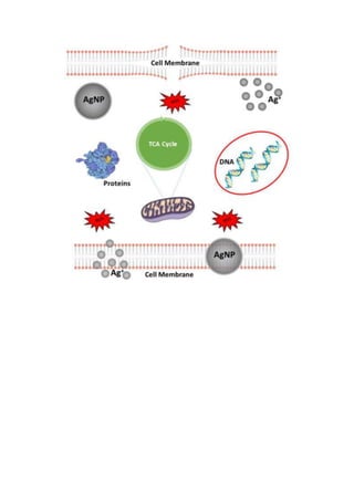
![Charging effects and surface potential variations of Cu-based nanowires
D. Nunes1,*
, T.R. Calmeiro1
, S. Nandy1
, J.V. Pinto1
, A. Pimentel1
, P. Barquinha1
, P.A.
Carvalho2,3
, E. Fortunato1
and R. Martins1,*
1
i3N/CENIMAT, Department of Materials Science, Faculty of Science and Technology,
Universidade NOVA de Lisboa, Campus de Caparica, 2829-516 Caparica, Portugal
2
SINTEF Materials and Chemistry, PB 124 Blindern, NO-0314 Oslo, Norway
3
CeFEMA, Instituto Superior Técnico, Universidade de Lisboa, 1049-001 Lisboa, Portugal
*daniela.gomes@fct.unl.pt and rm@uninova.pt
Abstract
Copper-based nanowires have attracted a growing interest in advanced materials and
nanotechnology research [1-3] owing to the high electrical conductivity of copper [4] and the p-type
semiconductor behavior of both cuprous oxide (Cu2O) [1, 5-8] and cupric oxide (CuO) [9, 10], which
present energy band gaps of, respectively, 2.17 eV [1] and 1.4 eV [11]. These materials are interesting
for a plethora of nano-optoelectronic applications [12-14], ranging from solar cells [15] to gas sensors
[9]. The present work reports charging effects and surface potential variations in pure copper, cuprous
oxide and cupric oxide nanowires observed by electrostatic force microscopy (EFM) and Kelvin probe
force microscopy (KPFM). The copper nanowires were produced by wet synthesis, oxidation into Cu2O
nanowires was achieved through microwave irradiation and CuO nanowires were obtained via furnace
annealing in atmospheric conditions. Structural characterization of the nanowires was carried out by X-
ray diffraction, scanning electron microscopy, transmission electron microscopy (Figure 1) and energy
dispersive X-ray spectroscopy. During the EFM experiments the electrostatic field of the positive probe
charged negatively the Cu-based nanowires, which in turn polarized the SiO2 dielectric substrate. Both
the probe/nanowire capacitance as well as the substrate polarization increased with the applied bias.
Cu2O and CuO nanowires behaved distinctively during the EFM measurements in accordance to their
band gap energies. The work functions (WF) of the Cu-based nanowires, obtained by KPFM
measurements (contact potential difference (CPD) profiles), yielded WFCuO > WFCu > WFCu2O (Figure 2).
References
[1] D. Nunes, A. Pimentel, P. Barquinha, P.A. Carvalho, E. Fortunato, R. Martins, Journal of Materials
Chemistry C, 2 (2014) 6097-6103.
[2] H. Guo, N. Lin, Y. Chen, Z. Wang, Q. Xie, T. Zheng, N. Gao, S. Li, J. Kang, D. Cai, D.-L. Peng, Sci.
Rep., 3 (2013).
[3] A.R. Rathmell, S.M. Bergin, Y.-L. Hua, Z.-Y. Li, B.J. Wiley, Advanced Materials, 22 (2010) 3558-
3563.
[4] M. Mohl, P. Pusztai, A. Kukovecz, Z. Konya, J. Kukkola, K. Kordas, R. Vajtai, P.M. Ajayan, Langmuir,
26 (2010) 16496-16502.
[5] L. Xiong, S. Huang, X. Yang, M. Qiu, Z. Chen, Y. Yu, Electrochimica Acta, 56 (2011) 2735-2739.
[6] V. Figueiredo, E. Elangovan, G. Gonçalves, P. Barquinha, L. Pereira, N. Franco, E. Alves, R.
Martins, E. Fortunato, Applied Surface Science, 254 (2008) 3949-3954.
[7] V. Figueiredo, E. Elangovan, G. Gonçalves, N. Franco, E. Alves, S.H.K. Park, R. Martins, E.
Fortunato, physica status solidi (a), 206 (2009) 2143-2148.
[8] Z. Zhang, R. Dua, L. Zhang, H. Zhu, H. Zhang, P. Wang, ACS Nano, 7 (2013) 1709-1717.](https://image.slidesharecdn.com/nanopt2016conferencebook-160222125923/85/NanoPt2016-Conference-Book-204-320.jpg)
![[9] N.D. Hoa, N. Van Quy, H. Jung, D. Kim, H. Kim, S.-K. Hong, Sensors and Actuators B: Chemical,
146 (2010) 266-272.
[10] X. Zhang, G. Wang, W. Zhang, N. Hu, H. Wu, B. Fang, The Journal of Physical Chemistry C, 112
(2008) 8856-8862.
[11] J.T. Chen, F. Zhang, J. Wang, G.A. Zhang, B.B. Miao, X.Y. Fan, D. Yan, P.X. Yan, Journal of
Alloys and Compounds, 454 (2008) 268-273.
[12] L. Liao, B. Yan, Y.F. Hao, G.Z. Xing, J.P. Liu, B.C. Zhao, Z.X. Shen, T. Wu, L. Wang, J.T.L. Thong,
C.M. Li, W. Huang, T. Yu, Applied Physics Letters, 94 (2009) -.
[13] X. Duan, C. Niu, V. Sahi, J. Chen, J.W. Parce, S. Empedocles, J.L. Goldman, Nature, 425 (2003)
274-278.
[14] G. Larrieu, X.L. Han, Vertical nanowire array-based field effect transistors for ultimate scaling,
Nanoscale, 5 (2013) 2437-2441.
[15] S. Anandan, X. Wen, S. Yang, Materials Chemistry and Physics, 93 (2005) 35-40.
Figures
Figure 1. SEM and TEM images of nanowires (a and d) Cu, (b and e) Cu2O produced by microwave
irradiation, and (c and f) CuO nanowires oxidized by furnace annealing in air.
Figure 2. Surface potential images of a Cu (a), Cu2O (b) and CuO (c) nanowires obtained from KPFM
measurements. Topography images of each nanowire are presented as insets. The CPD profiles from
images (a) to (c) are presented from (d) to (e).](https://image.slidesharecdn.com/nanopt2016conferencebook-160222125923/85/NanoPt2016-Conference-Book-205-320.jpg)
![One Step Synthesis and Polyacrylic Acid Functionalization of Multifunctional Eu-doped NaGdF4
Nanoparticles with Selected Size for Optical and MRI Imaging.
Nuria O. Nuñez
*[a]
, María García
[a]
, Jorge García-Sevillano
[b]
, Sara Rivera-Fernández
[c]
, Jesús M de la
Fuente
[c], [d], [e]
and Manuel Ocaña
[a]
[a]
Instituto de Ciencia de Materiales de Sevilla, CSIC, Américo Vespucio 49, 41092, Isla de la Cartuja,
Sevilla, Spain
[b]
Dpto. Física de Materiales, C-04, Universidad Autónoma de Madrid, Spain
[c]
Instituto de Nanociencia de Aragon, Universidad de Zaragoza, Mariano Esquillor s/n, Zaragoza,
50018, Zaragoza, Spain
[d]
Fundación ARAID, Zaragoza, Spain
[e]
Institute of Nano Biomedicine and Engineering, Key Laboratory for Thin Film and Microfabrication
Technology of the Ministry of Education, Research Institute of Translation Medicine, Shanghai Jiao
Tong University, Dongchuan Road 800, 200240 Shanghai, People’s Republic of China.
E-mail: nurianu@icmse.csic.es
Abstract
Nowadays, much attention is been paid in the biomedical field to the development of multifunctional
nanoparticles suitable for both, optical and magnetic resonance (MRI) imaging applications
[1-5]
because
they combine the high sensitivity of optical imaging for in vitro applications with the excellent spatial
resolution and depth for in vivo application associated to the MRI imaging.
[6-8]
In this work, we have
developed a simple method for the synthesis of uniform Eu-doped NaGdF4 nanospheres with optimised
luminescent properties and their functionalization with carboxylate (one-pot procedure) groups provided
by polyacrylic acid polymer (PAA).
[9]
The size of the nanospheres could be altered in the 60-95 nm
range by adjusting the amount of polyacrylic acid added. The luminescent and magnetic relaxivity
properties of the functionalized nanoparticles along with their negligible cytotoxicity and high colloidal
stability in 2-morpholinoethanesulfonic acid solutions, make them potential candidates for
biotechnological applications as “in vitro” optical biolabels and for application as MRI contrast agent
(Figure 1).
References
[1] G. Tian, Z. Gu, X. Liu, L. Zhou, W. Yin, L. Yan, S. Jin, W. Ren, G. Xing, S. Li and Y. Zhao, J. Phys.
Chem. C, 115 (2011) 23790.
[2] J. W. Mulder, A. W. Griffioen, G. J. Strijkers, D. P. Cormode, K. Nicolay and Z. A. Fayad,
Nanomedicine, 2 (2007) 307.
[3] J. Ryu, H. Y. Park, K. Kim, H. Kim, J. H. Yoo, M. Kang, K. Im, R. Grailhe and R. Song, J. Phys.
Chem.C,114 (2010) 21077.
[4] M. He, P. Huang, C. Zhang, H. Hu, C. Bao, G. Gao, R. He and D. Cui, Adv. Funct. Mater., 21 (2011)
4470.
[5] L. Zhou, Z. Gu, X. Liu, W. Yin, G. Tian, L. Yan, S. Jin, W. Ren, G. Xing, W. Li, X. Chang, Z. Hu and
Y. Zhao, J. Mater. Chem., 22 (2012) 966.
[6] G. K. Das, B. C. Heng, S. C. Ng, T. White, J. S Loo, L. D’Silva, P. Padmanabhan, K. K Bhakoo, S.
T. Selvan and T. Y. Tan, Langmuir, 26 (2010) 8959.
[7] X. Yu, Y. Shan, G. Li and K. Chen, J. Mater. Chem., 21 (2011) 8104.
[8] N. J. J. Johnson, W. Oakden, G. J. Stanisz, R. S. Prosser and F. C. J. M. van Veggel, Chem.
Mater., 23 (2011) 3714.
[9] N. Nuñez, J. M. de la Fuente, S. Rivera and M. Ocaña, Dalton Transactions, 42 (2013) 10725.](https://image.slidesharecdn.com/nanopt2016conferencebook-160222125923/85/NanoPt2016-Conference-Book-206-320.jpg)
![From the nano to the micro range: particle size method development
A. M. Paiva, S. Silva, S. S. Pinto, C. Cacela
Hovione FarmaCiencia SA. Sete Casas 2674-506, Loures. Portugal.
mgpaiva@hovione.com
Particle size (PS) is one of the most important quality attributes to monitor during process development
and production in the pharmaceutical industry [1]. Some of the processes being developed at Hovione
are multi-step procedures with different particle size ranges. With the purpose of controlling this critical
quality attribute (CQA), accurate and precise methods need to be developed for the different stages of
the processes. The work herein presented describes the development of two methods that were needed
to characterize and monitor both nano and micro particles of a given manufacturing process.
Two different PS methods were developed in order to support two manufacturing steps: a Laser
Diffraction method (LD) for the micro PS range control and a Dynamic Laser Diffraction (DLS) method
for the nano PS range assessment. In order to support method development, particles were further
characterized by optical microscopy.
For the LD method, a Mastersizer 2000 (Malvern Instruments Ltd) equipped with a Hydro 2000S
dispersion unit was used. The development included the selection of an adequate dispersant and the
determination of the optimal preparation conditions to obtain a stable suspension composed only of
primary particles. A repeatable and accurate LD method was successfully developed (Figure 1), which
median particle size of 37 µm.
This method proved to be accurate in a range between 5 and 40 µm (Table 1).
The DLS method was developed using a Zetasizer Nano Range equipment (Malvern Instruments Ltd).
A single method was developed for the characterization of the Active Pharmaceutical Ingredient (API)
suspension and for the The optimal API concentration was found by
successive dilutions in water with the aim of having a stable nanoparticle suspension, enabling an
accurate light scattering. The method developed allowed the characterization of the nanoparticles of the
API suspension (around 35 nm) and of the suspension (35-90 nm) (Figure 2).
Table 1. Particle size (micro and nanoparticles) data regarding several process steps and batches
Product
API suspension
(nm)
Intermediate 1
(µm)
Intermediate
2 (nm)
Drug Product
reference values (nm)
Batches
1
34
33 40 34
2 33 37 33
3 38 42 33
4 7 40 33
5 6 41 34
6 6 45 33
7 6 37 33](https://image.slidesharecdn.com/nanopt2016conferencebook-160222125923/85/NanoPt2016-Conference-Book-208-320.jpg)
![Two successful PS methods were developed and applied to the characterization of a new product in a
range between 35 nm to 40 µm. These methods were crucial to support the new manufacturing
process. As shown above in Table 1, a good correlation between the initial PS of the API suspension
and the PS of the final drug product was attained.
A)
Particle Size Distribution
0.01 0.1 1 10 100 1000 3000
Particle Size (µm)
0
0.5
1
1.5
2
2.5
3
3.5
4
4.5
5
5.5
6
6.5
7
7.5
8
8.5
9
9.5
10
10.5
11
11.5
12
12.5
13
13.5
14
14.5
Volume(%)
B)
Figure 1. A) Particle size distribution overlay curves obtained during LD method repeatability
assessment. B) Microscopy image of the
Figure 2. Particle size distribution curve obtained during DLS method assessment .
Moreover, the DLS method also enabled the prediction of the PS after the downstream processing. As
this is a multi-step process, the fact that the PS can be predicted earlier on, prevents possible issues
that might occur in the downstream processing due to unsuitable properties of the particles.
References
[1] Ana F.T. Silva et al., European Journal of Pharmaceutics and Biopharmaceutics, 85 (2013)
1006.](https://image.slidesharecdn.com/nanopt2016conferencebook-160222125923/85/NanoPt2016-Conference-Book-209-320.jpg)
![Dispersion and re-agglomeration phenomena of polymer-
functionalized graphite nanoflakes upon melt-mixing
M. C. Paiva, R. M. Santos, C. Vilaverde, E. Cunha and J. A. Covas
Institute for Polymers and Composites/I3N, University of Minho, Campus de Azurém, 4800-058
Guimarães, Portugal
mcpaiva@dep.uminho.pt
Abstract
Graphite nanoflake (GnF) powders are provided in the form of agglomerates of nanoflakes, stabilized by
Van der Waals interactions. The production of composites with relevant properties (mechanical, electrical,
thermo-electrical, barrier, etc.) requires that these agglomerates are well dispersed in the polymer melt,
and that they remain so during cooling to the solid state, as well as during re-melting, as frequently required
to obtain the final part. It is known that the nanoparticle surface chemistry and interaction with the polymer
matrix affects the dispersion in the polymer melt and the composite final properties. However, the overall
process is influenced by the processing parameters, particle surface chemistry and chemical nature of the
polymer matrix, and thus controversial conclusions are often reported in the literature.
In this work [1] GnF were chemically functionalized with pyrrolidine groups via 1,3 dipolar cycloaddition,
and then grafted with polypropylene-graft-maleic anhydride (PP-g-MA). The functionalization was
characterized by thermogravimetric analysis and X-ray photoelectron spectroscopy. The kinetics of
dispersion in polypropylene (PP) was studied using a prototype small-scale mixer that generates a strong
extensional flow under controlled conditions, and compared with the behavior of as-received GnF. The
prototype mixer, depicted in Figure 1, permits sample collection along its axis, thus allowing monitoring the
evolution of dispersion along the process. PP nanocomposites with 2 and 10 wt. % of as-received and PP-
grafted GnF were prepared under identical conditions. The progression of nanoparticle dispersion along
the mixer was analyzed by monitoring the nanoparticle agglomerate size at the micron level by optical and
scanning electron microscopies. The effect of nanoparticle dispersion on the polymer morphology was
studied by differential scanning calorimetry and X-ray diffraction. The electrical conductivity of the
composites was measured. GnF re-agglomeration effects upon melt relaxation were analyzed.
It was observed that, regardless of filler loading, there is a significant decrease of the agglomerate size
along the prototype length, showing that extensional flow efficiently induces the dispersion of graphite
nanoflakes. When the polymer melt is allowed to relax, a prominent increase of agglomerate area is
observed, suggesting that re-agglomeration took place. The morphology and/or cohesion of these re-
formed agglomerates seem to be different from that of the initial agglomerates fed into the dispersion
equipment, affecting its subsequent dispersion rate in a second mixing process. Surface modification of
GnF with polymer enhances the stability of dispersion and delays re-agglomeration.
References
[1] R. M. Santos, C. Vilaverde, E. Cunha, M. C. Paiva and J. A. Covas, Soft Matter, accepted for publication,
doi:10.1039/C5SM01366F.](https://image.slidesharecdn.com/nanopt2016conferencebook-160222125923/85/NanoPt2016-Conference-Book-210-320.jpg)
![Functional Characterization of α-Lactalbumin Nanotubes to Transport Food Additives
Clara Fuciños
1,2
, Pablo Fuciños
3
, Martín Míguez
1
, María L. Rúa
1
, António A. Vicente
2
, Lorenzo
Pastrana
3
1
Biotechnology Group, Department of Analytical Chemistry and Food Science, University of Vigo, As
Lagoas s/n, 32004 Ourense, Spain
2
Centre of Biological Engineering, University of Minho, Campus de Gualtar s/n, 4710-057 Braga,
Portugal
3
The International Iberian Nanotechnology Laboratory (INL), Avenida Mestre José Veiga s/n, 4715-330
Braga, Portugal
lorenzo.pastrana@inl.int
Abstract
Partial hydrolysis of α-lactalbumin (α-LA) induced by a serine endoprotease from Bacillus licheniformis
(BLP) results in the formation of nanotubes in the presence of a divalent ion, which promotes salt-bridge
formation between two deionized carboxylic groups [1]. Because of their GRAS status, protein
nanotubes will be useful for food applications, such as thickener agents or vehicle for controlled release
of bioactive molecules [2]. Despite of that, to our knowledge, only a paper describing the application of
BSA nanotubes to incorporate curcumin as bioactive compound has been published [3].
Functional characterization of α-LA nanotubes was evaluated in this work by determining their ability to
encapsulate and retain caffeine under different chemical conditions that could compromise the stability
of the nanotubes. Caffeine (MW: 194.19 g mol
-1
), used as active component of energy drinks, yields at
high concentrations a disgusting bitterness that has to be masked by adding sugars and flavors. To
avoid this inconvenient, this study proposes the encapsulation of caffeine into α-LA nanotubes previous
to the addition to the food product. As it would be desirable that caffeine remained protected into the
nanotubes during the food production and conservation, nanotubes' stability and caffeine release from
them was tested in usual environmental conditions that might occur for these food products.
For that purpose, temperatures from refrigeration to pasteurization (8-80 ºC) were evaluated, combined
with pH values from 2-8, the presence of chelating agents (EDTA, usually added as antioxidant agent at
maximum concentrations of 75 g mL
-1
), and the presence of salt (NaCl, usually added as preservative
and flavoring at concentrations around 1000 g mL
-1
). Caffeine-loaded nanotubes were prepared at
1.5/20 caffeine/α-LA (w/w) ratio, which was previously proven as highly effective for caffeine
encapsulation. %EE (encapsulation efficiency) was near 100% and %LC (loading capacity) was around 10%.
A 2
4
full-factorial design (Box, Hunter, and Hunter 2
K-p
type) was used to analyzed if the four variables
above exposed (T, pH, EDTA, NaCl) had significant effect on nanotube disassembly and caffeine
release. For each experiment of the design, caffeine release kinetics from nanotubes were performed.
The maximum percentage of caffeine released from α-LA nanotubes (%Caffree max) ranged from ∼ 40% to
100%. TEM micrographs and RP-HPLC chromatograms showed high correlation with the %Caffree max-
values. Thus, those conditions in which caffeine release was higher are corresponding to nanotubes
more degraded. Conversely, those conditions in which the release was lower are corresponding to
intact nanotubes or less degraded (Figure 1).
After neglecting the insignificant terms (p > 0.10), the fitted equation which describes the %Caffree max
released from nanotubes is the following:
! "#$%&’’() *+ , -./01 2 3/-4 5 33/46 2 3/.. 78 5 93:/0; 2 3/.. 7<= 5 ;/-: 2 3/.. 78 7<= 5
9-/31 2 3/.. 78 7>?8@ 5 90/-4 2 3/.. 7<= 7>?8@ 5 -/10 2 3/.. 78 7<= 7>?8@
(1)
The model obtained was statistically significant (α = 0.10), and the lack of fit was not significant. The
high r
2
value (r
2
= 0.8327 and r
2
adjusted = 0.7555) indicated good correlation between the adjusted and
predicted values, which supports the statistical validity and significance of the equation obtained.
The effect of T and pH on %Caffree max was significant (p < 0.05) and NaCl did not have any significant (p
> 0.05) effect on %Caffree max. The interaction of EDTA with T and pH had significant effect on %Caffree
max (p < 0.05). In absence of EDTA (Figure 2A) the effect of T on %Caffree max was intense, appearing
more free caffeine with increasing temperatures. Free caffeine also increased by reducing pH but the
effect was less intense than that observed with T. By introducing EDTA (Figure 2B and 2C) in the
release solution the effect of pH was increased at low T, and %Caffree max was clearly higher at low pH.](https://image.slidesharecdn.com/nanopt2016conferencebook-160222125923/85/NanoPt2016-Conference-Book-212-320.jpg)
![At high T the effect of the other parameters disappeared, probably because nanotubes' stability was too
low and additional effects could be neglected.
Within the domain evaluated, the minimum %Caffree max was ∼ 43.65 (i.e. ∼ 56.35% remained retained)
and occurred at 8 ºC, pH 7.5 and 75 g mL
-1
of EDTA. Therefore, those conditions that will help to
maintain caffeine encapsulated into α-LA nanotubes, during food processing and storing until their
consumption, are refrigeration temperatures at neutral or alkaline conditions. In acidic conditions the
absence of chelating agents would be preferable.
References
[1] Graveland-Bikker, J. F., Ipsen, R., Otte, J., & De Kruif, C. G., Langmuir, 20:16 (2004), 6841.
[2] Ipsen, R., & Otte, J., Biotechnology Advances, 25:6 (2007), 602.
[3] Sadeghi, R., Kalbasi, A., Emam-jomeh, Z., Razavi, S. H., Kokini, J., & Moosavi-Movahedi, A. A.,
Journal of Nanoparticle Research, 15:11 (2013), 1931.
[4] Gunasekaran, S., Ko, S., & Xiao, L., Journal of Food Engineering, 83:1 (2007), 31.
Figures
A
Low Caffeine Release High Caffeine Release
B C D
0 20 40 60
0
200
400
600
800
1000
Time (min)
mAU
0 20 40 60
0
200
400
600
800
1000
Time (min)
mAU
A
B
C
D
Low Caffeine Release High Caffeine Release
Figure 1. Transmission electron micrographs (TEM) and chromatograms of samples with α-LA
nanotubes subjected to different environmental conditions that may occur in food products: A) 8 ºC, pH
7.5, 0 g mL
-1
EDTA, 1000 g mL
-1
NaCl; B) 8 ºC, pH 7.5, 75 g mL
-1
EDTA, 0 g mL
-1
NaCl; C) 8 ºC,
pH 2, 75 g mL
-1
EDTA, 1000 g mL
-1
NaCl; D) 80 ºC, pH 2, 0 g mL
-1
EDTA, 1000 g mL
-1
NaCl. Scale
bar of the images is 200 nm.
Figure 2. Response surfaces corresponding to the combined effect of temperature (T) and pH and
EDTA on the maximum percentage of free caffeine released from nanotubes (%Caffree max) at A) 0 g
mL
-1
, B) 37.5 g mL
-1
, and C) 75 g mL
-1
of EDTA, according to Equation 1.](https://image.slidesharecdn.com/nanopt2016conferencebook-160222125923/85/NanoPt2016-Conference-Book-213-320.jpg)
![Gold-nanoparticles for MDR1 silencing in DOX treated Colon Cancer Cells
Pedro Pedrosa, Alexandra Fernandes & Pedro Viana Baptista
UCIBIO, DCV, Faculdade de Ciências e Tecnologia, Universidade NOVA de Lisboa,
Portugal
pm.pedrosa@campus.fct.unl.pt
Abstract
Many cancers develop resistance to chemotherapeutic agents, which become a major obstacle to
effective chemotherapy. Multidrug resistance (MDR) mechanisms may rely on the up-regulation of
membrane ATP-dependent efflux pumps that excrete drugs to the extracellular medium, decreasing
their intracellular concentration [1]. One of most studied examples is P-glycoprotein (MDR1) whose
overexpression is described in many cancers, including colorectal and hepatocellular carcinomas,
leukemia and lymphoma, where it confers cross-resistance to a variety of cytotoxic agents [1].
Targeting MDR mechanisms with iRNA capable to successfully silence gene expression, sensitizes
tumor cells to cytotoxic drugs. iRNA requires effective vectors for the silencing moieties (e.g. siRNA,
hairpin ssDNA) that can sustain degradation in circulation and deliver them intracellularly with minimal
toxicity on target cells [2,3].
Previously we reported that gold nanoparticles (AuNPs) functionalized with hairpin antisense ssDNA
oligonucleotides have equivalent silencing capacity and cellular toxicity than lipofectamine vectorized
siRNA [4,5]. In this work we used AuNPs functionalized with hairpin antisense oligonucleotides directed
at silencing MDR1 to increase sensitisation of colorectal carcinoma (HCT116) cells to doxorubicin. We
believe such systems will pave the way for combinatory strategies to overcome MDR in the clinics.
References
[1] Holohan, C., et al., Nat Rev Cancer, 10 (2013) 714-26.
[2] Wu, Y., et al., Colloids Surf B Biointerfaces, 138 (2016) 60-9.
[3] Nourbakhsh M et al., Iran J Basic Med Sci., 18 (2015) 385-92.
[4] Conde, J., et al., Biomaterials, 34 (2013) 2516-23.
[5] Conde, J., et al., Nanotoxicology, 5 (2014) 521-32.](https://image.slidesharecdn.com/nanopt2016conferencebook-160222125923/85/NanoPt2016-Conference-Book-214-320.jpg)
![Immobilization of Gold Nanoparticles and Trametes Versicolor Laccase Nanobioconjugates on
Membranes for the Development of Biosensors
Miguel Peixoto de Almeida, Marta Belda, Emma Calle, Eulália Pereira
UCIBIO/REQUIMTE, Departamento de Química e Bioquímica, Faculdade de Ciências, Universidade do
Porto, 4169-007 Porto, Portugal
mpda@fc.up.pt
The use of enzymes in environment-related processes, can be split in two main fields. The first is
(bio)remediation, where enzymes have the primary role and actively degrade hazardous compounds in
less or non-toxic products. The second is (bio)sensing, which is fundamental to determine when, where
and in what extension actions should be taken. The incorporation of enzymes in sensors are an excellent
way to assess the presence of one or more dangerous compounds and determine their concentration. [1]
The enhancement of this type of sensors can result in lowering the detection and quantification levels,
which, for example, results in earlier detection in a scenery of crescent contamination.
Gold nanoparticles (AuNPs) are today widely used in many areas, and the environment is no
exception. [2] The incorporation of this nanomaterial in biosensors can significantly improve the signal,
either by increasing the electric conduction between the enzyme active center and the electrode due to
the improvement in the direct electron transfer (DET) phenomena, which is highly relevant in the case of
electrochemical biosensors [3], or by increasing the activity of the enzyme itself, as has been proven in
some cases, with the formation of bionanoconjugates (BNCs) [4].
For purposes of recycling/reutilization or use in flow processes, there is a great interest in
immobilizing the enzymes and the AuNPs or the assembled BNCs. If the synthesis of AuNPs is well
established and easy to preform and, in the other hand, the enzymes commonly used in these biosensors
are relatively cheap and easy to obtain, it should not be the immobilization process to add high complexity
or costs to the process. So, in this work we tried to assemble bionanoconjugates directly in cellulose-
based and other materials representing different structures and surface chemistries: five common
membrane materials like hydrophilic mixed cellulose esters (HMCE), polytetrafluoroethylene (PTFE),
hydrophilic polyvinylidene fluoride (HPVDF), regenerated cellulose (RC), nitrocellulose (NC) and two
more simple options: filter paper (FP) and copy paper (CP).
Three of these seven materials were chosen for the BNCs assembling, schematized in figure 1,
being these three the ones with higher 15 nm AuNPs loading (HMCE, NC and FP, respectively A, E and
F in figure 2). The other main element of the BNCs is laccase from Trametes versicolor (LTv), an enzyme
from the class of the phenol oxidases, capable of many applications, namely the sensing of hazardous
compounds like bisphenol A, aminophenols and others [5].
The set of obtained data from activity tests shows that there is an increase of activity in the
presence of gold nanoparticles in the supporting material. This observation can be justified by small
changes in the enzyme conformation due to electrostatic interactions between the nanoparticles and the
protein, as suggested before in our group for other phenol oxidase (mushroom tyrosinase) [4]. Since the
support is enriched with AuNPs, as can be seen in SEM images in figure 3 together with the strong red
coloration shown in figure 1, although it is not enough to transform these non-conductive materials in good
conductors, is certainly a step in that direction, which can facilitate electrochemical applications. The
enzymatic activity enhancement was verified for all the three cellulose-based materials tested, however
HMCE and NC offer better resistance to manipulation than FP. In the other hand, FP is a very cheap
product when compared to HMCE and NC and yet relatively well controlled in its composition and purity.
This work can be interpreted as a promising first step towards cheap and disposable nanobiosensors.
References
[1] Rao MA, Scelza R, Acevedo F, Diez MC, Gianfreda L, Chemosphere, 107 (2014) 145-162
[2] Peixoto de Almeida M, Pereira E, Baptista P, Gomes I, Figueiredo S, Soares L, Franco R, Gold
Nanoparticles in Analytical Chemistry, 66 (2014) 529-567](https://image.slidesharecdn.com/nanopt2016conferencebook-160222125923/85/NanoPt2016-Conference-Book-215-320.jpg)
![[3] Christenson A, Dimcheva N, Elena, Ferapontova EE, Gorton L, Ruzgas T, Stoica L, Shleev S,
Yaropolov AI, Haltrich D, Thorneley RNF, Aust SD, Electroanalysis, 16 (2004) 1074-1092
[4] Cortez J, Vorobieva E, Gralheira D, Osório I, Soares L, Vale N, Pereira E, Gomes P, Franco R, Journal
of Nanoparticle Research, 13 (2011) 1101-1113
[5] Fernández-Fernández M, Sanromán MA, Moldes D, Biotechnology Advances, 31 (2013) 1808-1825
Acknowledgements
The authors are grateful to Fundação para a Ciência e a Tecnologia (FCT) and Fundo Europeu de
Desenvolvimento Regional (FEDER), in the context of the COMPETE program, for financial support
through project UID/MULTI/04378/2013, project PTDC/CTM-NAN/2912/2014, and fellowship
SFRH/BD/95983/2013 (for MPA).
Figures
Figure 1. Schematic representation of bionanoconjugates assembling on supporting materials.
A B C D E F G
Figure 2. Pictures of seven nanoparticle-enriched supporting materials. A – hydrophilic mixed cellulose
esters, B – polytetrafluoroethylene, C – hydrophilic polyvinylidene fluoride, D – regenerated cellulose, E
– nitrocellulose, F – filter paper, G – copy paper.
A B C
Figure 3. Back-scattering SEM images (50000x magnification) of the three selected nanoparticle-enriched
supporting materials. A – hydrophilic mixed cellulose esters, B – nitrocellulose, C – filter paper.](https://image.slidesharecdn.com/nanopt2016conferencebook-160222125923/85/NanoPt2016-Conference-Book-216-320.jpg)
![Fabrication of biodegradable microneedles for peptide delivery
Liliana R Pires, Rizwan Gill, Hélder Fonseca, Rosana Dias, Paulo Freitas, João Gaspar
INL International Iberian Nanotechnology Laboratory, Av Mestre Veiga, Braga, Portugal
liliana.pires@inl.int
Abstract (Arial 10)
Microneedles have been extensively investigated in the recent years as mean to mediate the delivery of
drugs and/or peptides to the epidermal and/or intradermal space, overcoming the skin stratum corneum
barrier. These devices hold the potential of allowing self-administration and painless application.
Microneedles can be designed to dissolve in the skin, assuring biodegradability and safe disposal
without biohazardous waste [1]. Particularly in the field of vaccination, the intradermal administration of
antigens through the application of microneedle devices showed improved efficiency comparing to
conventional injection procedures, being currently under clinical trials [2]. In this study we aim at
designing and fabricating fully biodegradable polymeric microneedles that allow the sustained release of
biologically active peptides to the intradermal space.
The approach used is to prepare a Si needle that acts as the master for a mold fabricated afterwards.
Silicon microneedle masters were firstly prepared using a sequential isotropic-anisotropic-isotropic deep
reactive ion etching (DRIE) process, previously developed for sub-5-µm needles [3] and extended here
to structures in the range of 100-500 µm. A silicon wafer (700-750 µm thick) with silicon dioxide mask
was patterned using lithography. Microneedle shape was determined by DRIE. Wafers were diced into 2
x 2 cm pieces and characterized by scanning electron microscopy (SEM). Poly(dimethylsiloxane)
(PDMS) molds were prepared as previously described [4] after silanization of the silicon masters to
facilitate removal of the molded materials. To obtain the polymeric microneedles mixtures of poly(vinyl
acetate) (PVA) and poly(vinyl pyrrolidone) (PVP) were poured onto the prepared PDMS molds. Vacuum
was applied to fill the molds and subsequently the solution was allowed to dry (24hrs). Solid
microneedle patches were peeled off from the molds and analyzed by optical microscopy. Different
PVA/PVP ratios were tested in order to optimize microneedle degradability, drug release and ability to
pierce the skin.
Silicon needles were obtained by microfabrication techniques (Figure 1 A). Results show PDMS molds
replicating the silicon master shape (Figure 1 B). Polymeric microneedles were successfully prepared
showing around 400 µm height and 200 µm width (Figure 1 C). An aspect considered
suitable for microneedle perforation of the skin. The preparation of sharper needles is currently being
pursued.
References
[1] Lee JW, Park J-H,Prausnitz MR. Biomaterials, 29 (2008) 2113.
[2] Koutsonanos DG, Vassilieva EV, Stavropoulou A, Zarnitsyn VG, Esser ES, Taherbhai MT, Prausnitz
MR, Compans RW,Skountzou I. Scientific Reports, 2 (2012) 357.
[3] Held J, Gaspar J, Ruther P, Hagner M, Cismak A, Heilmann A, and Paul O, Journal Micromechanics
Microengineering, 20 (2010) 025024.
[4] Dieguez L; Winter MA; Pocock KJ; Bremmell KE, Thierry B. The Analyst 140 (2015) 3565.
Figures](https://image.slidesharecdn.com/nanopt2016conferencebook-160222125923/85/NanoPt2016-Conference-Book-217-320.jpg)
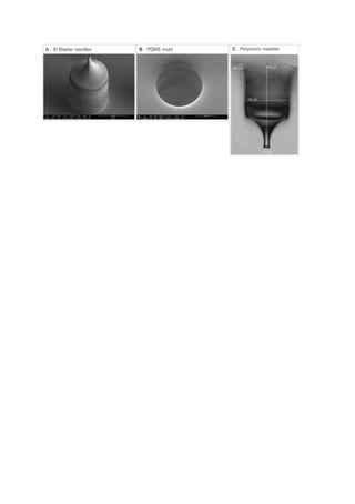
![Layer-by-Layer Films Containing Peptides of the Cry1Ab16 Toxin from Bacillus thuringiensis for
Nanodevices Development
Alexandra Plácido1
, Emanuel Airton de Oliveira Farias2, Mariela M. Marani3, Andreanne G.
Vasconcelos2, Ana C. Mafud4, Yvonne P. Mascarenhas4, Carla Eiras2,5, José Roberto S. A. Leite2,
Cristina Delerue-Matos1
1REQUIMTE/LAQV, Instituto Superior de Engenharia do Porto, Instituto Politécnico do Porto, Rua Dr.
António Bernardino de Almeida, 431, 4200-072, Porto, Portugal; 2Núcleo de Pesquisa em
Biodiversidade e Biotecnologia, BIOTEC, Universidade Federal do Piauí, UFPI, 64202020, Parnaíba,
Piauí, Brazil; 3IPEEC-CENPAT-CONICET
t, Argentina; 4Instituto de Física de
São Carlos, Universidade de São Paulo, USP, 13566-590, São Carlos, SP, Brazil; 5Laboratório de
Materiais Avançados, LIMAV, Engenharia de Materiais, Centro de Tecnologia, CT, Universidade
Federal do Piauí, UFPI, 64049550, Teresina, Piauí, Brazil.
E-mail: alexandra.placido@gmail.com
Abstract
Among thin film production techniques, layer-by-layer (LbL) self-assembly stands out because of its
versatility, which has enabled applications in the fields of biomaterials, biosensors, drug/gene delivery,
tissue engineering, implantable materials, diagnostics, electronics, energy, and optics [1]. Peptides are
potential candidates to meet the needs of the modern world in relation to diagnosis, disease monitoring,
quality control in industry, and more recently, detection of genetically modified organisms (GMOs) and
food safety through the development of biosensors [2]. Cry1Ab16 is a toxin of crystalline insecticidal
proteins that has been widely used in GMOs to gain resistance to pests. For the first time, in this study,
peptides derived from the immunogenic Cry1Ab16 toxin were immobilized as LbL films. Given the
concern about food and environmental safety, a peptide with immunogenic potential, PcL342-354C, was
selected for characterization of its electrochemical, optical, and morphological properties. The results
obtained by cyclic voltammetry (CV) showed that the peptide have an irreversible oxidation process in
electrolyte of 0.1 mol L-1 potassium phosphate buffer (PBS) at pH 7.2. It was also observed that the
electrochemical response of the peptide is governed mainly by charge transfer. In an attempt to
maximize the electrochemical signal of peptide, it was intercalated with natural (agar, alginate and
chitosan) or synthetic polymers (polyethylenimine (PEI) and Poly(sodium 4-styrenesulfonate) (PSS)).
The presence of synthetic polymers on the film increased the electrochemical signal of PcL342-354C up
to 100 times. Images by Atomic Force Microscopy showed that the immobilized PcL342-354C formed
self-assembled nanofibers with diameters ranging from 100 to 200 nm on the polymeric film (Fig. 1). By
UV-Visible spectroscopy (UV-Vis) it was observed that the ITO/PEI/PSS/PcL342-354C film grows
linearly up to the fifth layer, thereafter tending to saturation. X-ray diffraction confirmed the presence on
the films of crystalline ITO and amorphous polypeptide phases. In general, the ITO/PEI/PSS/PcL342-
354C film characterization proved that this system is an excellent candidate for applications in
electrochemical sensors and other biotechnological applications for GMOs and environmental
indicators.
References
[1] M.F. Zampa, I.M. Araújo, V. Costa, C.H.N. Costa, J.R. Santos, V. Zucolotto, C. Eiras, J.R.S. Leite,
Leishmanicidal activity and immobilization of dermaseptin 01 antimicrobial peptides in ultrathin films for
nanomedicine applications, Nanomedicine, 5 (2009) 352-358.
[2] A. Plácido, J.S. Amaral, J. Costa, T.J.R. Fernandes, M.B.P.P. Oliveira, C. Delerue-Matos, I. Mafra,
Novel Strategies for Genetically Modified Organism Detection, in: E.I. Academic Press (Ed.) Genetically
Modified Organisms in Food Production - Safety, Regulation and Public Health, Waltham, MA, USA,
2016, pp. 119-131.
Acknowledgments
This work was partially supported by grants from the Brazilian funding agencies (Fundacao de Amparo
a Pesquisa do Piaui) FAPEPI, (Comissão de Aperfeiçoamento de Pessoal do Nível Superior)) CAPES,
and (Conselho Nacional de Desenvolvimento Científico e Tecnológico) CNPq. In addition, financial
support was received from CONICET and ANPCyT.
hrough grant number PEst-C/EQB/LA0006/2011. Alexandra Plácido is
gratefully to FCT by her grant SFRH/BD/97995/2013, financed by POPH QREN Tipologia 4.1](https://image.slidesharecdn.com/nanopt2016conferencebook-160222125923/85/NanoPt2016-Conference-Book-219-320.jpg)
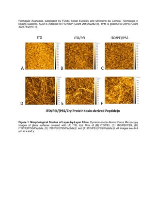
![Microfluidics with in-situ SAXS: from manipulation of soft materials to the study of out-of-
equilibrium phenomena
Bruno F. B. Silva
1
, Miguel Zepeda-Rosales
2
, Youli Li
2
, Ulf Olsson
3
and Cyrus R. Safinya
4
1
International Iberian Nanotechnology Laboratory, 4715-330 Braga, Portugal;
2
Materials Research
Laboratory, University of California, Santa Barbara, CA 93106, USA;
3
Division of Physical Chemistry ,
Centre for Chemistry and Chemical Engineering, Lund University, P.O. Box 124, SE-221 00 Lund,
Sweden;
4
Department of Materials, Department of Physics, and Molecular, Cellular & Developmental
Biology Department, University of California, Santa Barbara, CA 93106, USA
bruno.silva@inl.int
Abstract
Soft materials encompass a wide variety of nano- and meso-structured materials, and by definition are
deformable by thermal stresses and fluctuations. These materials are ubiquitous in biology (e.g. in the
membranes and cytoskeleton of cells) and technological applications (e.g. in drug delivery formulations
and liquid crystal displays). Nevertheless, despite their equilibrium properties having been the subject of
considerable attention, much remains unknown about their out-of-equilibrium behavior and dynamics. In
this talk, I will show how microfluidic chips (devices that involve precise control and manipulation of
fluids under sub-millimeter confinement), can be used to manipulate soft materials at the nano-scale.
This allows us to study fundamental out-of-equilibrium processes (e.g. coupling of structure and flow,
dissipation), as well as control and build complex out-of-equilibrium structures of technological interest.
In a first example, we use this manipulation ability to create well-defined flowing interfaces to
study the interplay between shear-flow forces and the structure of liquid crystals and surfactant
monolayers [1]. By use of a microfocused x-ray beam applied in-situ on the microfluidic device we are
able to determine the orientation field of the liquid crystal molecules, and how this orientation is
influenced by the flow conditions and chemical nature of the interfaces. In a second example, I will show
how microfluidics with in-situ SAXS can be used to study the kinetic evolution of phase transitions, more
specifically, the lamellar-to-microemulsion transition in surfactant-oil-water systems (Fig. 1).
References
[1] B.F.B. Silva, M.Z. Rosales, N. Venkateswaran, B.J. Fletcher, L.G. Carter, T. Matsui, T.M. Weiss, J.
Han, Y. Li, U. Olsson, C.R. Safinya, Langmuir, 31 (2015) 4361-4371
Figure 1: Schematic of the described experiment. The lamellar phase is flowed in the middle
microchannel (100x100 µm cross section), and mixed with either water or pentanol, flowing from the
side channels. The three flow rates and their ratios determine the final composition of the system
(chosen to match the microemulsion regions of the phase diagram) and the observation time.](https://image.slidesharecdn.com/nanopt2016conferencebook-160222125923/85/NanoPt2016-Conference-Book-221-320.jpg)
![RECONSTRUCTION PECULIARITY IN CO-PRECIPITATED Mg/Al AND
Mg/Al/Ce LAYERED DOUBLE HYDROXIDES
A. Smalenskaite1
, A. N. Salak2
, M. G. S. Ferreira2
, A. Kareiva1
1Department of Inorganic Chemistry, Vilnius University, Naugarduko 24, LT-03225 Vilnius, Lithuania
2Department of Materials and Ceramic Engineering/CICECO, University of Aveiro, 3810-193 Aveiro, Portugal
E-mail: aurelija.smalenskaite@gmail.com
Abstract
The layered double hydroxides (LDHs) are anionic compounds made up of positively charged brucite-like
layers with an interlayer gallery containing charge compensating anions and water molecules. The metal
cations occupy the centers of shared octahedral whose vertices contain hydroxide ions that connect to form
infinite two-dimensional sheets [1].
mixed metal oxides (MMO) [2], which have high adsorption capacity. Their high adsorption capacity and high
anion exchange capacity are comparable to those of anion exchange resins. This facilitates LDH application
as adsorption materials, catalyst precursors and catalyst supports [3]. MMO are able to recover the original
layered structure, a propert If MMO are put into aqueous solution, in the
presence of anions, the layered structure is recovered with anions incorporated in the interlayer. A more
irregular structure of agglomerated flake-like platelets has been observed after reconstruction [5] ( Figure 1).
Figure 1. Schematic representation of flake-like LDH formation process
Cerium based inhibitor creates a passive insoluble oxide layer that stops the oxygen diffusion from the
aggressive environment to the surface [6]. In this study, the intercalation of cerium in the Mg/Al layered
double hydroxide was investigated, for the first time to our knowledge. The simple co-precipitation method
was used for the fabrication of Mg/Al and Mg/Al/Ce specimens. LDH samples were synthesized by adding a
mixture of Mg(NO3)2
.6 H2O and Al(NO3)3
.9 H2O with molar ratio (3:1) drop by drop to the solution of
NaHCO3 and NaOH (1:2) under vigorous stirring. The pH of the solution was kept between 7 and 9 during
the synthesis. The formation of the MMO was achieved by heating The
MMO powders were
oxide in 40 mL of water). Synthesis of Mg/Al/Ce compounds were performed in the same way as Mg/Al LDH,
but the pH of the solution during the synthesis was 10. The effect of Ce3+ ion concentration on phase
structure of Mg3Al1-xCex system was studied. The Ce3+ concentration in crystal lattice was changed from 0.05
to 2 mol%. The influence of Ce3+ ions content and synthesis conditions on phase composition, crystal size
and morphology of Mg3Al1-xCex will be discussed. All synthesized samples were analysed and characterized
using X-ray diffraction (XRD) analysis, scanning electron microscopy (SEM) coupled with energy-dispersive
X-ray spectroscopy (EDX).](https://image.slidesharecdn.com/nanopt2016conferencebook-160222125923/85/NanoPt2016-Conference-Book-222-320.jpg)
![References
[1] X. Bi, H. Zhang, L. Dou. Pharmaceutics 6 (2014) 298-332.
[2] J. S. Valente, M. S. Cantu, M. Lima, F. Figueras. Chem. Mater. 21 (2009) 5809 5818.
[3] J. T. Feng, Y. J. Lin, D. G. Evans, X. Duan, D. Q. Li. J. Catal. 266 (2009) 351 358.
[4] V. Rives. Eds. Layered Double Hydroxides: Present and Future; Nova Science Publishers, Inc.New York,
(2001).
[5] F. Winter, X. Xia, B. P. C. Hereijgers, J. H. Bitter, A. J. van Dillen, M. Muhler, K. P. J. de Jong, J. Phys.
Chem. B 110 (2006) 9211 9218.
[6] M. L. Zheludkevich, R. Serra, M. F. Montemor, K. A. Yasakau, I. M. Miranda Salvado, M. G. S. Ferreira.
Electrochim. Acta 51 (2005) 208-217.
Acknowledgements
-Curie grant agreement No
645660.](https://image.slidesharecdn.com/nanopt2016conferencebook-160222125923/85/NanoPt2016-Conference-Book-223-320.jpg)
![Bismuth substitution for magnesium and aluminium effects in Mg/Al/Bi layered double
hydroxide.
Denis Sokol1,
*, Andrei N. Salak2
, Mario G. S. Ferreira2
, Aivaras Kareiva1
1
Department of Inorganic Chemistry, Vilnius University, Naugarduko 24, LT-03225 Vilnius, Lithuania
2
Department of Materials and Ceramic Engineering/CICECO, University of Aveiro, 3810-193 Aveiro,
Portugal
*E-mail: denis.sokol@chf.vu.lt
Layered double hydroxides (LDHs), hydrotalcite-type compounds (HTC) or two-dimensional (2D)
anionic clays are the commonly used names to describe a class of layered materials based on the
brucite (Mg(OH)2) crystal structure and having a general chemical formula of [M
II
1-x M
III
x(OH)2]
x+
(A
m-
)x/m 2O. The structure of LDHs is comprised of positively charged metal hydroxide layers [M
II
1-x
MIII
x(OH)2]x+
and negatively charged anions (Am-
)x/m in the interlayer space. The molecules of H2O are
usually present in the interlayer space [1, 2].
As shown in Figure 1, LDHs are composed of brucite-like layers in which a fraction of the divalent metal
cations M2+
(e.g., Mg2+
, Fe2+
, Co2+
, Cu2+
, Ni2+
, or Zn2+
) coordinated octahedrally by hydroxyl groups
have been replaced isomorphously by the trivalent metal cations M3+
(e.g., Al3+
, Cr3+
,Ga3+
, In3+
, Mn3+
or
Fe3+
), giving positively charged layers [3].
In the last decades, layered double hydroxides where magnesium or aluminium cations have been
replaced by same charge and similar ionic radii possessing metal ions have been synthesized and
investigated. However, the publications about LDHs with bismuth containing layered double hydroxide
are not found.
In this study, the Mg/Al/Bi HTC type compounds with different magnesium and aluminium substitution
level by bismuth were synthesized via novel alkoxy-free sol-gel and co-precipitation (low super
saturation) methods. Their mixed metal oxides (MMO) were obtained after thermal treatment of Mg/Al/Bi
layered double hydroxides, those MMO where reconstructed in water back to the layered structure. By
tuning the ratio of Mg:Al:Bi, the solvent composition, reaction and treatment temperature, the bismuth
substituted Mg/Al/Bi LDH and appropriate MMO were successfully synthesized. The influence of
bismuth substitution level, bismuth ionic radii, temperature and reconstruction pH on the phase
composition of final product is discussed. All synthesized samples by two synthetic techniques were
analyzed and characterized using X-ray diffraction (XRD) analysis, scanning electron microscopy (SEM)
coupled with energy-dispersive X-ray spectroscopy (EDX) and thermogravimetric (TG) analysis.
References
[1] F. Cavani, F. Trifiro, A. Vaccari, Catalysis Today, 11 (1991) 173-301.
[2] K.-H. Goh, Z. Dong, Water research 42 (2008) 1343-1368.
[3] G. Fan, F. Li, D.G. Evans, X. Duan, Chem. Soc. Rev, 43 (2014) 7040-7066.
Acknowledgements
This work was supported by project TUMOCS. This project has received funding from the European
-Curie grant
agreement No 645660.](https://image.slidesharecdn.com/nanopt2016conferencebook-160222125923/85/NanoPt2016-Conference-Book-224-320.jpg)
![Critical Current (Ic) Calculation for SHNO Devices using the experimentally
measured Spin hall angle ( SH) in Ta/CoFeB bilayer
M. Tarequzzaman1, 2
, M. Decker3
, J. D. Costa1
, B. Lacoste1
, T. Boehnert1
, E. Paz1
, C. H. Back3
,
R. Ferreira1
and P. P. Freitas1, 2
1International Iberian Nanotechnology Laboratory (INL), Ave. Mestre Jose Veiga, 4715-330, Braga,
Portugal
2Physics Department, Instituto Superior Tecnico (IST) - Technical University of Lisbon, 1000-029,
Lisbon, Portugal
3Institut für Experimentelle Physik, Universität Regensburg, D-93040 Regensburg, Germany
*tareq.zaman@inl.int
Spin transfer torque nano-oscillator (STNOs) driven by Spin hall effect (SHE) known as spin hall
nano-oscillators (SHNOs), opens up a new era of spin based electronic devices. SHNOs have several
advantages over conventional STNOs. In particular, pure spin current can be used to oscillate the
free layer (magnetic layer) rather than using dc current through the devices that can destroy the thin
barrier used in the devices.[1, 2] SHE occurs when an un-polarized current flows in a strong spin orbit
coupling matierials (non-magnetic), up and down spin are scattered in opposite directions thus
generating a pure spin current transverse to the applied current. The amplitude of spin current density
(Js SH = ) ( ), where Je is current
density applied to the strong spin orbit coupling materials. However, SHNO devices are to be
functional, we need to achieve two important parameter, high current density (1E11 A/m2) in the Ta
layer without destroying the MgO barriers and spin hall angle.[3]
In this work, two sets of X Ta / 3.0 CoFeB (thickness in nanometer) bilayer has been deposited
changing the thickness of Ta layer (X= 10 nm and 30 nm) using a Singulus TIMARIS PVD system.
Three different (2 µm, 4 µm and 6 µm) stripes has been patterned using direct laser lithography (DWL)
and followed by ion beam etching. The Room temperature time-resolved magneto-optical Kerr effect
(TR-MOKE) measurement setup has been used for the modulation of damping measurement. From
the measurement analysis of the FMR linewidth vs. the applied charge current, we have calculated
the spin Hall a SH). An analysis of spin hall angle has been shown in See Fig.1. From the and
analysis and solving the theoretical model, we calculated the applied current needed in Tantalum
layer in order to oscillate the free layer of magnetic tunnel junction (MTJ) based spin hall nano-
oscillator devices. (See table.1)
Reference:
[1] V. E. Demidov, H. Ulrichs, S. V. Gurevich, S. O. Demokritov, S. S. Tiberkevich, A. N. Slavin, A.
Zholud and S. Urazhdin, Nature Communication, 5:3179, 2014,
[2] L. Liu, C. F. Pai, D. C. Ralph, and R. A. Buhrman, PRL, 186602 (2012), 109.
[3] L. Liu, T. Moriyama, D. C. Ralph, and R. A. Buhrman, PRL, 036601 (2011), 106.](https://image.slidesharecdn.com/nanopt2016conferencebook-160222125923/85/NanoPt2016-Conference-Book-226-320.jpg)
![Equation: = [( ) µ0* Ms* t * (Hc + Meff)] / ( ),
Critical Current in Ta layer to oscillate free layer (mA) Ic = Area of the Ta layer *
Spin
hall
angle
SH=
Js/Je
Ta
thickn
ess
(nm)
Widt
h
(µm)
Area
(cm2
)
Nano-
pillar
dimen
sion
(nm2
)
NP Area
(cm2
)
Critical
current to
oscillate
free layer
(STT)(amp)
Current
density to
Oscillate
free layer
(amp/cm2
)
JcSH
=Jc(ST
T)x 0.5
(Spin
polarizati
SH
Critical
Current in
Ta layer
(mA)
0.03 30 1.0 3.00E-10 50*50 2.5E-11 1.29E-04 5.16E+06 8.60E+07 25.80
0.03 30 1.0 3.00E-10 50*150 7.5E-11 3.87E-04 5.16E+06 8.60E+07 25.80
0.03 30 1.0 3.00E-10 75*150 1.125E-10 5.81E-04 5.16E+06 8.60E+07 25.80
-15 -10 -5 0 5 10 15
2.7
2.8
2.9
3.0
3.1
3.2
3.3
3.4
3.5
Applied Current (mA)
Linewidth(mT)
Table1: Calculation of critical current to oscillate free layer.
Fig.1: The linewidth of the FMR measurement for different applied currents,
with the external field in two different direction.](https://image.slidesharecdn.com/nanopt2016conferencebook-160222125923/85/NanoPt2016-Conference-Book-227-320.jpg)
![Diameter modulated magnetic nanowires by combined strategies of electrochemical anodization
and atomic layer deposition
J. M. Teixeira1
, F. Lu1, V. Vega1, B, Hernando1, V.M. Prida1
1Departamento de Física, Universidad de Oviedo, Calvo Sotelo s/n, 33007-Oviedo, Asturias, Spain
mesquitajose@uniovi.es
Abstract (Arial 10)
One dimensional (1-D) nanostructures such as nanowires, nanotubes and nanorods are the smallest
dimensional structures that can be used for both efficient magneto-transport of electrons and optical
excitation, because of their high surface-to-volume ratio and tunable electron transport properties due to
quantum confinement effect. These two factors make them critical to the functional properties exhibited
by these 1-D systems and their potential for integration in high-density nanoscale devices including
sensors, opto-electronics and magnetics [1].
Since its discovery in the past nineties [2], highly hexagonally self-ordered nanoporous alumina
membranes have been employed as patterned templates for the electroplating growth of metallic and
magnetic nanowire arrays [3]. On the other hand, the thin film technique of atomic layer deposition
(ALD) possesses the unique ability of coating the surfaces of complex substrates conformally,
particularly highly porous ones [4].
More recently, it has overcome a huge scientific interest on the development and fabrication of metallic
and magnetic nanowires and nanotubes exhibiting either geometrical or compositional modulations
along their length [5-12]. These kind of advanced modulated nanostructures can exhibit novel
interesting phenomena, because these modulations can act as pinning centers of the magnetic domain
walls [8], which convert them in outstanding candidates for spintronic applications and magnetic data
storage devices like memories .
Here we report on a novel approach to the template-assisted synthesis of diameter modulated magnetic
nanowire arrays, by electrodeposition growth inside the well-ordered and geometrically tuneable
designed nanopores of alumina templates. In a first process, the diameter modulated nanopores of the
alumina templates are produced by employing a combined strategy of electrochemical anodization
together with atomic layer deposition techniques. This combined procedure allows for the successive
functionalization of well-ordered nanopores, firstly synthesized through mild anodization process, by thin
layers of SiO2 conformal coatings, in order to increase the resistance of the nanopores to the acidic
media and thus enabling selective chemical etching to achieve the tailoring of several diameter
modulations along the length of each nanopore. The so obtained peculiar porous structure is then
replicated by means of potentiostatic electrodeposition growth of nickel nanowires inside the
geometrically modulated nanopores of the alumina template, leading to well ordered and geometrically
modulated Ni nanowire arrays, as it can be seen in Figure 1. The structure, morphology and
composition of the nanowires is analyzed by SEM, TEM, EDX and SAED, while the effect of the
diameter modulation of the nanowires on their magnetic properties is studied by the VSM hysteresis
loops measured at room temperature and under a maximum field of 3 T applied along the parallel and
perpendicular directions respect to the nanowires axis. The magnetic behavior of the diameter
modulated Ni nanowire arrays will be compared with the one of homogeneous Ni nanowires.
References
[1] A. K. Wanekaya, W. Chen, N. V. Myung, A. Mulchandani, Electroanalysis 18 (2006) 533.
[2] H. Masuda, K. Fukuda, Science 268 (1995) 1466.
[3] K. Nielsch, F. Müller, A.P. Li, U. Gösele, Advanced Materials 12 (2000) 582.
[4] Y. Wu, L. Assaud, C. Kryschi, B. Capon, C. Detavernier, L. Santinaccic, J. Bachmann, J. Mater.
Chem. A 3 (2015) 5971.
[5]Z. Fan, R. Kapadia, P. W. Leu, X. Zhang, Y.-L. Chueh, K. Takei, K. Yu, A. Jamshidi, A. A. Rathore, D.
J. Ruebusch, M. Wu, A. Javey, Nano Lett. 10 (2010) 3823.
[6] Lee, K. Schwirn, M. Steinhart, E. Pipel, R. Scholz, U. Gösele, Nature Nanotechnology 3 (2008) 234.
[7] K. Pitzschel, J. M. Montero-Moreno, J. Escrig, O. Albrecht, K. Nielsch, J. Bachmann, ACS Nano 3
(2009) 3463.
[8] K. Pitzschel, J. Bachmann, S. Martens, J.M. Montero-Moreno, J. Kimling, G. Meier, J. Escrig, K.
Nielsch, D. Görlitz, Journal of Applied Physics 109 (2011) 033907.](https://image.slidesharecdn.com/nanopt2016conferencebook-160222125923/85/NanoPt2016-Conference-Book-228-320.jpg)
![[9] V. M Prida, J. García, L. Iglesias, V. Vega, D. Görlitz, K. Nielsch, E. D. Barriga-Castro, R. Mendoza-
Reséndez, A. Ponce, C. Luna, Nanoscale Research Letters 8 (2013) 263.
[10] M. Shaker-Salem, P. Sergelius, R. M. Corona, J. Escrig, D. Görlitz, K. Nielsch, Nanoscale 5 (2013)
3941.
[11] I. Minguez-Bacho, S. Rodríguez-López, M. Vázquez, M. Hernández-Vélez, K. Nielsch,
Nanotechnology 25 (2014) 145301.
[12] O Iglesias-Freire, C. Bran, E. Berganza, I. Mínguez-Bacho, C. Magén, M. Vázquez, A. Asenjo,
Nanotechnology 26 (2015) 395702.
Figures
SiO2
(a) (b) (c)
Figure 1: a) Scanning electron micrograph of free-standing diameter modulated Ni nanowires
after being released from the alumina template by selective chemical etching; b) magnification of
an isolated Ni nanowire displaying its dimensions in both zones around the modulation of the
diameter; c) enlargement of the selected area marked in red in b), near the border of the
nanowire showing the thickness of the SiO2 layer deposited by ALD covering the Ni nanowire
surface.
TOPIC: Nanomaterials ORAL](https://image.slidesharecdn.com/nanopt2016conferencebook-160222125923/85/NanoPt2016-Conference-Book-229-320.jpg)
![Freestanding conjugated microporous polymer nanomembranes for gas separation
Manuel Tsotsalas, Peter Lindemann,1 Sergey Shishatskiy,2 Volker Abetz,2,3 Peter Krolla-Sidenstein,1
André Beyer,4 Armin Gölzhäuser,4 Veronica Mugnaini,1 H. Gliemann,1 S. Brase,5,6 and C. Woll,1
1
Institute of Functional Interfaces (IFG), Karlsruhe Institute of Technology (KIT), Hermann-von-
Helmholtz-Platz 1, 76344 Eggenstein-Leopoldshafen, Germany;
2
Institute of Polymer Research, Helmholtz-Zentrum Geesthacht, 21502 Geesthacht, Germany;
3
Institute of Physical Chemistry, University of Hamburg, 20148 Hamburg, Germany
4
Faculty of Physics, Bielefeld University, 33615 Bielefeld, Germany;
5
Institute for Organic Chemistry (IOC), Karlsruhe Institute of Technology (KIT), Fritz-Haber-Weg 6,
76131 Karlsruhe, Germany;
6
Institute of Toxicology and Genetics (ITG), Karlsruhe Institute of Technology (KIT), Hermann-von-
Helmholtz-Platz 1, 76344 Eggenstein-Leopoldshafen, Germany.
manuel.tsotsalas@kit.edu
Abstract
Conjugated microporous polymers (CMPs) are materials of exceptionally low densities, yet high thermal
and chemical stability, which are particularly appealing for applications in catalysis, gas storage,
separation and sensing as well as applications in biology and medicine.[1] Porosity in this class of
materials is obtained by preventing efficient packing of rigid, sterically demanding building blocks.
Among the large variety of CMP applications, two-dimensional nanomembranes with a thickness below
10 nm exhibiting tunable pore sizes that can act as molecular sieves have a particularly large potential,
since they are predicted to be ideal separation membranes with many advantages over bulk
membranes.[2] However, the inert nature of CMP materials causes severe, intrinsic challenges in their
processing to yield large scale membranes. Indeed, CMPs are, in contrast to most polymers, not soluble
in organic solvents and as a result, common processing techniques to fabricate polymer films from a
solution such as spin coating, cannot be applied. Here we present the synthesis of CMP coatings with
tunable composition and thickness using rigid building blocks grown in a layer-by-layer fashion. A
crucial element of our strategy has been the use of sacrificial substrates in order to obtain free-standing
CMP membranes (see Figure 1 for a scanning electron microscope image (SEM) of a CMP membrane).
The approach described here allows fabrication of POP membranes with thicknesses as low as few
nanometers. In addition we will show that the POP membranes can be applied as highly selective gas
separation membranes [3] and their surface can be functionalized post-synthetically.[4]
References
[1] (a) J.-X. Jiang et al. Top. Curr. Chem. 2010, 293, 1; (b) R. Dawson et al. Progress in Polym. Sci.
2012, 37, 530; (c) J. R. Holst et al. Macromolecules 2010, 43, 8531.
[2] N. McKeown et al. Macromolecules 2010, 43, 5163.
[3] P. Lindemann, M. Tsotsalas, S. Shishatskiy, V. Abetz, P. Krolla-Sidenstein, C. Azucena, L.
Monnereau, A. Beyer, A. Gölzhäuser, H. Gliemann, S. Bräse, C. Wöll. Chem Mater 2014, 26, 7189.
[4] P. Lindemann, A. Schade, Laure Monnereau, W. Feng, K. Batra, H. Gliemann, P. A. Levkin, S.
Bräse, C. Wöll, M. Tsotsalas (submitted)
Figures
Figure 1. SEM image of a freestanding MOP Nanomembranes via layer-by-layer synthesis on sacrificial
substrates.](https://image.slidesharecdn.com/nanopt2016conferencebook-160222125923/85/NanoPt2016-Conference-Book-230-320.jpg)
![Layered Double Hydroxides: towards a new type of Nano-Magnets
Daniel E.L. Vieira, Andrei N. Salak, Mário G.S. Ferreira
Department of Materials and Ceramic Engineering, CICECO- Aveiro Institute of Materials,
University of Aveiro, 3810-193 Aveiro, Portugal
Danielevieira@ua.pt
Abstract
Most of the materials that combine magnetic, ferroelectric and ferroelastic properties are
composed of three-dimensional chains of oxygen octahedra. Usually, these octahedra are corner-
linked; however, there are solids that have edge-linked or face-linked configurations.
Layered double hydroxides (LDHs) are materials whose crystalline structure is built up from
linked oxygen octahedra containing metal cations [1]. The general formula of the most common LDHs
can be represented as [M2+
1-xM3+
x(OH)2]x+(Az-)x/z·nH2O where n is an amount of crystal water per formula
unit. LDHs are composed of positively charged parallel hydroxide layers and change-compensating
anions Az- intercalated into interlayer space. The interlayer distance can vary over a wide range
depending on nature and orientation of the intercalated anions as well as on amount of crystal water.
The use of LDHs with different cationic and anionic content for various applications has been
reported [2]. In particular, LDH materials containing magnetic transition metals (Fe, Ni, among others)
can be potentially used as sustainable and recycled catalysts, adsorbents and ion exchangers. It has
recently been shown that same specific distortions that modify magnetic order could appear in a
framework of the face-linked octahedra [3]. This can be considered as a theoretical basis to discover
new magnetic systems including those based on LDHs.
Magnetic properties of some LDHs containing Co2+, Ni2+, Fe3+ were studied [4,5]. It was
demonstrated that the onset temperature for spontaneous magnetization (2-15 K) depends on the
interlayer distance. However, no long-range magnetic ordering was revealed and those LDHs were
considered as spin glasses. It should be noticed that no attempt to order paramagnetic cations in
oxygen octahedral layers has been made.
It has been shown that the presence of paramagnetic atoms in LDH structure allows an alignment
of the anisotropic particles in external magnetic field even at ambient temperature. A successful
magnetic-field-assisted assembly of Co-Fe LDH films has been reported [6].
Here we present the preliminary results on preparation of 2D nano-magnets via combination of
three approaches: formation of long-range ordering of paramagnetic cations in an LDH layer, creation of
a magnetic order across the layer through paramagnetic anionic complexes, and arrangement of the
flake-shaped LDH particles in an external magnetic field. LDHs with Cu2+ and Co2+ as bivalent cations
and with Fe3+, Al3+ and Mn3+ as trivalent cations were prepared. The main method used to prepare
these LDHs was co-precipitation. In the cases when this route was inefficient, LDHs were formed
through sol-gel followed by rehydration of the previously calcined material in an appropriate solution (so
called reconstruction).
Chemical composition, crystal structure and microstructure of the obtained LDH nanoparticles
were characterized using x-ray diffraction and scanning electron microscopy. The magnetic properties
were measured using a superconducting quantum interference device (SQUID) magnetometer.
The financial support of FCT-Portugal through project PTDC/CTM-NAN/2418/2014
NANOCONCOR is gratefully acknowledged. This work was also supported by project TUMOCS. This
-Curie grant agreement No 645660.
References
[1] X. Duan, D.G. Evans, Layered double hydroxides, series Structure & Bonding, Springer-Verlag,
Berlin Heidelberg 119 (2006) 234 p.
[2 12 (2002) 3191.
[3] K.I. Kugel, D.I. Khomskii, A.O. Sboychakov, S.V. Streltsov, Phys. Rev. B 91 (2015) 155125.
[4] M. Intissar, R. Segni, C. Payen, J.P. Besse, F. Leroux, J. Solid State Chem. 167 (2002) 508.
[5] E. Coronado, J.R. Galan-Mascaros, C. Marti-Gastaldo, A. Ribera, E. Palacios, M. Castro, R. Burriel,
Inorg. Chem. 47 (2008) 9103.
[6] M. Shao, M. Wei, D.G. Evans, X. Duan, Chem. Commun. 47 (2011) 3171.](https://image.slidesharecdn.com/nanopt2016conferencebook-160222125923/85/NanoPt2016-Conference-Book-231-320.jpg)
![Green synthesis of copper nanoparticles based on grape stalk waste
and spent coffee as reducing agents
N. Gerits
1
, F. Torre
2
, J. Poch
3
, N. Fiol
2
, I. Villaescusa
2
1
Groep Management & Technologie, Campus Diepenbeek, UC Leuven-Limburg,
Agoralaan gebouw B, bus 3 • 3590 Diepenbeek , Belgium
2
Chemical Engineering Department, Escola Politècnica Superior, Universitat de Girona, Mª
Aurèlia Capmany, 61, 17071 Girona, Spain
3
Applied Informatics and Mathematics Department, Escola Politècnica Superior, Universitat
de Girona, Mª Aurèlia Capmany, 61, 17071 Girona , Spain
isabel.villaescusa@udg.edu
Copper plays a significant role in the electronics industry. This element has an excellent
electrical conductivity, good compatibility with other materials, catalytic behaviour and its prize
is relatively low. In recent years, the production of copper and copper oxide nanoparticles
has attracted scientist attention because they exhibit physical properties that are useful for a
wide range of applications in diverse fields, including, microelectronics, catalysis,
antimicrobial products, etc.
Chemical synthesis of copper nanoparticles has been carried out using a variety of reducing
systems that includes NaBH4, Cu, Ni, Co complexes and macrocyclic ligands. Taking into
account that the solvents and the reducing agents used for nanoparticles production are toxic,
in recent years great efforts have been made to find more sustainable methods and less toxic
reagents to carry out nanoparticles synthesis. It has been reported that plants possess
components that can act as reducing agent and stabilizers for nanoparticles production.
Noble metals nanoparticles with high reduction potentials have been successfully synthesized
by using different plants extracts. Nevertheless, the ”green” synthesis of nanoparticles of
metals with lower reduction potentials still presents a high challenge for scientists in the
coming years.
The aim of the present work is to investigate the usefulness of grape stalk waste [1] and spent
coffee [2] for the green synthesis of copper nanoparticles. The analysis of the composition of
these two wastes showed that they possess reducing agents like polyphenolic compounds
and sugars.
The extract containing the reducing agents was obtained by putting into contact the wastes at
a given particle size with Milli-Q water. The effect of temperature (20-100
o
C) and contact time
(0-120 minutes) on polyphenolic compounds and reducing sugars content were the variables
studied. Statistical analysis put into evidence that temperature has a positive effect only on
the polyphenols extraction and contact time on the content of both reducing agents.
Copper nanoparticles were obtained by putting into contact the extracts with a solution of
copper(II)sulphate. The variables studied were temperature, contact time and agitation. The
instrumental analytical techniques used to follow the formation and the detection of copper
nanoparticles were UV/Vis and Scanning Electron Microscopy (SEM) coupled with Energy
Dispersive X-ray (EDX). Results of these analyses showed: (i) A hinted peak between 400-
600 nm in the UV/Vis spectra that could be compatible with the presence of copper
nanoparticles in all solutions in which grape stalk extract acted as reducing agent. This peak](https://image.slidesharecdn.com/nanopt2016conferencebook-160222125923/85/NanoPt2016-Conference-Book-233-320.jpg)
![was not observed in the case of nanoparticles based on spent coffee (ii) Most of the
nanoparticles were aggregations as observed by (SEM) (iii) Copper could not be detected in
isolated nanoparticles by (EDX) coupled with SEM.
References
[1] D. Pujol, C. Liu, N. Fiol, M. À. Olivella, J. Gominho, I. Villaescusa, and H. Pereira, Ind.
Crops Prod., vol. 50 (2013), 494–500.
[2] D. Pujol, C. Liu, J. Gominho, M. À. Olivella, N. Fiol, I. Villaescusa, and H. Pereira, Ind.
Crops Prod., vol. 50 (2013), 423–429.](https://image.slidesharecdn.com/nanopt2016conferencebook-160222125923/85/NanoPt2016-Conference-Book-234-320.jpg)

The document is a program for the Nanopt 2016 international conference held in Braga, Portugal, featuring over 200 attendees, keynote speakers, and a variety of presentations on advancements in nanotechnology and nanoscience. It outlines contributions from sponsors and exhibitors, highlighting their roles in supporting the event and showcasing their scientific instruments and services. The event aims to foster collaboration and innovation among researchers, industry professionals, and students in the field.


