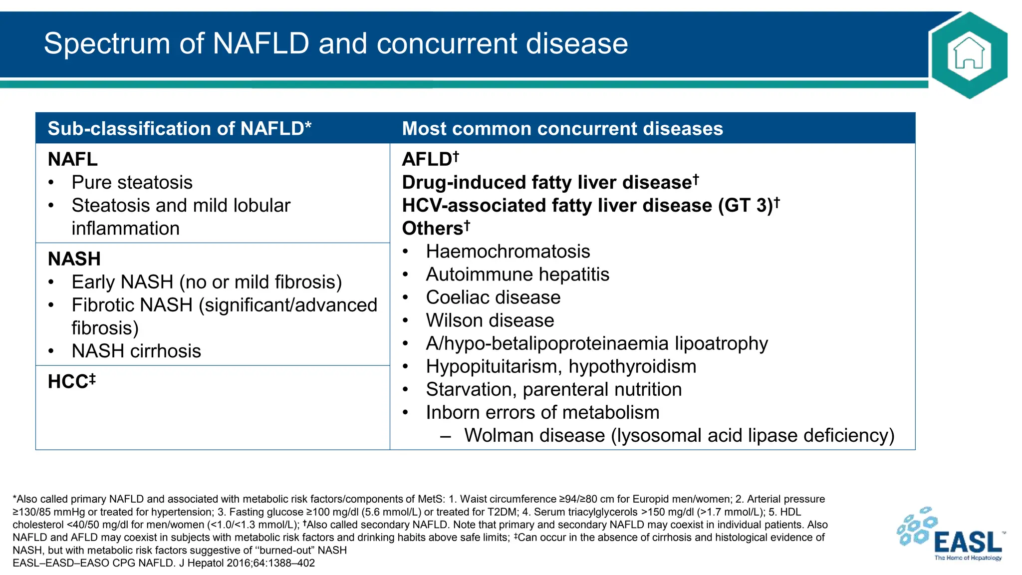
Non-alcoholic fatty
liver disease
Background
Definitions of NAFLD, NAFL and NASH
*According to histological analysis or proton density fat fraction or >5.6% by proton MRS or quantitative fat/water-selective MRI;
†Daily alcohol consumption of ≥30 g for men and ≥20 g for women
EASL–EASD–EASO CPG NAFLD. J Hepatol 2016;64:1388–402
NAFLD
• Excessive hepatic fat accumulation with IR
• Steatosis in >5% of hepatocytes*
• Exclusion of secondary causes and AFLD†
NASH
NAFL
• Pure steatosis
• Steatosis and mild lobular inflammation
Cirrhotic
F4 fibrosis
Fibrotic
≥F2 to ≥F3 fibrosis
Early
F0/F1 fibrosis
HCC
Definitive diagnosis of NASH requires a liver biopsy
Spectrum of NAFLD and concurrent disease
*Also called primary NAFLD and associated with metabolic risk factors/components of MetS: 1. Waist circumference ≥94/≥80 cm for Europid men/women; 2. Arterial pressure
≥130/85 mmHg or treated for hypertension; 3. Fasting glucose ≥100 mg/dl (5.6 mmol/L) or treated for T2DM; 4. Serum triacylglycerols >150 mg/dl (>1.7 mmol/L); 5. HDL
cholesterol <40/50 mg/dl for men/women (<1.0/<1.3 mmol/L); †Also called secondary NAFLD. Note that primary and secondary NAFLD may coexist in individual patients. Also
NAFLD and AFLD may coexist in subjects with metabolic risk factors and drinking habits above safe limits; ‡Can occur in the absence of cirrhosis and histological evidence of
NASH, but with metabolic risk factors suggestive of ‘‘burned-out” NASH
EASL–EASD–EASO CPG NAFLD. J Hepatol 2016;64:1388–402
Sub-classification of NAFLD* Most common concurrent diseases
NAFL
• Pure steatosis
• Steatosis and mild lobular
inflammation
AFLD†
Drug-induced fatty liver disease†
HCV-associated fatty liver disease (GT 3)†
Others†
• Haemochromatosis
• Autoimmune hepatitis
• Coeliac disease
• Wilson disease
• A/hypo-betalipoproteinaemia lipoatrophy
• Hypopituitarism, hypothyroidism
• Starvation, parenteral nutrition
• Inborn errors of metabolism
– Wolman disease (lysosomal acid lipase deficiency)
NASH
• Early NASH (no or mild fibrosis)
• Fibrotic NASH (significant/advanced
fibrosis)
• NASH cirrhosis
HCC‡
Multiple organs are likely to be involved in NAFLD
Nobili, V et al. J Hepatol 2013;58:121829
Copyright © 2013 European Association for the Study of the Liver Terms and Conditions
• Pathogenesis of NAFLD probably involves inter-organ crosstalk
– Adipose tissue, pancreas, gut, and liver
hepatic IR and inflammation
Byrne CD, Targher G. J Hepatol 2015;62:S47–64
Copyright © 2014 European Association for the Study of the Liver Terms and Conditions
Screening, prevalence and incidence
1. Vernon G, et al. Aliment Pharmacol Ther 2011;34:27485; 2. Younossi ZM, et al. Medicine 2012;91:31927;
EASL–EASD–EASO CPG NAFLD. J Hepatol 2016;64:1388–402
• NAFLD is the most common liver disorder in Western countries, affecting 17–46% of adults1
– Part of metabolic syndrome (MetS) and its components, which also increase the risk of more advanced
disease
– NAFLD is also present in 7% of normal-weight (lean) individuals2
Recommendations
Patients with IR and/or metabolic risk factors (i.e. obesity or MetS) should undergo
procedures for the diagnosis of NAFLD
A 1
Screen individuals with steatosis for secondary causes of NAFLD, including a careful
assessment of alcohol intake. Always consider the interaction between moderate amounts
of alcohol and metabolic factors in fatty liver
A 1
Identify other chronic liver diseases that may coexist with NAFLD as these might result in
more severe liver injury
B 1
Grade of evidence Grade of recommendation
Screening, prevalence and incidence
*Aged >50 years, T2DM, MetS
EASL–EASD–EASO CPG NAFLD. J Hepatol 2016;64:1388–402
• Value of screening for NAFLD in the community is limited
– High direct and indirect costs
– Low predictive value of non-invasive tests
– Risks associated with liver biopsy
– Lack of effective treatments
• Diagnosis of NASH provides important diagnostic information
– Points to increased risk of fibrosis progression, cirrhosis and possibly HCC
Recommendations
All individuals with steatosis should be screened for features of MetS, independent of
liver enzymes. All individuals with persistently abnormal liver enzymes should be
screened for NAFLD
A 1
In subjects with obesity or MetS, screening for NAFLD should be part of routine work-up.
In high-risk individuals* case finding of advanced disease is advisable
A 2
Grade of evidence Grade of recommendation
Pathogenesis: lifestyle and genes
1. Barrera F, George J. Clin Liver Dis 2014;18:91–112;
EASL–EASD–EASO CPG NAFLD. J Hepatol 2016;64:1388–402
• A Western diet/lifestyle has been associated with weight gain and obesity, and NAFLD1
Recommendation
Unhealthy lifestyles play a role in the development and progression of NAFLD.
The assessment of dietary and physical activity habits is part of comprehensive NAFLD
screening
A 1
Grade of evidence Grade of recommendation
Obesity
NAFLD
High calorie intake
Excess carbohydrates
High fructose intake
Sedentary behaviour
Pathogenesis: lifestyle and genes
*Grade of evidence B, grade of recommendation 2
1. Anstee QM, et al. Nat Rev Gastroenterol Hepatol 2013;10:330–44;
EASL–EASD–EASO CPG NAFLD. J Hepatol 2016;64:1388–402
• Several genetic modifiers of NAFLD have been identified1
– A minority have been robustly validated
• PNPLA3 I148M and TM6SF2 E167K carriers have a higher liver fat content*
– Increased risk of NASH
– NAFLD not systematically associated with features of IR
Recommendation
Genotyping may be considered in selected patients and clinical studies but is not
recommended routinely
B 2
Grade of evidence Grade of recommendation
Progressive liver disease in NAFLD
Byrne CD, Targher G. J Hepatol 2015;62:S47–64
Copyright © 2014 European Association for the Study of the Liver Terms and Conditions
Natural history of NAFLD over 8–13 years
de Alwis NMW, Day CP. J Hepatol 2008;48:S104–12
Copyright © 2008 European Association for the Study of the Liver Terms and Conditions
Steatosis
NASH 
F1F2
fibrosis
HCC
Death/
LTx Cirrhosis
Advanced
F3
fibrosis
1240%
510%
050%
8%
13%
2550%
14%
25%
7%
Liver biopsy
*Should not be used for initial diagnosis
EASL–EASD–EASO CPG NAFLD. J Hepatol 2016;64:1388–402
• Liver biopsy is essential for the diagnosis of NASH
– Clinical, biochemical or imaging measures cannot distinguish NASH from steatosis
• NAFL encompasses
– Steatosis alone plus ONE of lobular or portal inflammation OR ballooning
• NASH requires
– Steatosis AND
– Lobular or portal inflammation AND
– Ballooning
• NAS scoring indicates disease severity*
Recommendations
NASH has to be diagnosed by a liver biopsy showing steatosis, hepatocyte ballooning
and lobular inflammation
A 1
Grade of evidence Grade of recommendation
Non-invasive assessment of steatosis
EASL–EASD–EASO CPG NAFLD. J Hepatol 2016;64:1388–402
• Steatosis should be documented whenever NAFLD is suspected
– Predicts future T2DM, cardiovascular events and arterial hypertension
– Quantification of fat content is of limited clinical relevance
• Except as a surrogate of treatment effectiveness
Recommendations
US is the preferred first-line diagnostic procedure for imaging of NAFLD, as it
provides additional diagnostic information
A 1
Whenever imaging tools are not available or feasible serum biomarkers and scores are an
acceptable alternative for the diagnosis of steatosis
B 2
A quantitative estimation of liver fat can only be obtained by 1H-MRS. This technique is of
value in clinical trials and experimental studies, but is expensive and not recommended in
the clinical setting
A 1
Grade of evidence Grade of recommendation
Non-invasive assessment of fibrosis
EASL–EASD–EASO CPG NAFLD. J Hepatol 2016;64:1388–402
• Fibrosis is the most important prognostic factor in NAFLD
– Correlates with liver-related outcomes and mortality
– Advanced fibrosis indicates thorough investigation
Recommendations
Biomarkers, fibrosis scores, and transient elastography, are acceptable non-invasive
procedures to identify those at low risk of advanced fibrosis/cirrhosis
A 2
Biomarkers/scores PLUS transient elastography might confer additional diagnostic
accuracy and reduce need for liver biopsy
B 2
Monitoring of fibrosis progression may rely on biomarkers/scores and transient
elastography, although this strategy requires validation
C 2
The identification of advanced fibrosis or cirrhosis by serum biomarkers/scores and/or
elastography is less accurate and needs to be confirmed by liver biopsy, according to the
clinical context
B 2
In selected patients at high risk of liver disease progression, monitoring should include a
repeat biopsy after 5-year follow-up
C 2
Grade of evidence Grade of recommendation
Potential algorithm for non-invasive assessment: prediction rules and
blood-based biomarkers
*Estimated prevalence for low-, intermediate- and high-risk groups
Vilar-Gomez E, Chalasani N. J Hepatol 2018;68:30515
Copyright © 2017 European Association for the Study of the Liver Terms and Conditions
Non-invasive assessment of paediatric NAFLD
Roberts EA. J Hepatol 2007;46:113342
Copyright © 2007 European Association for the Study of the Liver Terms and Conditions
EASL–EASD–EASO CPG NAFLD. J Hepatol 2016;64:1388–402
• NAFLD should always be suspected in
obese children
– Exclude other causes
– Evaluate elevated aminotransferase levels
and liver hyperechogenicity
• Due to the poor sensitivity in overweight/obese
children, non-invasive markers and imaging
techniques are the first diagnostic step
Recommendations
In children, predictors of fibrosis, including elastometry, ARFI imaging and serum
biomarkers might help reduce the number of biopsies
B 2
Grade of evidence Grade of recommendation
Common related metabolic disorders
*Gender-adjusted; †ethnicity-adjusted
EASL–EASD–EASO CPG NAFLD. J Hepatol 2016;64:1388–402
• NAFLD is closely associated with:
– IR in the liver as well as adipose and muscle tissue
– MetS
• Three of: impaired fasting glucose or T2DM, hypertriglyceridaemia, low HDL-C,* increased waist
circumference,† high blood pressure
All components of MetS correlate with liver fat content:
Evaluate risk of NAFLD in patients with MetS
Evaluate MetS in patients with NAFLD
Common related metabolic disorders
*E.g. US-defined steatosis with normal body weight
EASL–EASD–EASO CPG NAFLD. J Hepatol 2016;64:1388–402
• In individuals without diabetes, HOMA-IR can be considered as a surrogate for IR
HOMA-IR:
Fasting glucose (mmol/L) + insulin (mU/ml)
22.5
Recommendations
HOMA-IR can be recommended if proper reference values have been established A 1
HOMA-IR is of limited use for NAFLD diagnosis in patients with metabolic risk factors. It
could confirm altered insulin sensitivity, thereby favouring a diagnosis of IR-associated
NAFLD in cases of diagnostic uncertainty*
B 2
During follow-up, HOMA-IR might help identify patients at risk of NASH or fibrosis
progression in selected cases. Improvement of HOMA-IR during weight loss may indicate
metabolic improvement
C 2
Grade of evidence Grade of recommendation
Common related metabolic disorders: obesity
EASL–EASD–EASO CPG NAFLD. J Hepatol 2016;64:1388–402
• BMI and waist circumference are positively related to NAFLD
– Predictors of advanced disease, particularly in the elderly
Recommendations
Follow up is mandatory in obesity, which is the major phenotype and risk condition for
NAFLD, driven by IR, and also increases the risk of advanced disease
A 1
Most lean individuals with NAFLD display IR and altered body fat distribution even
though they have less severe metabolic disturbance than overweight NAFLD. Follow-up is
nonetheless required because of possible disease progression
B 2
Grade of evidence Grade of recommendation
Common related metabolic disorders: T2DM
EASL–EASD–EASO CPG NAFLD. J Hepatol 2016;64:1388–402
• Irrespective of liver enzymes, diabetes risk and T2DM are closely associated with:
– Severity of NAFLD
– Progression to NASH
– Presence of advanced fibrosis
– Development of HCC
Recommendations
In individuals with NAFLD, screening for diabetes is mandatory, by fasting or random
blood glucose or HbA1c…
A 1
…and if available, by the standardized 75 g OGTT in high-risk groups B 1
Look for NAFLD in patients with T2DM, irrespective of liver enzyme levels, due to high
risk of disease progression
A 2
Grade of evidence Grade of recommendation
Diagnosis: protocol for evaluation of NAFLD
*According to a priori probability or clinical evaluation
EASL–EASD–EASO CPG NAFLD. J Hepatol 2016;64:1388–402
• Incidental discovery of steatosis indicates comprehensive evaluation
– Family and personal history of NAFLD-associated diseases
– Exclusion of secondary causes of steatosis
Level Variable
Initial evaluation 1. Alcohol intake: <20 g/day (women), <30 g/day (men)
2. Personal and family history of diabetes, hypertension and CVD
3. BMI, waist circumference, change in body weight
4. Hepatitis B/hepatitis C virus infection
5. History of steatosis-associated drugs
6. Liver enzymes (ALT, AST, GGT)
7. Fasting blood glucose, HbA1c, OGTT, (fasting insulin [HOMA-IR])
8. Complete blood count
9. Serum total and HDL cholesterol, triacylglycerol, uric acid
10. Ultrasonography (if suspected for raised liver enzymes)
Extended*
evaluation
1. Ferritin and transferrin saturation
2. Tests for coeliac and thyroid diseases, polycystic ovary syndrome
3. Tests for rare liver diseases (Wilson, autoimmune disease, AATD)
Diagnosis: diagnostic flow-chart
*Steatosis biomarkers: Fatty Liver Index, SteatoTest, NAFLD Fat score;
†Liver tests: ALT AST, GGT; ‡Any increase in ALT, AST or GGT;
§Serum fibrosis markers: NAFLD Fibrosis Score, FIB-4, Commercial tests (FibroTest, FibroMeter, ELF);
‖Low risk: indicative of no/mild fibrosis; medium/high risk: indicative of significant fibrosis or cirrhosis
EASL–EASD–EASO CPG NAFLD. J Hepatol 2016;64:1388–402
• Metabolic work-up must carefully
assess all components of MetS
• Obesity/T2DM or raised liver enzymes
in patients with metabolic risk factors
should prompt non-invasive screening
to predict steatosis, NASH and fibrosis
Steatosis absent
Normal
liver enzymes
Follow-up/
3–5 years
Ultrasound/
liver enzymes
Steatosis present
Normal
liver enzymes
Follow-up/
2 years
Liver enzymes,
fibrosis
biomarkers
Serum fibrosis
markers§
Low risk‖ Medium/
high risk‖
Metabolic risk factors present
Ultrasound (steatosis biomarkers)*/
liver enzymes†
Abnormal
liver enzymes‡
Specialist referral
Identify other chronic liver diseases
In-depth assessment of disease severity
Decision to perform liver biopsy
Initiate monitoring/therapy
Natural history and complications: progression
1. Sing S et al. Clin Gastroenterol Hepatol 2015;13:643–54;
EASL–EASD–EASO CPG NAFLD. J Hepatol 2016;64:1388–402
• In general, NAFLD is a slowly progressive disease, both in adults and in children
– Rate of progression corresponds to 1 fibrosis stage every 14 years in NAFL and every 7 years
in NASH
– Rate of progression is doubled by arterial hypertension1
– Progression of fibrosis is more rapid in about 20% of cases1
• Paediatric NAFLD is of concern
– Potential for severe liver-related complications later in life
– NASH-related cirrhosis has been reported as early as 8 years of age
Recommendations
NASH patients with fibrosis associated with hypertension should receive closer monitoring
because of a higher risk of disease progression
B 1
Grade of evidence Grade of recommendation
Natural history and complications: CVD
EASL–EASD–EASO CPG NAFLD. J Hepatol 2016;64:1388–402
• Prevalence and incidence of CVD is higher in NAFLD than in matched controls
– Driven by the association between NAFLD and MetS components
• CVD should be identified in NAFLD, regardless of traditional risk factors
• CVD and metabolic risk factors are also reported in adolescents and children with NAFLD
Recommendations
Screening of the cardiovascular system is mandatory in all individuals with NAFLD
because CV complications frequently dictate the outcome
A 1
Grade of evidence Grade of recommendation
Putative connection between NAFLD, CVD and CKD
Byrne CD, Targher G. J Hepatol 2015;62:S47–64
Copyright © 2014 European Association for the Study of the Liver Terms and Conditions
Natural history and complications: HCC
EASL–EASD–EASO CPG NAFLD. J Hepatol 2016;64:1388–402
• Cumulative incidence of NAFLD-associated HCC varies according to study population
• Large number of NAFLD cases at risk of HCC makes systematic surveillance
largely impracticable
– PNPLA3 rs738409 C>G gene polymorphism is associated with increased HCC risk
– However, HCC surveillance in NAFLD is not yet considered cost effective
Recommendations
Although NAFLD is a risk factor for HCC, which may also develop in the pre-cirrhotic
stage, and the risk is further increased by the presence of the PNPLA3 rs738409 C>G
polymorphism, no recommendation can be currently made on the timing of surveillance
and its cost effectiveness
B 1
Grade of evidence Grade of recommendation
Treatment: diet and lifestyle changes
EASL–EASD–EASO CPG NAFLD. J Hepatol 2016;64:1388–402
• Epidemiology suggests a close relationship between an unhealthy lifestyle and NAFLD
• Diet and lifestyle changes are mandatory in all patients
– Modest weight loss reduces liver fat, improves hepatic IR, and can result in NASH regression
– Weight loss of 7% is associated with histological improvement
Recommendations
Structured programmes aimed at lifestyle changes towards healthy diet and habitual
physical activity are advisable in NAFLD
C 2
Patients without NASH or fibrosis should receive counselling for healthy diet and physical
activity but no pharmacotherapy
B 2
In overweight/obese NAFLD, a 7–10% weight loss is the target of most lifestyle
interventions, and results in improvement of liver enzymes and histology
B 1
Grade of evidence Grade of recommendation
Treatment: diet and lifestyle changes
EASL–EASD–EASO CPG NAFLD. J Hepatol 2016;64:1388–402
• A pragmatic, individually tailored approach is required
– Dietary restriction PLUS
– Progressive increase in aerobic exercise/resistance training
Recommendations
Dietary recommendations should consider energy restriction and exclusion of NAFLD-
promoting components (processed food, and food and beverages high in added fructose).
The macronutrient composition should be adjusted according to the Mediterranean diet
B 1
Both aerobic exercise and resistance training effectively reduce liver fat. The choice
of training should be tailored based on patients’ preferences to be maintained in the
long-term
B 2
Grade of evidence Grade of recommendation
Components of a lifestyle approach to NAFLD
EASL–EASD–EASO CPG NAFLD. J Hepatol 2016;64:1388–402
Comprehensive
lifestyle approach
Energy restriction
• Calorie restriction
• 710% weight loss target
• Long-term maintenance approach
Macronutrient composition
• moderate fat
• Moderate carbohydrate
• Low-carbohydrate ketogenic diets
or high protein
Fructose intake
• Avoid fructose-containing
food and drink
Daily alcohol intake
• Strictly below 30 g men
and 20 g women
Coffee consumption
• No liver-related limitations
Physical activity
• 150200 min/week moderate intensity
in 35 sessions
• Resistance training to promote
musculoskeletal fitness and improve
metabolic factors
Treatment: pharmacotherapy
*Age > 50 years, diabetes, MetS, increased ALT
EASL–EASD–EASO CPG NAFLD. J Hepatol 2016;64:1388–402
• Treatment should be indicated in:
– Progressive NASH
– Early-stage NASH with risk of fibrosis progression*
– Active NASH with high necroinflammatory activity
• Treatment should reduce NASH-related mortality and progression to cirrhosis or HCC
– Resolution of NASH-defining lesions accepted as surrogate endpoint
• Safety and tolerability are prerequisites
– Extensive comorbidities associated with significant polypharmacy and increased likelihood of DDIs
Recommendations
Pharmacotherapy should be reserved for patients with NASH, particularly for those
with significant fibrosis (stage F2 and higher). Patients with less severe disease, but at
high risk of disease progression could also be candidates for treatment
B 1
Grade of evidence Grade of recommendation
Treatment: pharmacotherapy
*Age > 50 years, diabetes, MetS, increased ALT
EASL–EASD–EASO CPG NAFLD. J Hepatol 2016;64:1388–402
• Treatment should be indicated in:
– Progressive NASH
– Early-stage NASH with risk of fibrosis progression*
– Active NASH with high necroinflammatory activity
• Treatment should reduce NASH-related mortality and progression to cirrhosis or HCC
– Resolution of NASH-defining lesions accepted as surrogate endpoint
• Safety and tolerability are prerequisites
– Extensive comorbidities associated with significant polypharmacy and increased likelihood of DDIs
No drugs are approved for NASH
No specific therapy can be recommended
Any drug treatment is off label
Recommendations
Pharmacotherapy should be reserved for patients with NASH, particularly for those
with significant fibrosis (stage F2 and higher). Patients with less severe disease, but at
high risk of disease progression could also be candidates for treatment
B 1
Grade of evidence Grade of recommendation
Treatment: pharmacotherapy
*Most efficacy data, but off-label outside T2DM; †Better safety and tolerability than pioglitazone in the short-term;
‡No recommendations can be made in patients with normal baseline ALT
EASL–EASD–EASO CPG NAFLD. J Hepatol 2016;64:1388–402
• Insulin sensitizers
– Little evidence of histological efficacy with metformin
– PPAR agonist pioglitazone better than placebo
• Improved all histological features except fibrosis
• Achieved resolution of NASH more often
• Antioxidants
– Vitamin E may improve steatosis, inflammation and ballooning and resolve NASH in some patients
• Concerns about long-term safety exist
Recommendations
While no firm recommendations can be made, pioglitazone* or vitamin E† or their
combination could be used for NASH
B 2
The optimal duration of therapy is unknown; in patients with increased ALT at baseline,
treatment should be stopped if there is no reduction in aminotransferases after 6 months
of therapy‡
C 2
Grade of evidence Grade of recommendation
Treatment: pharmacotherapy
EASL–EASD–EASO CPG NAFLD. J Hepatol 2016;64:1388–402
• Lipid-lowering agents
– Statins have not been adequately tested in NASH
Recommendations
Statins may be confidently used to reduce LDL cholesterol and prevent cardiovascular
risk, with no benefits or harm to liver disease.
Similarly, n-3 polyunsaturated fatty acids reduce both plasma and liver lipids, but there are
no data to support their use specifically for NASH
B 1
Grade of evidence Grade of recommendation
MOA of pharmacological treatments for NAFLD
Konerman MA, et al. J Hepatol 2018;68:362–75
Copyright © 2017 European Association for the Study of the Liver Terms and Conditions
Treatment: paediatric NAFLD
EASL–EASD–EASO CPG NAFLD. J Hepatol 2016;64:1388–402
• Diet and exercise training reduce steatosis, but do not affect ballooning, inflammation,
and fibrosis
• The long-term outcome of paediatric NASH remains poor
– Drugs have shown beneficial effects but fibrotic lesions are refractory to treatment
Recommendations
Diet and physical activity improve steatosis and hepatic inflammation in paediatric NAFLD,
but no beneficial effects on fibrosis have ever been demonstrated. No safe drug
treatment has proven effective on fibrosis in paediatric NAFLD
B 1
Grade of evidence Grade of recommendation
Treatment: surgery
EASL–EASD–EASO CPG NAFLD. J Hepatol 2016;64:1388–402
• Bariatric surgery is an option in patients unresponsive to lifestyle changes and pharmacotherapy
– Reduces weight and metabolic complications
– Stable results in the long term
• NAFLD-associated cirrhosis is one of the top three indications for LTx
Recommendations for bariatric surgery
Bariatric surgery reduces liver fat and is likely to reduce NASH progression; prospective data
have shown an improvement in all histological lesions of NASH, including fibrosis
B 1
Recommendations for liver transplant
LTx is an accepted procedure in patients with NASH and end-stage liver disease. Overall
survival is comparable to other indications, despite a higher cardiovascular mortality. Patients
with NASH and liver failure and/or HCC are candidates for liver transplantation
A 1
Grade of evidence Grade of recommendation

NAFattyLiverDisease-EASL-CPG-Slide-Deck.pptx

  • 1.
  • 2.
  • 3.
    Definitions of NAFLD,NAFL and NASH *According to histological analysis or proton density fat fraction or >5.6% by proton MRS or quantitative fat/water-selective MRI; †Daily alcohol consumption of ≥30 g for men and ≥20 g for women EASL–EASD–EASO CPG NAFLD. J Hepatol 2016;64:1388–402 NAFLD • Excessive hepatic fat accumulation with IR • Steatosis in >5% of hepatocytes* • Exclusion of secondary causes and AFLD† NASH NAFL • Pure steatosis • Steatosis and mild lobular inflammation Cirrhotic F4 fibrosis Fibrotic ≥F2 to ≥F3 fibrosis Early F0/F1 fibrosis HCC Definitive diagnosis of NASH requires a liver biopsy
  • 4.
    Spectrum of NAFLDand concurrent disease *Also called primary NAFLD and associated with metabolic risk factors/components of MetS: 1. Waist circumference ≥94/≥80 cm for Europid men/women; 2. Arterial pressure ≥130/85 mmHg or treated for hypertension; 3. Fasting glucose ≥100 mg/dl (5.6 mmol/L) or treated for T2DM; 4. Serum triacylglycerols >150 mg/dl (>1.7 mmol/L); 5. HDL cholesterol <40/50 mg/dl for men/women (<1.0/<1.3 mmol/L); †Also called secondary NAFLD. Note that primary and secondary NAFLD may coexist in individual patients. Also NAFLD and AFLD may coexist in subjects with metabolic risk factors and drinking habits above safe limits; ‡Can occur in the absence of cirrhosis and histological evidence of NASH, but with metabolic risk factors suggestive of ‘‘burned-out” NASH EASL–EASD–EASO CPG NAFLD. J Hepatol 2016;64:1388–402 Sub-classification of NAFLD* Most common concurrent diseases NAFL • Pure steatosis • Steatosis and mild lobular inflammation AFLD† Drug-induced fatty liver disease† HCV-associated fatty liver disease (GT 3)† Others† • Haemochromatosis • Autoimmune hepatitis • Coeliac disease • Wilson disease • A/hypo-betalipoproteinaemia lipoatrophy • Hypopituitarism, hypothyroidism • Starvation, parenteral nutrition • Inborn errors of metabolism – Wolman disease (lysosomal acid lipase deficiency) NASH • Early NASH (no or mild fibrosis) • Fibrotic NASH (significant/advanced fibrosis) • NASH cirrhosis HCC‡
  • 5.
    Multiple organs arelikely to be involved in NAFLD Nobili, V et al. J Hepatol 2013;58:121829 Copyright © 2013 European Association for the Study of the Liver Terms and Conditions • Pathogenesis of NAFLD probably involves inter-organ crosstalk – Adipose tissue, pancreas, gut, and liver
  • 6.
    hepatic IR andinflammation Byrne CD, Targher G. J Hepatol 2015;62:S47–64 Copyright © 2014 European Association for the Study of the Liver Terms and Conditions
  • 7.
    Screening, prevalence andincidence 1. Vernon G, et al. Aliment Pharmacol Ther 2011;34:27485; 2. Younossi ZM, et al. Medicine 2012;91:31927; EASL–EASD–EASO CPG NAFLD. J Hepatol 2016;64:1388–402 • NAFLD is the most common liver disorder in Western countries, affecting 17–46% of adults1 – Part of metabolic syndrome (MetS) and its components, which also increase the risk of more advanced disease – NAFLD is also present in 7% of normal-weight (lean) individuals2 Recommendations Patients with IR and/or metabolic risk factors (i.e. obesity or MetS) should undergo procedures for the diagnosis of NAFLD A 1 Screen individuals with steatosis for secondary causes of NAFLD, including a careful assessment of alcohol intake. Always consider the interaction between moderate amounts of alcohol and metabolic factors in fatty liver A 1 Identify other chronic liver diseases that may coexist with NAFLD as these might result in more severe liver injury B 1 Grade of evidence Grade of recommendation
  • 8.
    Screening, prevalence andincidence *Aged >50 years, T2DM, MetS EASL–EASD–EASO CPG NAFLD. J Hepatol 2016;64:1388–402 • Value of screening for NAFLD in the community is limited – High direct and indirect costs – Low predictive value of non-invasive tests – Risks associated with liver biopsy – Lack of effective treatments • Diagnosis of NASH provides important diagnostic information – Points to increased risk of fibrosis progression, cirrhosis and possibly HCC Recommendations All individuals with steatosis should be screened for features of MetS, independent of liver enzymes. All individuals with persistently abnormal liver enzymes should be screened for NAFLD A 1 In subjects with obesity or MetS, screening for NAFLD should be part of routine work-up. In high-risk individuals* case finding of advanced disease is advisable A 2 Grade of evidence Grade of recommendation
  • 9.
    Pathogenesis: lifestyle andgenes 1. Barrera F, George J. Clin Liver Dis 2014;18:91–112; EASL–EASD–EASO CPG NAFLD. J Hepatol 2016;64:1388–402 • A Western diet/lifestyle has been associated with weight gain and obesity, and NAFLD1 Recommendation Unhealthy lifestyles play a role in the development and progression of NAFLD. The assessment of dietary and physical activity habits is part of comprehensive NAFLD screening A 1 Grade of evidence Grade of recommendation Obesity NAFLD High calorie intake Excess carbohydrates High fructose intake Sedentary behaviour
  • 10.
    Pathogenesis: lifestyle andgenes *Grade of evidence B, grade of recommendation 2 1. Anstee QM, et al. Nat Rev Gastroenterol Hepatol 2013;10:330–44; EASL–EASD–EASO CPG NAFLD. J Hepatol 2016;64:1388–402 • Several genetic modifiers of NAFLD have been identified1 – A minority have been robustly validated • PNPLA3 I148M and TM6SF2 E167K carriers have a higher liver fat content* – Increased risk of NASH – NAFLD not systematically associated with features of IR Recommendation Genotyping may be considered in selected patients and clinical studies but is not recommended routinely B 2 Grade of evidence Grade of recommendation
  • 11.
    Progressive liver diseasein NAFLD Byrne CD, Targher G. J Hepatol 2015;62:S47–64 Copyright © 2014 European Association for the Study of the Liver Terms and Conditions
  • 12.
    Natural history ofNAFLD over 8–13 years de Alwis NMW, Day CP. J Hepatol 2008;48:S104–12 Copyright © 2008 European Association for the Study of the Liver Terms and Conditions Steatosis NASH  F1F2 fibrosis HCC Death/ LTx Cirrhosis Advanced F3 fibrosis 1240% 510% 050% 8% 13% 2550% 14% 25% 7%
  • 13.
    Liver biopsy *Should notbe used for initial diagnosis EASL–EASD–EASO CPG NAFLD. J Hepatol 2016;64:1388–402 • Liver biopsy is essential for the diagnosis of NASH – Clinical, biochemical or imaging measures cannot distinguish NASH from steatosis • NAFL encompasses – Steatosis alone plus ONE of lobular or portal inflammation OR ballooning • NASH requires – Steatosis AND – Lobular or portal inflammation AND – Ballooning • NAS scoring indicates disease severity* Recommendations NASH has to be diagnosed by a liver biopsy showing steatosis, hepatocyte ballooning and lobular inflammation A 1 Grade of evidence Grade of recommendation
  • 14.
    Non-invasive assessment ofsteatosis EASL–EASD–EASO CPG NAFLD. J Hepatol 2016;64:1388–402 • Steatosis should be documented whenever NAFLD is suspected – Predicts future T2DM, cardiovascular events and arterial hypertension – Quantification of fat content is of limited clinical relevance • Except as a surrogate of treatment effectiveness Recommendations US is the preferred first-line diagnostic procedure for imaging of NAFLD, as it provides additional diagnostic information A 1 Whenever imaging tools are not available or feasible serum biomarkers and scores are an acceptable alternative for the diagnosis of steatosis B 2 A quantitative estimation of liver fat can only be obtained by 1H-MRS. This technique is of value in clinical trials and experimental studies, but is expensive and not recommended in the clinical setting A 1 Grade of evidence Grade of recommendation
  • 15.
    Non-invasive assessment offibrosis EASL–EASD–EASO CPG NAFLD. J Hepatol 2016;64:1388–402 • Fibrosis is the most important prognostic factor in NAFLD – Correlates with liver-related outcomes and mortality – Advanced fibrosis indicates thorough investigation Recommendations Biomarkers, fibrosis scores, and transient elastography, are acceptable non-invasive procedures to identify those at low risk of advanced fibrosis/cirrhosis A 2 Biomarkers/scores PLUS transient elastography might confer additional diagnostic accuracy and reduce need for liver biopsy B 2 Monitoring of fibrosis progression may rely on biomarkers/scores and transient elastography, although this strategy requires validation C 2 The identification of advanced fibrosis or cirrhosis by serum biomarkers/scores and/or elastography is less accurate and needs to be confirmed by liver biopsy, according to the clinical context B 2 In selected patients at high risk of liver disease progression, monitoring should include a repeat biopsy after 5-year follow-up C 2 Grade of evidence Grade of recommendation
  • 16.
    Potential algorithm fornon-invasive assessment: prediction rules and blood-based biomarkers *Estimated prevalence for low-, intermediate- and high-risk groups Vilar-Gomez E, Chalasani N. J Hepatol 2018;68:30515 Copyright © 2017 European Association for the Study of the Liver Terms and Conditions
  • 17.
    Non-invasive assessment ofpaediatric NAFLD Roberts EA. J Hepatol 2007;46:113342 Copyright © 2007 European Association for the Study of the Liver Terms and Conditions EASL–EASD–EASO CPG NAFLD. J Hepatol 2016;64:1388–402 • NAFLD should always be suspected in obese children – Exclude other causes – Evaluate elevated aminotransferase levels and liver hyperechogenicity • Due to the poor sensitivity in overweight/obese children, non-invasive markers and imaging techniques are the first diagnostic step Recommendations In children, predictors of fibrosis, including elastometry, ARFI imaging and serum biomarkers might help reduce the number of biopsies B 2 Grade of evidence Grade of recommendation
  • 18.
    Common related metabolicdisorders *Gender-adjusted; †ethnicity-adjusted EASL–EASD–EASO CPG NAFLD. J Hepatol 2016;64:1388–402 • NAFLD is closely associated with: – IR in the liver as well as adipose and muscle tissue – MetS • Three of: impaired fasting glucose or T2DM, hypertriglyceridaemia, low HDL-C,* increased waist circumference,† high blood pressure All components of MetS correlate with liver fat content: Evaluate risk of NAFLD in patients with MetS Evaluate MetS in patients with NAFLD
  • 19.
    Common related metabolicdisorders *E.g. US-defined steatosis with normal body weight EASL–EASD–EASO CPG NAFLD. J Hepatol 2016;64:1388–402 • In individuals without diabetes, HOMA-IR can be considered as a surrogate for IR HOMA-IR: Fasting glucose (mmol/L) + insulin (mU/ml) 22.5 Recommendations HOMA-IR can be recommended if proper reference values have been established A 1 HOMA-IR is of limited use for NAFLD diagnosis in patients with metabolic risk factors. It could confirm altered insulin sensitivity, thereby favouring a diagnosis of IR-associated NAFLD in cases of diagnostic uncertainty* B 2 During follow-up, HOMA-IR might help identify patients at risk of NASH or fibrosis progression in selected cases. Improvement of HOMA-IR during weight loss may indicate metabolic improvement C 2 Grade of evidence Grade of recommendation
  • 20.
    Common related metabolicdisorders: obesity EASL–EASD–EASO CPG NAFLD. J Hepatol 2016;64:1388–402 • BMI and waist circumference are positively related to NAFLD – Predictors of advanced disease, particularly in the elderly Recommendations Follow up is mandatory in obesity, which is the major phenotype and risk condition for NAFLD, driven by IR, and also increases the risk of advanced disease A 1 Most lean individuals with NAFLD display IR and altered body fat distribution even though they have less severe metabolic disturbance than overweight NAFLD. Follow-up is nonetheless required because of possible disease progression B 2 Grade of evidence Grade of recommendation
  • 21.
    Common related metabolicdisorders: T2DM EASL–EASD–EASO CPG NAFLD. J Hepatol 2016;64:1388–402 • Irrespective of liver enzymes, diabetes risk and T2DM are closely associated with: – Severity of NAFLD – Progression to NASH – Presence of advanced fibrosis – Development of HCC Recommendations In individuals with NAFLD, screening for diabetes is mandatory, by fasting or random blood glucose or HbA1c… A 1 …and if available, by the standardized 75 g OGTT in high-risk groups B 1 Look for NAFLD in patients with T2DM, irrespective of liver enzyme levels, due to high risk of disease progression A 2 Grade of evidence Grade of recommendation
  • 22.
    Diagnosis: protocol forevaluation of NAFLD *According to a priori probability or clinical evaluation EASL–EASD–EASO CPG NAFLD. J Hepatol 2016;64:1388–402 • Incidental discovery of steatosis indicates comprehensive evaluation – Family and personal history of NAFLD-associated diseases – Exclusion of secondary causes of steatosis Level Variable Initial evaluation 1. Alcohol intake: <20 g/day (women), <30 g/day (men) 2. Personal and family history of diabetes, hypertension and CVD 3. BMI, waist circumference, change in body weight 4. Hepatitis B/hepatitis C virus infection 5. History of steatosis-associated drugs 6. Liver enzymes (ALT, AST, GGT) 7. Fasting blood glucose, HbA1c, OGTT, (fasting insulin [HOMA-IR]) 8. Complete blood count 9. Serum total and HDL cholesterol, triacylglycerol, uric acid 10. Ultrasonography (if suspected for raised liver enzymes) Extended* evaluation 1. Ferritin and transferrin saturation 2. Tests for coeliac and thyroid diseases, polycystic ovary syndrome 3. Tests for rare liver diseases (Wilson, autoimmune disease, AATD)
  • 23.
    Diagnosis: diagnostic flow-chart *Steatosisbiomarkers: Fatty Liver Index, SteatoTest, NAFLD Fat score; †Liver tests: ALT AST, GGT; ‡Any increase in ALT, AST or GGT; §Serum fibrosis markers: NAFLD Fibrosis Score, FIB-4, Commercial tests (FibroTest, FibroMeter, ELF); ‖Low risk: indicative of no/mild fibrosis; medium/high risk: indicative of significant fibrosis or cirrhosis EASL–EASD–EASO CPG NAFLD. J Hepatol 2016;64:1388–402 • Metabolic work-up must carefully assess all components of MetS • Obesity/T2DM or raised liver enzymes in patients with metabolic risk factors should prompt non-invasive screening to predict steatosis, NASH and fibrosis Steatosis absent Normal liver enzymes Follow-up/ 3–5 years Ultrasound/ liver enzymes Steatosis present Normal liver enzymes Follow-up/ 2 years Liver enzymes, fibrosis biomarkers Serum fibrosis markers§ Low risk‖ Medium/ high risk‖ Metabolic risk factors present Ultrasound (steatosis biomarkers)*/ liver enzymes† Abnormal liver enzymes‡ Specialist referral Identify other chronic liver diseases In-depth assessment of disease severity Decision to perform liver biopsy Initiate monitoring/therapy
  • 24.
    Natural history andcomplications: progression 1. Sing S et al. Clin Gastroenterol Hepatol 2015;13:643–54; EASL–EASD–EASO CPG NAFLD. J Hepatol 2016;64:1388–402 • In general, NAFLD is a slowly progressive disease, both in adults and in children – Rate of progression corresponds to 1 fibrosis stage every 14 years in NAFL and every 7 years in NASH – Rate of progression is doubled by arterial hypertension1 – Progression of fibrosis is more rapid in about 20% of cases1 • Paediatric NAFLD is of concern – Potential for severe liver-related complications later in life – NASH-related cirrhosis has been reported as early as 8 years of age Recommendations NASH patients with fibrosis associated with hypertension should receive closer monitoring because of a higher risk of disease progression B 1 Grade of evidence Grade of recommendation
  • 25.
    Natural history andcomplications: CVD EASL–EASD–EASO CPG NAFLD. J Hepatol 2016;64:1388–402 • Prevalence and incidence of CVD is higher in NAFLD than in matched controls – Driven by the association between NAFLD and MetS components • CVD should be identified in NAFLD, regardless of traditional risk factors • CVD and metabolic risk factors are also reported in adolescents and children with NAFLD Recommendations Screening of the cardiovascular system is mandatory in all individuals with NAFLD because CV complications frequently dictate the outcome A 1 Grade of evidence Grade of recommendation
  • 26.
    Putative connection betweenNAFLD, CVD and CKD Byrne CD, Targher G. J Hepatol 2015;62:S47–64 Copyright © 2014 European Association for the Study of the Liver Terms and Conditions
  • 27.
    Natural history andcomplications: HCC EASL–EASD–EASO CPG NAFLD. J Hepatol 2016;64:1388–402 • Cumulative incidence of NAFLD-associated HCC varies according to study population • Large number of NAFLD cases at risk of HCC makes systematic surveillance largely impracticable – PNPLA3 rs738409 C>G gene polymorphism is associated with increased HCC risk – However, HCC surveillance in NAFLD is not yet considered cost effective Recommendations Although NAFLD is a risk factor for HCC, which may also develop in the pre-cirrhotic stage, and the risk is further increased by the presence of the PNPLA3 rs738409 C>G polymorphism, no recommendation can be currently made on the timing of surveillance and its cost effectiveness B 1 Grade of evidence Grade of recommendation
  • 28.
    Treatment: diet andlifestyle changes EASL–EASD–EASO CPG NAFLD. J Hepatol 2016;64:1388–402 • Epidemiology suggests a close relationship between an unhealthy lifestyle and NAFLD • Diet and lifestyle changes are mandatory in all patients – Modest weight loss reduces liver fat, improves hepatic IR, and can result in NASH regression – Weight loss of 7% is associated with histological improvement Recommendations Structured programmes aimed at lifestyle changes towards healthy diet and habitual physical activity are advisable in NAFLD C 2 Patients without NASH or fibrosis should receive counselling for healthy diet and physical activity but no pharmacotherapy B 2 In overweight/obese NAFLD, a 7–10% weight loss is the target of most lifestyle interventions, and results in improvement of liver enzymes and histology B 1 Grade of evidence Grade of recommendation
  • 29.
    Treatment: diet andlifestyle changes EASL–EASD–EASO CPG NAFLD. J Hepatol 2016;64:1388–402 • A pragmatic, individually tailored approach is required – Dietary restriction PLUS – Progressive increase in aerobic exercise/resistance training Recommendations Dietary recommendations should consider energy restriction and exclusion of NAFLD- promoting components (processed food, and food and beverages high in added fructose). The macronutrient composition should be adjusted according to the Mediterranean diet B 1 Both aerobic exercise and resistance training effectively reduce liver fat. The choice of training should be tailored based on patients’ preferences to be maintained in the long-term B 2 Grade of evidence Grade of recommendation
  • 30.
    Components of alifestyle approach to NAFLD EASL–EASD–EASO CPG NAFLD. J Hepatol 2016;64:1388–402 Comprehensive lifestyle approach Energy restriction • Calorie restriction • 710% weight loss target • Long-term maintenance approach Macronutrient composition • moderate fat • Moderate carbohydrate • Low-carbohydrate ketogenic diets or high protein Fructose intake • Avoid fructose-containing food and drink Daily alcohol intake • Strictly below 30 g men and 20 g women Coffee consumption • No liver-related limitations Physical activity • 150200 min/week moderate intensity in 35 sessions • Resistance training to promote musculoskeletal fitness and improve metabolic factors
  • 31.
    Treatment: pharmacotherapy *Age >50 years, diabetes, MetS, increased ALT EASL–EASD–EASO CPG NAFLD. J Hepatol 2016;64:1388–402 • Treatment should be indicated in: – Progressive NASH – Early-stage NASH with risk of fibrosis progression* – Active NASH with high necroinflammatory activity • Treatment should reduce NASH-related mortality and progression to cirrhosis or HCC – Resolution of NASH-defining lesions accepted as surrogate endpoint • Safety and tolerability are prerequisites – Extensive comorbidities associated with significant polypharmacy and increased likelihood of DDIs Recommendations Pharmacotherapy should be reserved for patients with NASH, particularly for those with significant fibrosis (stage F2 and higher). Patients with less severe disease, but at high risk of disease progression could also be candidates for treatment B 1 Grade of evidence Grade of recommendation
  • 32.
    Treatment: pharmacotherapy *Age >50 years, diabetes, MetS, increased ALT EASL–EASD–EASO CPG NAFLD. J Hepatol 2016;64:1388–402 • Treatment should be indicated in: – Progressive NASH – Early-stage NASH with risk of fibrosis progression* – Active NASH with high necroinflammatory activity • Treatment should reduce NASH-related mortality and progression to cirrhosis or HCC – Resolution of NASH-defining lesions accepted as surrogate endpoint • Safety and tolerability are prerequisites – Extensive comorbidities associated with significant polypharmacy and increased likelihood of DDIs No drugs are approved for NASH No specific therapy can be recommended Any drug treatment is off label Recommendations Pharmacotherapy should be reserved for patients with NASH, particularly for those with significant fibrosis (stage F2 and higher). Patients with less severe disease, but at high risk of disease progression could also be candidates for treatment B 1 Grade of evidence Grade of recommendation
  • 33.
    Treatment: pharmacotherapy *Most efficacydata, but off-label outside T2DM; †Better safety and tolerability than pioglitazone in the short-term; ‡No recommendations can be made in patients with normal baseline ALT EASL–EASD–EASO CPG NAFLD. J Hepatol 2016;64:1388–402 • Insulin sensitizers – Little evidence of histological efficacy with metformin – PPAR agonist pioglitazone better than placebo • Improved all histological features except fibrosis • Achieved resolution of NASH more often • Antioxidants – Vitamin E may improve steatosis, inflammation and ballooning and resolve NASH in some patients • Concerns about long-term safety exist Recommendations While no firm recommendations can be made, pioglitazone* or vitamin E† or their combination could be used for NASH B 2 The optimal duration of therapy is unknown; in patients with increased ALT at baseline, treatment should be stopped if there is no reduction in aminotransferases after 6 months of therapy‡ C 2 Grade of evidence Grade of recommendation
  • 34.
    Treatment: pharmacotherapy EASL–EASD–EASO CPGNAFLD. J Hepatol 2016;64:1388–402 • Lipid-lowering agents – Statins have not been adequately tested in NASH Recommendations Statins may be confidently used to reduce LDL cholesterol and prevent cardiovascular risk, with no benefits or harm to liver disease. Similarly, n-3 polyunsaturated fatty acids reduce both plasma and liver lipids, but there are no data to support their use specifically for NASH B 1 Grade of evidence Grade of recommendation
  • 35.
    MOA of pharmacologicaltreatments for NAFLD Konerman MA, et al. J Hepatol 2018;68:362–75 Copyright © 2017 European Association for the Study of the Liver Terms and Conditions
  • 36.
    Treatment: paediatric NAFLD EASL–EASD–EASOCPG NAFLD. J Hepatol 2016;64:1388–402 • Diet and exercise training reduce steatosis, but do not affect ballooning, inflammation, and fibrosis • The long-term outcome of paediatric NASH remains poor – Drugs have shown beneficial effects but fibrotic lesions are refractory to treatment Recommendations Diet and physical activity improve steatosis and hepatic inflammation in paediatric NAFLD, but no beneficial effects on fibrosis have ever been demonstrated. No safe drug treatment has proven effective on fibrosis in paediatric NAFLD B 1 Grade of evidence Grade of recommendation
  • 37.
    Treatment: surgery EASL–EASD–EASO CPGNAFLD. J Hepatol 2016;64:1388–402 • Bariatric surgery is an option in patients unresponsive to lifestyle changes and pharmacotherapy – Reduces weight and metabolic complications – Stable results in the long term • NAFLD-associated cirrhosis is one of the top three indications for LTx Recommendations for bariatric surgery Bariatric surgery reduces liver fat and is likely to reduce NASH progression; prospective data have shown an improvement in all histological lesions of NASH, including fibrosis B 1 Recommendations for liver transplant LTx is an accepted procedure in patients with NASH and end-stage liver disease. Overall survival is comparable to other indications, despite a higher cardiovascular mortality. Patients with NASH and liver failure and/or HCC are candidates for liver transplantation A 1 Grade of evidence Grade of recommendation

Editor's Notes

  • #3 NAFL, non-alcoholic fatty liver; NAFLD, non-alcoholic fatty liver disease; NASH, non-alcoholic steatohepatitis
  • #4 AFLD, alcoholic fatty liver disease; HCC, hepatocellular carcinoma; IR, insulin resistance; MRI, magnetic resonance imaging; MRS, magnetic resonance spectroscopy; NAFL, non-alcoholic fatty liver; NASH, non-alcoholic steatohepatitis
  • #5 AFLD, alcoholic fatty liver disease; GT, genotype; HCC, hepatocellular carcinoma; HCV, hepatitis C virus; HDL, high-density lipoprotein; MetS, metabolic syndrome; NAFL, non-alcoholic fatty liver; NASH, non-alcoholic steatohepatitis; T2DM, type 2 diabetes mellitus
  • #6 DAMP, damage-associated molecular pattern; FFA, free fatty acid; HSC, hepatic stellate cell; PAMP, pathogen-associated molecular pattern Image sourced from Nobili et al. J Hepatol 2013;58:1218-1229
  • #7 AGAT, acylglycerol- cyltransferase; AGPAT, acylglycerol phosphate acyltransferase; CGI-58, comparative gene identification-58; CYP 2E1, cytochrome P450 2E1; DAG, di-acylglycerol; DGAT, di-acylglycerol acyltransferase; IKKb, IjB kinase b; IR, insulin resistance; IRS-2-P, insulin receptor substrate-2 phosphorylation; JNK, c-Jun N-terminal kinase; LPA, lysophosphatidic acid; GPAT, glycerol-3-phosphate acyltransferase; LCFA, long-chain fatty acid; MAG, mono-acyl glycerol; mTOR, mechanistic target of rapamycin; mTORC-2, mechanistic target of rapamycin complex-2; PA, phosphatidic acid; PAP, phosphatidate phosphohydrolase; PKCe, protein kinase Ce; TAG, tri-acylglycerol; TGRLP, triglyceride-rich lipoprotein; VLDL, very-low-density lipoprotein Lipid-induced mechanisms contributing to hepatic insulin resistance and inflammation in NAFLD Synthesis of lipid intermediates from long-chain fatty acids (LCFAs), e.g., ceramide, lyso-phosphatidic acid, phosphatidic acid (PA), di-acylglycerol (DAG), tri-acylglycerol (TAG) and very-low-density lipoprotein (VLDL) secretion. Synthesis of various species of DAG in particular may promote hepatic insulin resistance and inflammation. Synthesis of ceramide may also increase resistance to insulin action by decreasing efficient insulin signaling. (AGAT, acyl glycerol acyl transferase; AGPAT, acylglycerol phosphate acyltransferase; CGI-58, Comparative Gene Identification-58; Cytochrome P450 2E1; CYP 2E1; DGAT, di-acyl glycerol acyl transferase; IKKβ, IκB kinase β; IRS-2-P, insulin receptor substrate-2 phosphorylation; JNK, c-Jun N-terminal kinase; LPA, lysophosphatidic acid; GPAT, glycerol-3-phosphate acyltransferase; MAG, mono-acyl glycerol; mTOR, mechanistic target of rapamycin; mTORC-2, mechanistic target of rapamycin complex-2; PA, phosphatidic acid; PAP, phosphatidate phosphohydrolase; PKCε, protein kinase Cε; TGRLPs, triglyceride-rich lipoproteins). From: Byrne CD and Targher G. J Hepatol 2015;62:S47-S64 Copyright © 2014 European Association for the Study of the Liver Terms and Conditions
  • #8 IR, insulin resistance
  • #9 HCC, hepatocellular carcinoma; MetS, metabolic syndrome; NASH, non-alcoholic steatohepatitis; T2DM, type 2 diabetes mellitus
  • #11 IR, insulin resistance; NASH, non-alcoholic steatohepatitis
  • #12 DAG, di-acylglycerol; Di-P PA, di-palmitoyl phosphatic acid; HCC, hepatocellular carcinoma; LCFA, long-chain fatty acid; TAG, tri-acylglycerol The influences of visceral ectopic fat accumulation, adipose tissue inflammation, type 2 diabetes, diet and intestinal dysbiosis to promote the development of progressive liver disease in NAFLD Visceral ectopic fat accumulation, which often occurs with inflammation and type 2 diabetes, causes resistance to insulin action and hepatic necro-inflammation (by Kupffer cell activation) with activation of hepatic stellate cells and increased production of collagen matrix and progression of liver disease. Progression of liver disease over an ill-defined period of time causes advanced liver fibrosis, cirrhosis and, in some cases, hepatocellular carcinoma. Poor diet (particularly high fat and high fructose intakes) along with genetic factors (e.g., PNPLA3 polymorphisms) may also play a role in NAFLD progression increasing hepatic lipid accumulation and increasing risk of liver fibrosis. Alternations in the diet may cause dysbiosis of the gut microbiota with hepato-toxic effects from secondary bile acids. Dysbiosis may alter the production of short-chain fatty acids (from fermentation of dietary carbohydrate), and increase the production of lipopolysaccharide into the portal circulation (from egress of intestinal bacteria caused by increased intestinal permeability). Such effects create a pro-inflammatory hepatic stimulus that increases risk of progression of NAFLD. (DAG, di-acylglycerol; HCC, hepatocellular carcinoma; LCFA, long-chain fatty acid; PNPLA3, patatin-like phospholipase domain-containing 3). From: Byrne CD, Targher G. J Hepatol 2015;62:S47–64 Copyright © 2014 European Association for the Study of the Liver Terms and Conditions
  • #13 HCC, hepatocellular carcinoma; NASH, non-alcoholic steatohepatitis; LTx, liver transplantation
  • #14 NAFL, non-alcoholic fatty liver; NASH, non-alcoholic steatohepatitis; NAS, NAFLD Activity Score Image from Journal of Hepatology 2013 vol. 59 :131–137
  • #15 1H-MRS, proton magnetic resonance spectroscopy; T2DM, type 2 diabetes mellitus; US, ultrasound
  • #17 ALT, alanine aminotransferase; ELF, enhanced liver fibrosis; GGT, gamma-glutamyl transferase; MRE, magnetic resonance elastography; NFS, NAFLD fibrosis score; NPV, negative predictive value; PPV, positive predictive value; VCTE, vibration-controlled transient elastography Citation for image: Vilar-Gomez and Chalasani J Hepatol 2018: 68:305–15
  • #18 ALT, alanine aminotransferase; ARFI, acoustic radiation force impulse; CNS, central nervous system Progression to pediatric NAFLD: what are the possible contributory factors? In this cartoon the normal child (circle; red triangular liver) becomes obese (ellipse indicating central obesity) with a fatty liver (yellow liver) and later more obese with a damaged liver (yellow heterogeneous liver). Initial factors are diet elicting hyperinsulinemia; secondary factors include genetic and environmental (contributing to less physical activity). Determining the relative contribution of direct hepatocellular damage and inflammation in the progression from simple steatosis to chronic liver damage requires more data; likewise the mechanistic importance of decreased serum adiponectin needs further investigation. In principle, this disease mechanism can apply to the normal child who develops fatty liver without becoming technically overweight/obese (circle; yellow liver).
  • #19 HDL-C, high-density lipoprotein cholesterol; IR, insulin resistance; MetS, metabolic syndrome; T2DM, type 2 diabetes mellitus
  • #20 HOMA-IR, Homeostatic Model Assessment of Insulin Resistance; IR, insulin resistance; US, ultrasound
  • #21 BMI, body mass index; IR, insulin resistance
  • #22 HbA1c, glycated haemoglobin; HCC, hepatocellular carcinoma; NASH, non-alcoholic steatohepatitis; OGTT, oral glucose tolerance test; T2DM, type 2 diabetes mellitus
  • #23 AATD, α1-antitrypsin deficiency; ALT, alanine aminotransferase; AST, aspartate aminotransferase; BMI, body mass index; CVD, cardiovascular disease; GGT, gamma-glutamyl transpeptidase; HbA1c, glycated haemoglobin; HDL, high-density lipoprotein; HOMA-IR, Homeostatic Model Assessment of Insulin Resistance; OGTT, oral glucose tolerance test
  • #24 ALT, alanine aminotransferase; AST, aspartate aminotransferase; ELF, enhanced liver fibrosis; GGT, gamma-glutamyl transpeptidase; MetS, metabolic syndrome; NASH, non-alcoholic steatohepatitis; T2DM, type 2 diabetes mellitus
  • #25 NAFL, non-alcoholic fatty liver; NASH, non-alcoholic steatohepatitis
  • #26 CV, cardiovascular; CVD, cardiovascular disease; MetS, metabolic syndrome
  • #27 CKD, chronic kidney disease; CRP, C-reactive protein; CVD, cardiovascular disease; FGF-21, fibroblast growth factor-21; HDL, high-density lipoprotein; LDL-C, low-density lipoprotein cholesterol; PAI-1, plasminogen activator inhibitor-1; TNF, tumour necrosis factor Schematic representation of the putative mechanisms underlying the contribution of NAFLD to the increased risk of CVD, CKD and other structural and arrhythmic cardiac complications. The complex and intertwined interactions among NAFLD, abdominal obesity and insulin resistance make it extremely difficult to dissect out the specific role of the liver and the underlying mechanisms responsible for the association between NAFLD and the risk of developing CVD, CKD and other structural cardiac complications (i.e., aortic valve sclerosis, cardiac dysfunction/hypertrophy, congestive heart failure and atrial fibrillation). NAFLD might be associated with such complications either as a consequence of shared cardio-metabolic risk factors and co-morbidities or as a marker of ectopic fat accumulation in other organs. For instance, myocardial steatosis and increased pericardial fat volume as well as fatty kidney and increased renal sinus fat volume may exert local adverse effects that result in structural and functional derangements of the myocardium and kidneys. However, in this dangerous and intricate scenario, growing evidence indicates that NAFLD is not only a simple marker of vascular/cardiac and kidney damage but also may play a part in the pathophysiology of CVD, CKD and other cardiac complications. Indeed, NAFLD may directly contribute to the development and progression of these vascular/cardiac complications through the hepatic production of lipids, atherogenic lipoproteins, the induction of hepatic/peripheral insulin resistance and dysglycaemia (i.e., increased hepatic glucose production), and the systemic release of numerous potentially pathogenic mediators (i.e., pro-inflammatory biomarkers, pro-oxidant molecules, and pro-coagulant and pro-fibrogenic factors). (CRP, C-reactive protein; FGF-21, fibroblast growth factor-21; HDL, high-density lipoprotein; LDL-C, low-density lipoprotein cholesterol; PAI-1, plasminogen activator inhibitor-1; TNF, tumour necrosis factor). From: Byrne CD and Targher G. J Hepatol 2015;62:S47-S64 Copyright © 2014 European Association for the Study of the Liver Terms and Conditions
  • #28  HCC, hepatocellular carcinoma
  • #29 IR, insulin resistance; NASH, non-alcoholic steatohepatitis
  • #32 ALT, alanine aminotransferase; DDI, drug-drug interaction; HCC, hepatocellular carcinoma; MetS, metabolic syndrome; NASH, non-alcoholic steatohepatitis
  • #33 ALT, alanine aminotransferase; DDI, drug-drug interaction; HCC, hepatocellular carcinoma; MetS, metabolic syndrome; NASH, non-alcoholic steatohepatitis
  • #34 ALT, alanine aminotransferase; NASH, non-alcoholic steatohepatitis; PPAR, peroxisome proliferator-activated receptor; T2DM, type 2 diabetes mellitus
  • #35 LDL, low-density lipoprotein; NASH, non-alcoholic steatohepatitis
  • #36 ACC, Acetyl-CoA Carboxylase; AOC, amine oxidase, copper containing; ASK, Apoptosis signal-regulating kinase; DNL, de novo lipogenesis; ER, endoplasmic reticulum; FGF, fibroblast growth factor; FFA, free fatty acids; FXR, Farnesoid X receptor; IL, interleukin; LPS, lipopolysaccharide; MOA, mechanism of action; ROS, reactive oxygen species; SIM, simtuzumab; SHP, small heterodimer partner; SREBP, Sterol regulatory element binding proteins; TGF, Transforming growth factor; TLR, toll like receptor; TNF, tumor necrosis factor; TR, thyroid receptor; UPR, unfolded protein response; Jun N-terminal kinases; VLDL, very-low-density lipoprotein Mechanism of action of pharmacologic treatments for NAFLD and NASH. From: Konerman MA et al. J Hepatol 2018;68:362–375 https://doi.org/10.1016/j.jhep.2017.10.015
  • #37 NASH, non-alcoholic steatohepatitis
  • #38 HCC, hepatocellular carcinoma; LTx; liver transplantation; NASH, non-alcoholic steatohepatitis