Methylmalonic acidemia (MMA) is a rare genetic disorder that affects protein metabolism. It results in the buildup of methylmalonic acid in the body. Key symptoms include kidney dysfunction, pancreatitis, intellectual disabilities, and potentially coma or death if left untreated. This case report describes an examination of a 20-year-old Hispanic male diagnosed with MMA at age 1. He presented with reduced vision, nystagmus, optic nerve pallor, and signs consistent with retinitis pigmentosa sine pigmento secondary to his MMA. He was educated on vision rehabilitation and referred for testing to confirm the diagnosis and rule out other issues. His ocular exam revealed several complications characteristic of MMA including low
cataract is clouding of the lens inside the eye..
it is very useful topic in medical filed...in this presentation all content is included about cataract like causes, symptoms and treatment...its very useful in your study.
This document discusses Retinitis Pigmentosa (RP), a group of inherited retinal disorders characterized by progressive dysfunction of rod photoreceptors and subsequent degeneration of cone photoreceptors. It causes night blindness and visual field loss. RP has several subtypes and can be inherited in autosomal dominant, recessive or X-linked patterns. Diagnosis involves visual field testing, electroretinography and fundus examination. There is no cure but vitamin supplements and low vision aids can help manage symptoms.
Refractive changes in Diabetic Mellitus (Optometry Management)Manal AlRomeih
A 41-year-old female presented with difficulty reading small prints and using her cellphone for 6 months. She was diagnosed with hyperopic presbyopia but returned with worsening vision. Her blood sugar was found to be elevated, diagnosing her with diabetes mellitus. Diabetes can cause refractive changes, cataracts, and retinopathy due to physiological changes in the eye. A full ocular examination is important for managing diabetic patients.
Age related macular degeneration (AMD) is a leading cause of vision loss among older adults that results from the degeneration of photoreceptors in the macula. While there is no cure for AMD, studies have found that increasing intake of the carotenoids lutein and zeaxanthin through diet or supplementation can help delay progression of the disease and potentially reverse visual impairment. The average American consumes only 1-3mg of lutein and zeaxanthin per day, but increasing intake to 10mg daily has been shown to benefit AMD prevention and treatment. Lutein and zeaxanthin accumulate in high concentrations in the macula where they help protect against light damage and reduce oxidative stress.
Age related macular degeneration (AMD) is a leading cause of vision loss among older adults that results from the degeneration of photoreceptors in the macula. While there is no cure for AMD, studies have found that increasing intake of the carotenoids lutein and zeaxanthin through diet or supplementation can help delay progression of the disease and potentially reverse visual impairment. The average American consumes only 1-3mg of lutein and zeaxanthin per day, but increasing intake to 10mg daily has been shown to be beneficial. Lutein and zeaxanthin accumulate in the macula and help protect against light-induced damage by reducing oxidative stress.
This document discusses glaucomatous optic atrophy (GOA), an irreversible end-stage condition caused by glaucoma that results in severe vision loss. GOA can be caused by uncontrolled primary or secondary glaucoma over many years. The case report describes a patient who developed GOA in one eye due to non-compliance with treatment for chronic open-angle glaucoma, resulting in advanced cupping and pallor of the optic nerve. Aggressive treatment including multiple medications and laser surgery was able to slow progression in the other eye but vision could not be restored in the affected eye. Genetic factors may also contribute to glaucoma types like normal tension glaucoma that can lead to GOA.
This document presents a case of congenital glaucoma in an 8-year-old female. Examination under anesthesia revealed increased intraocular pressure and a hazy cornea in the left eye, while the right eye was normal. A provisional diagnosis of congenital glaucoma in the left eye was made. Medical treatment was initiated but surgery was declined. The patient was followed up regularly with good intraocular pressure control on medication. The document then reviews definitions, epidemiology, genetics, classifications, theories of development and clinical features of congenital glaucoma.
This study examined long-term changes in visual acuity and refractive error for 61 patients with amblyopia who were treated between 1983-1993. For patients with strabismic and anisometropic amblyopia, mean visual acuity improved after treatment but regressed slightly at long-term follow-up, while refractive error showed a myopic shift. For patients with isoametropic amblyopia, mean visual acuity and refractive error significantly improved at long-term follow-up. The study found that visual acuity gains from amblyopia treatment are generally maintained long-term, though some regression occurs, and refractive error tends to become more myopic over time regardless of amblyopia type.
A 39-year-old male presented with a three-year history of decreased and fluctuating vision in his left eye. Examination revealed essential iris atrophy in the left eye, which was causing fluctuating intraocular pressure and visual acuity due to corneal changes. The patient has been managed with hypertonic saline drops and has not experienced further pressure spikes, though his vision remains fluctuating.
Diabetic retinopathy management an advanced approach gonzalezDiopsys, Inc.
Electroretinography (ERG) provides objective information about retinal function that can help doctors better manage diabetic retinopathy. Specifically, flicker ERG testing measures cone cell response and can identify early retinal changes, stage disease severity, and evaluate treatment effectiveness. By detecting functional changes not seen with visual acuity tests alone, ERG allows for improved decision-making regarding treatment options and goals of care for the millions of Americans with diabetic retinopathy.
This case report describes a 33-year-old female patient with macular degeneration who sought treatment with acupuncture in addition to her conventional care. She had previously received injections of anti-VEGF drugs but was unsatisfied with the side effects and dependence on medication. Over four months of acupuncture treatment, the patient's visual acuity improved and she discontinued invasive biomedical interventions. The report analyzes the patient's condition in terms of traditional Chinese medicine patterns including liver and kidney yin deficiency, liver qi stagnation and spleen qi deficiency, and qi and blood stagnation.
Bilateral lens capsule rupture in a patient with previously undiagnosed alpor...Riyad Banayot
Ophthalmologists may be the first to consider the diagnosis of Alport’s Syndrome based on lens changes. Uncontrolled Blood pressure can delay surgery during which time IOP should be monitored closely. Results of lensectomies with foldable IOL implantation are successful. To our knowledge, this is the second report of a case of bilateral lens capsule rupture in a patient with previously undiagnosed Alport’s Syndrome.
This document discusses inborn errors of amino acid metabolism. It begins by defining inborn errors of metabolism as inherited metabolic disorders caused by enzymatic defects present from birth. It then discusses several specific inborn errors of amino acid metabolism, including phenylketonuria (PKU), alkaptonuria, tyrosinemia, and albinism. For each, it provides a brief overview of causes, symptoms, diagnosis, and treatment. The document concludes by discussing additional inborn errors of amino acid metabolism such as urea cycle defects, homocystinuria, maple syrup urine disease, hyperprolinemia, nonketotic hyperglycinemia, hyperoxaluria, and glycinuria.
Approach to a patient with bilateral vision lossNeurologyKota
This document discusses various causes of persistent bilateral visual loss in children and adults. In children, common non-progressive causes include congenital optic nerve anomalies such as optic nerve hypoplasia, morning glory disc anomaly, or optic nerve coloboma. In middle-aged or older adults, common causes of non-progressive bilateral visual loss include non-arteritic anterior ischemic optic neuropathy and glaucoma. Progressive bilateral visual loss can be caused by conditions affecting the optic nerves, chiasm, or retrochiasmal pathways such as pituitary tumors, aneurysms, or toxic/nutritional optic neuropathies. A thorough history, exam, and testing is needed to determine the underlying etiology in each case.
This document discusses optic nerve disease and optic neuropathy. It covers the clinical features, examination, investigations, and various causes of optic nerve disease including optic neuritis, nutritional optic neuropathy, anterior ischemic optic neuropathy, papilledema, and Leber's hereditary optic neuropathy. Examinations discussed include visual acuity testing, color vision testing, visual field testing, ophthalmoscopy, fluorescein angiography, and visual evoked potentials. Causes covered in more depth include optic neuritis, anterior ischemic optic neuropathy, nutritional optic neuropathy, papilledema, and Leber's hereditary optic neuropathy.
The document discusses visual impairment in children and its evaluation and diagnosis. It defines various types of visual impairment including reduced visual acuity and visual field loss. It describes examining a child's visual acuity, contrast sensitivity, and visual field. Common causes of visual impairment in children mentioned include congenital glaucoma, leucocoria (white eye reflex), cataracts, retinopathy of prematurity, and refractive errors. The evaluation process involves taking a history, performing eye examinations, and conducting investigations to confirm diagnoses. Treatment aims to detect issues early to prevent vision loss and may include optical devices, non-optical methods, and visual rehabilitation.
This is a powerpoint of presenting Joubert syndrome and related disorder. Its discusses its epidemiology, definition, presentation, clinical pearls and treatment.
Dry Eye and Ocular surface diseases in diabetes mellitusDhwanit Khetwani
RELATION OF DIABETES WITH DRY EYE AND OTHER OCULAR SURFACE DISEASES, MADE FOR THE PURPOSE PROTOCOL PRESENTATION. MADE BY DR DHWANIT KHETWANI OPHTHALMOLOGY RESIDENT
Glaucoma the-silent-thief of sight by Abdulwahab UsmanAbdulwahab Usman
This document summarizes a presentation about glaucoma given by Abdulwahab Usman. It defines glaucoma as a group of eye diseases characterized by progressive optic neuropathy and irreversible vision loss associated with increased intraocular pressure. It discusses the anatomy and physiology related to aqueous humor production and flow. It also classifies glaucoma, describes primary open angle glaucoma and primary angle closure glaucoma in more detail, and outlines nursing care and challenges in glaucoma management.
A case of Alport syndrome presented with bilateral anterior lenticonusBIJCROO
Purpose: The aim of this study was to report a rare case of Alport syndrome presented with bilateral anterior
lenticonus in a 16-year-old boy.
Case report: A 16-year-old boy presented with decreased vision, a hearing defect, anemia, and proteinuria. His
best corrected visual acuity was 6/18 in both eyes. Slit lamp biomicroscope showed anterior lenticonus in both
eyes. He was managed by correction of refractive error and urgent referral to a nephrologist.
Conclusion: It is easy to diagnose Alport syndrome clinically, and close communication among ophthalmologists,
otorhinolaryngologists, and nephrologists is crucial for effective management of this syndrome.
This document provides an overview of pediatric neuro-ophthalmology and various conditions that can cause visual impairment or loss in children. It discusses cortical visual impairment, congenital optic nerve anomalies, swollen optic discs, brain tumour related visual loss, optic atrophy, pupil abnormalities, and ocular motor nerve palsies. Evaluation and management are outlined for many of the conditions. The seminar aims to review specific topics like optic nerve hypoplasia, morning glory disc anomaly, optic disc coloboma, intracranial hypertension, optic neuritis, and brainstem gliomas among others.
This document discusses ophthalmic issues that commonly affect the elderly population. It notes that visual impairment is an important health problem in elderly individuals, with the most common causes being age-related cataract, age-related macular degeneration, and glaucoma. The document provides details on the pathogenesis and treatment of these common ocular conditions, as well as other geriatric ophthalmic problems including dry eye, presbyopia, corneal diseases, eyelid disorders, and ocular surface tumors. Regular eye exams are recommended for early detection and management of vision issues in senior citizens.
This document discusses neuroregression in children. It begins by outlining key points about neurometabolic disorders, including that they cause diverse neurological manifestations and require a systematic clinical, biochemical and imaging approach for diagnosis. It then discusses various inborn errors of metabolism classified by pathway and organelle. Clinical features of different conditions are provided, along with details about common neonatal and childhood presentations of neuroregression. The challenges in diagnosis and important clues are reviewed. Investigations and the objectives of evaluation are described. Broad management approaches and considerations for specific conditions like Hurler disease and Niemann-Pick disease type A are highlighted.
Retinal dystrophies can be divided into four groups: generalized photoreceptor dystrophies, macular dystrophies, generalized choroidal dystrophies, and hereditary vitreoretinopathies. Retinitis pigmentosa is the most common form of generalized photoreceptor dystrophy and is characterized by progressive vision loss starting with night blindness and constricting visual fields. It can be inherited in autosomal dominant, recessive or X-linked patterns. Other forms of generalized photoreceptor dystrophy discussed include atypical RP, cone dystrophy, and Bietti crystalline choreoretinal dystrophy.
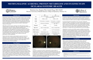
1. Kristina Lin, Eugene Pak, Linda Pang, OD, FAAO
Western University of Health Sciences, College of Optometry, Pomona, CA
Background
METHYLMALONIC ACIDEMIA: PROTEIN METABOLISM AND ITS EFFECTS ON
OCULAR & SYSTEMIC HEALTH
References
Conclusion
MMA is a protein metabolism disorder that can affect infants to adults and is
characterized by an accumulation of methylmalonic acid. Some long term
complications of MMA are chronic kidney dysfunction, pancreatitis, and intellectual
disabilities. If these issues are not addressed, it can lead to coma or death. It is important
to educate patients, family members, as well as healthcare providers, regarding the signs
of MMA. In addition, optometric management including support programs, community
resources, and vision rehabilitation can address patient’s visual needs and quality of life.
Optometrists need to be aware of ocular pathology associated metabolic disease in order
to recognize and properly manage the condition and optimize quality of life through low
vision rehabilitation.
1. Coulombe JT, Shih VE, Levy HL. Massachusetts metabolic disorders screening program. II. Methylmalonic aciduria. Pediatrics. Jan 1981;67(1):26-31.
2. Gerth C, Morel CF, Feigenbaum A, Levin AV. Ocular phenotype in patients with methylmalonic aciduria and homocystinuria, cobalamin C type. J. AAPOS.
2008; 12(6):591-596.
3. Hamel C. Retinitis pigmentosa. Orphanet J. of Rare Dis. 2006; 1(40):687-693.
4. Levrat V, Forest I, Fouilhoux A, et al. Carglumic acid: an additional therapy in the treatment of organic acidurias with hyperammonemia? Orphanet. J. Rare Dis.
2008; 3:2.
5. Ng J, Karr D, Reznick L, Pennesi M. Presentation and progression of the ocular manifestation of methylmalonic acidemia in children. Invest. Ophthalmol. Vis.
Sci. 2013; 54: 1332.
Case Report
Our patient was educated on the effects of Methylmalonic acidemia (MMA) on
functional vision and daily living. No spectacles were prescribed at the time due to lack
of improvement in visual acuity with trial frame refraction. We educated the patient and
his aunt about vision rehabilitation services and resources to maximize visual function.
Our patient was also referred for an electroretinogram (ERG) testing to confirm
diagnosis of retinitis pigmentosa sine pigmento. Subsequent follow-up visits included
prescribing optical devices and appropriate training.
Discussion
Our patient was diagnosed with suspected RP Sine Pigmento secondary to MMA. He
presented with ocular signs of arteriolar attenuation and waxy pallor of the optic disc.
However, because he did not present with bony spicules upon fundus examination, we
referred our patient for an ERG to confirm our diagnosis of suspected RP Sine Pigmento.
Our patient demonstrated several different ocular and systemic complications
characteristic of MMA. This includes loss of contrast sensitivity, progressive field loss,
decrease in vision, intellectual disability, and seizures. Young individuals suspected of
organic academia are recommended to receive a screening and evaluation of multiple
factors, such as pH, glucose, urea, electrolytes, bicarbonate, and ketones. Bone marrow
suppression can also occur, which can cause thrombocytopenia, neutropenia and
pancytopenia, so a complete blood count and differentials are used to aid in diagnosis.
Although there is no known cure for MMA, treatment is available to prevent further
complications. Systemic management consists of a low protein diet, avoiding various
medications, such as broad spectrum antibiotics, vitamins, and carglumic acid, especially
for individuals with ammonia levels exceeding 400 micromol/L in the blood.4 Current
literature shows very limited information regarding cases of MMA associated with
pigmentary retinopathy and optometric management. However, a review evaluating
children with MMA with homocystinuria, cobalamin C type suggested a specific
progression in retinal changes, with bull’s eye maculopathy, optic atrophy, and vascular
changes occurring with an increase in age.5
Methylmalonic acidemia (MMA) is a rare type of organic acidemia, which are disorders
that affect certain enzymes responsible in the breakdown of specific amino acids. MMA
may present ocular findings of nystagmus, reduced vision, and Retinitis Pigmentosa.3
Retinitis Pigmentosa (RP) is characterized by a decrease in visual acuity, progressive
field loss with an abnormal electroretinogram (ERG). RP usually presents with a triage
of arteriolar attenuation, waxy pallor of the optic disc and pigmented bone spicules. If
there are no signs of bone spicules present then the condition is called Retinitis
Pigmentosa Sine Pigmento.
MMA follows autosomal recessive inheritance with variable expression. It has an
estimated incidence of 1:48,000 infants.1 MMA causes a buildup of methylmalonic acid
in the body due to a deficiency of adenosylcobalamin-dependent enzyme
methylmalonyl-CoA mutase or vitamin B12 (also known as cobalamin). MMA is a
result of the mutation of the MMACHC gene causing an autosomal recessive disorder
called Cobalamin C type (cblC).2 CblC is the most common inborn error of Vitamin
B12 metabolism that results in the malformation of B12 cofactors, adenosylcobalamin
and methylcobalamin, directly affecting the visual system. These two cofactors are
essential to the formation of methionine, which is needed for rod and cone
photoreceptors to function appropriately.
Patients with the cblC mutation can either displayed a progressive cone-rod or a
nonprogressive rod-cone retinal dysfunction both presenting with a reduced
photoreceptor response on the ERG, making it look similar to RP.2 In addition to RP,
individuals diagnosed with MMA may experience moderate hepatomegaly, metabolic
acidosis (pH <7.30), ketoaciduria, hypoglycemia, seizures, failure to thrive,
developmental delay, and facial dysmorphism.
In December 2014, a twenty year old Hispanic male was presented to the Eye Care
Center for a vision rehabilitation Examination. The chief concern, brought forth by the
patient’s aunt, was longstanding reduced vision at distance. Medical history revealed he
was diagnosed with MMA at one year of age. When asked previous surgeries or
accidents, his aunt revealed he had a gastrostomy feeding tube insertion, a motor vehicle
accident at the age of six, during which vision changes were first noticed and was
subsequently diagnosed as legally blind. Our patient was positive for a history of heart
surgery in December 2013, and seizures following a liver and kidney transplant in March
2014. In addition, he has impaired cognitive function. Ocular history was positive for
optic nerve atrophy in both eyes with temporal pallor, intermittent exotropia and mild
myopia, which was diagnosed on October 2013 at Children’s Hospital Los Angeles. In
October 2015, he had developed a jerk nystagmus to the right. Our patient has an allergy
to Benadryl and protein
OD OS
VAs
dVA: 2/25M (20/250) dVA: 2/400ft (20/4000)
nVA OU: 0.4/4M (20/200)
Pupils PERRL (-) APD PERRL (-) APD
EOMs Full and jerky, (+) nystagmus Full and jerky, (+) nystagmus
CF Restricted in all quadrants
Restricted in all quadrants
OS>> OD
Tonometry 19 mmHg 19 mmHg
Optic Nerve Grade 3-4 temporal pallor Grade 3-4 temporal pallor
Retina- Vascular Low A/V Low A/V and attenuation
MARS Contrast Profound degree of loss Profound degree of loss
Amsler Grid
(+) metamorphosia in superior
and superior temporal fields
(+) metamorphosia in the
superior field
OD OS
Figure 1. Fundus photos right eye (left) and left eye (right). Fundus demonstrates arteriolar attenuation as well as optic
nerve pallor.