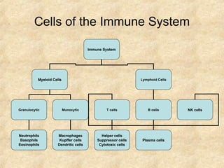
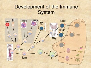
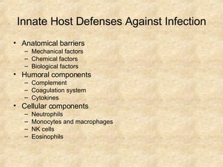
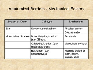
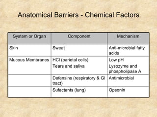
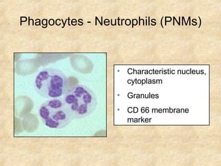
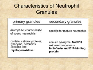
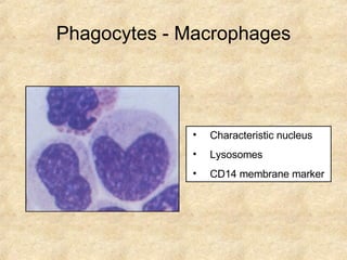
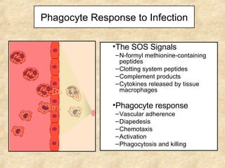
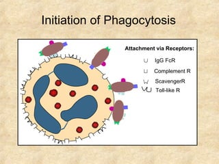
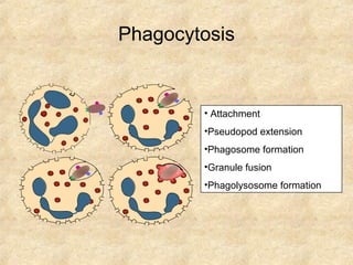
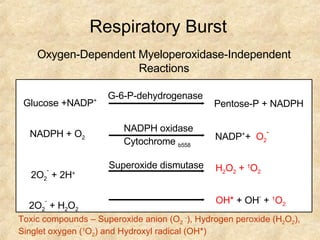
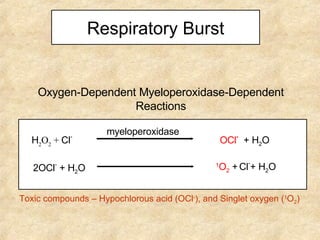
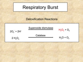
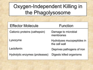
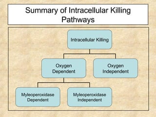
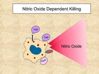
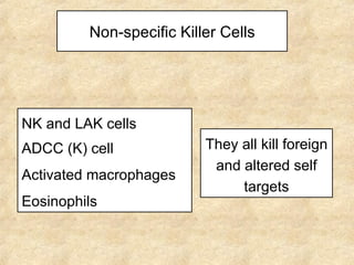
Eugene P. Mayer Office: Bldg. #2, Rm. B18 Phone: 733-3281 Email: MAYER@MED.SC.EDU
Overview of the Immune System Interactions between the two systems Immune System Innate (Nonspecific) 1 o  line of defense Adaptive (Specific) 2 o  line of defense Protects/re-exposure Cellular Components Humoral Components Cellular Components Humoral Components
Comparison   of Innate and Adaptive Immunity No memory   No time lag Not antigen specific A lag period Antigen specific Development of memory Innate Immunity Adaptive Immunity
Cells of the Immune System Immune System Myeloid Cells Lymphoid Cells Granulocytic Monocytic T cells B cells Neutrophils Basophils Eosinophils Macrophages Kupffer cells Dendritic cells Helper cells Suppressor cells Cytotoxic cells Plasma cells NK cells
Development of the Immune System ery pl mye neu m φ lym nk thy CD8 + CD4 + CTL TH2 TH1
Function of the Immune System (Self/Non-self Discrimination) To protect from pathogens Intracellular ( e.g.  viruses and some bacteria and parasites) Extracellular ( e.g.  most bacteria, fungi and parasites) To eliminate modified or altered self
Infection and Immunity Balance infection immunity Bolus of infection x virulence immunity Disease =
Effects of the Immune System Beneficial: Protection from Invaders Elimination of Altered Self Detrimental: Discomfort and collateral damage (inflammation) Damage to self (hypersensitivity or autoimmunity)
Innate (Nonspecific) Immunity
Overview of the Immune System Immune System Innate (Nonspecific) Adaptive (Specific) Cellular Components Humoral Components Cellular Components Humoral Components
Innate Host Defenses Against Infection Anatomical barriers Mechanical factors Chemical factors Biological factors Humoral components Complement Coagulation system Cytokines Cellular components Neutrophils Monocytes and macrophages NK cells Eosinophils
Anatomical Barriers - Mechanical Factors System or Organ Cell type Mechanism Skin Squamous epithelium Physical barrier Desquamation Mucous Membranes Non-ciliated epithelium ( e.g.  GI tract) Peristalsis Ciliated epithelium ( e.g.  respiratory tract) Mucociliary elevator Epithelium ( e.g.  nasopharynx) Flushing action of tears, saliva, mucus, urine
Anatomical Barriers - Chemical Factors System or Organ Component Mechanism Skin Sweat Anti-microbial fatty acids Mucous Membranes HCl (parietal cells) Tears and saliva Low pH Lysozyme and phospholipase A Defensins (respiratory & GI tract) Antimicrobial Sufactants (lung) Opsonin
Anatomical Barriers - Biological Factors System or Organ Component Mechanism Skin and mucous membranes Normal flora Antimicrobial substances Competition for nutrients and colonization
Humoral Components Component Mechanism Complement Lysis of bacteria and some viruses Opsonin Increase in vascular permeability Recruitment and activation of phagocytic cells Coagulation system Increase vascular permeability Recruitment of phagocytic cells Β -lysin from platelets – a cationic detergent Lactoferrin and transferrin Compete with bacteria for iron Lysozyme Breaks down bacterial cell walls Cytokines Various effects
Cellular Components Cell Functions Neutrophils Phagocytosis and intracellular killing Inflammation and tissue damage Macrophages Phagocytosis and intracellular killing Extracellular killing of infected or altered self targets Tissue repair Antigen presentation for specific immune response NK and LAK cells Killing of virus-infected and altered self targets Eosinophils Killing of certain parasites
Phagocytosis and Intracellular Killing
Phagocytes - Neutrophils (PNMs) Characteristic nucleus, cytoplasm  Granules CD 66 membrane marker
Characteristics of Neutrophil Granules primary granules   contain  cationic proteins, lysozyme, defensins, elastase and  myeloperoxidase secondary granules   contain lysozyme, NADPH oxidase components,  lactoferrin   and B12-binding protein azurophilic; characteristic of young neutrophils; specific for mature neutrophils
Characteristic nucleus Lysosomes CD14 membrane marker Phagocytes - Macrophages
Phagocyte Response to Infection The SOS Signals N-formyl methionine-containing peptides Clotting system peptides Complement products Cytokines released by tissue macrophages Phagocyte response Vascular adherence Diapedesis Chemotaxis Activation Phagocytosis and killing
Attachment via Receptors: IgG FcR ScavengerR Complement R Toll-like R Initiation of Phagocytosis
Phagocytosis Attachment Pseudopod extension Phagosome formation Granule fusion Phagolysosome formation
Respiratory Burst Oxygen-Dependent Myeloperoxidase-Independent Reactions Toxic compounds – Superoxide anion (O 2   - ), Hydrogen peroxide (H 2 O 2 ), Singlet oxygen ( 1 O 2 ) and Hydroxyl radical (OH*) Pentose-P + NADPH G-6-P-dehydrogenase Glucose +NADP + NADPH oxidase Cytochrome  b558 NADP + +  O 2 - NADPH + O 2 Superoxide dismutase H 2 O 2  +  1 O 2 2O 2 -  + 2H + 2O 2 -  + H 2 O 2 OH*  + OH -  +  1 O 2
Respiratory Burst Oxygen-Dependent Myeloperoxidase-Dependent Reactions myeloperoxidase OCl -   + H 2 O H 2 O 2  +  Cl - 2OCl -  + H 2 O 1 O 2   +   Cl - + H 2 O Toxic compounds – Hypochlorous acid (OCl - ), and Singlet oxygen ( 1 O 2 )
Respiratory Burst Detoxification Reactions H 2 O 2   +   O 2 Superoxide dismutase H 2 O  + O 2 Catalase 2O 2 -  + 2H + 2 H 2 O 2
Oxygen-Independent Killing in the Phagolysosome Effector Molecule Function Cationic proteins (cathepsin) Damage to microbial membranes Lysozyme Hydrolyses mucopeptides in the cell wall Lactoferrin Deprives pathogens of iron Hydrolytic enzymes (proteases) Digests killed organisms
Summary of Intracellular Killing Pathways Intracellular Killing Oxygen Dependent Oxygen Independent Myleoperoxidase Dependent Myleoperoxidase Independent
Nitric Oxide Dependent Killing IFN γ  TNF TNF Nitric Oxide Nitric Oxide
Non-specific Killer Cells NK and LAK cells ADCC (K) cell Activated macrophages Eosinophils They all kill foreign and altered self targets
Natural Killer (NK) cells also known as large granular lymphocytes (LGL) kill virus-infected or malignant cells identified by the presence of CD56 & CD16 and absence of CD3 activated by IL2 and IFN- γ  to become LAK cells
Lymphokine Activated Killer (LAK) cell IL2 IFN IFN IL2 kills malignant cells kills transformed and malignant cells
Regulation of NK Cell Function MHC I KIR KAR KAL No Killing Killing
K Cells morphologically undefined mediate ADCC have Fc receptor recognize antibody coated targets could be NK cells (IgG), macrophages (IgG), eosinophils (IgE) or other cells (IgG)

MIỄN DỊCH BÀI 1

  • 1.
    Eugene P. MayerOffice: Bldg. #2, Rm. B18 Phone: 733-3281 Email: MAYER@MED.SC.EDU
  • 2.
    Overview of theImmune System Interactions between the two systems Immune System Innate (Nonspecific) 1 o line of defense Adaptive (Specific) 2 o line of defense Protects/re-exposure Cellular Components Humoral Components Cellular Components Humoral Components
  • 3.
    Comparison of Innate and Adaptive Immunity No memory No time lag Not antigen specific A lag period Antigen specific Development of memory Innate Immunity Adaptive Immunity
  • 4.
    Cells of theImmune System Immune System Myeloid Cells Lymphoid Cells Granulocytic Monocytic T cells B cells Neutrophils Basophils Eosinophils Macrophages Kupffer cells Dendritic cells Helper cells Suppressor cells Cytotoxic cells Plasma cells NK cells
  • 5.
    Development of theImmune System ery pl mye neu m φ lym nk thy CD8 + CD4 + CTL TH2 TH1
  • 6.
    Function of theImmune System (Self/Non-self Discrimination) To protect from pathogens Intracellular ( e.g. viruses and some bacteria and parasites) Extracellular ( e.g. most bacteria, fungi and parasites) To eliminate modified or altered self
  • 7.
    Infection and ImmunityBalance infection immunity Bolus of infection x virulence immunity Disease =
  • 8.
    Effects of theImmune System Beneficial: Protection from Invaders Elimination of Altered Self Detrimental: Discomfort and collateral damage (inflammation) Damage to self (hypersensitivity or autoimmunity)
  • 9.
  • 10.
    Overview of theImmune System Immune System Innate (Nonspecific) Adaptive (Specific) Cellular Components Humoral Components Cellular Components Humoral Components
  • 11.
    Innate Host DefensesAgainst Infection Anatomical barriers Mechanical factors Chemical factors Biological factors Humoral components Complement Coagulation system Cytokines Cellular components Neutrophils Monocytes and macrophages NK cells Eosinophils
  • 12.
    Anatomical Barriers -Mechanical Factors System or Organ Cell type Mechanism Skin Squamous epithelium Physical barrier Desquamation Mucous Membranes Non-ciliated epithelium ( e.g. GI tract) Peristalsis Ciliated epithelium ( e.g. respiratory tract) Mucociliary elevator Epithelium ( e.g. nasopharynx) Flushing action of tears, saliva, mucus, urine
  • 13.
    Anatomical Barriers -Chemical Factors System or Organ Component Mechanism Skin Sweat Anti-microbial fatty acids Mucous Membranes HCl (parietal cells) Tears and saliva Low pH Lysozyme and phospholipase A Defensins (respiratory & GI tract) Antimicrobial Sufactants (lung) Opsonin
  • 14.
    Anatomical Barriers -Biological Factors System or Organ Component Mechanism Skin and mucous membranes Normal flora Antimicrobial substances Competition for nutrients and colonization
  • 15.
    Humoral Components ComponentMechanism Complement Lysis of bacteria and some viruses Opsonin Increase in vascular permeability Recruitment and activation of phagocytic cells Coagulation system Increase vascular permeability Recruitment of phagocytic cells Β -lysin from platelets – a cationic detergent Lactoferrin and transferrin Compete with bacteria for iron Lysozyme Breaks down bacterial cell walls Cytokines Various effects
  • 16.
    Cellular Components CellFunctions Neutrophils Phagocytosis and intracellular killing Inflammation and tissue damage Macrophages Phagocytosis and intracellular killing Extracellular killing of infected or altered self targets Tissue repair Antigen presentation for specific immune response NK and LAK cells Killing of virus-infected and altered self targets Eosinophils Killing of certain parasites
  • 17.
  • 18.
    Phagocytes - Neutrophils(PNMs) Characteristic nucleus, cytoplasm Granules CD 66 membrane marker
  • 19.
    Characteristics of NeutrophilGranules primary granules contain cationic proteins, lysozyme, defensins, elastase and myeloperoxidase secondary granules contain lysozyme, NADPH oxidase components, lactoferrin and B12-binding protein azurophilic; characteristic of young neutrophils; specific for mature neutrophils
  • 20.
    Characteristic nucleus LysosomesCD14 membrane marker Phagocytes - Macrophages
  • 21.
    Phagocyte Response toInfection The SOS Signals N-formyl methionine-containing peptides Clotting system peptides Complement products Cytokines released by tissue macrophages Phagocyte response Vascular adherence Diapedesis Chemotaxis Activation Phagocytosis and killing
  • 22.
    Attachment via Receptors:IgG FcR ScavengerR Complement R Toll-like R Initiation of Phagocytosis
  • 23.
    Phagocytosis Attachment Pseudopodextension Phagosome formation Granule fusion Phagolysosome formation
  • 24.
    Respiratory Burst Oxygen-DependentMyeloperoxidase-Independent Reactions Toxic compounds – Superoxide anion (O 2 - ), Hydrogen peroxide (H 2 O 2 ), Singlet oxygen ( 1 O 2 ) and Hydroxyl radical (OH*) Pentose-P + NADPH G-6-P-dehydrogenase Glucose +NADP + NADPH oxidase Cytochrome b558 NADP + + O 2 - NADPH + O 2 Superoxide dismutase H 2 O 2 + 1 O 2 2O 2 - + 2H + 2O 2 - + H 2 O 2 OH* + OH - + 1 O 2
  • 25.
    Respiratory Burst Oxygen-DependentMyeloperoxidase-Dependent Reactions myeloperoxidase OCl - + H 2 O H 2 O 2 + Cl - 2OCl - + H 2 O 1 O 2 + Cl - + H 2 O Toxic compounds – Hypochlorous acid (OCl - ), and Singlet oxygen ( 1 O 2 )
  • 26.
    Respiratory Burst DetoxificationReactions H 2 O 2 + O 2 Superoxide dismutase H 2 O + O 2 Catalase 2O 2 - + 2H + 2 H 2 O 2
  • 27.
    Oxygen-Independent Killing inthe Phagolysosome Effector Molecule Function Cationic proteins (cathepsin) Damage to microbial membranes Lysozyme Hydrolyses mucopeptides in the cell wall Lactoferrin Deprives pathogens of iron Hydrolytic enzymes (proteases) Digests killed organisms
  • 28.
    Summary of IntracellularKilling Pathways Intracellular Killing Oxygen Dependent Oxygen Independent Myleoperoxidase Dependent Myleoperoxidase Independent
  • 29.
    Nitric Oxide DependentKilling IFN γ  TNF TNF Nitric Oxide Nitric Oxide
  • 30.
    Non-specific Killer CellsNK and LAK cells ADCC (K) cell Activated macrophages Eosinophils They all kill foreign and altered self targets
  • 31.
    Natural Killer (NK)cells also known as large granular lymphocytes (LGL) kill virus-infected or malignant cells identified by the presence of CD56 & CD16 and absence of CD3 activated by IL2 and IFN- γ to become LAK cells
  • 32.
    Lymphokine Activated Killer(LAK) cell IL2 IFN IFN IL2 kills malignant cells kills transformed and malignant cells
  • 33.
    Regulation of NKCell Function MHC I KIR KAR KAL No Killing Killing
  • 34.
    K Cells morphologicallyundefined mediate ADCC have Fc receptor recognize antibody coated targets could be NK cells (IgG), macrophages (IgG), eosinophils (IgE) or other cells (IgG)