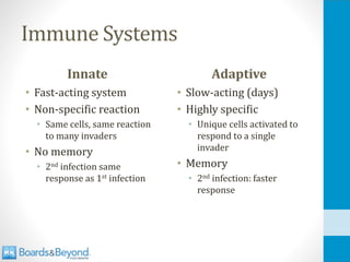
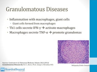
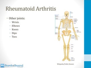
Immune Systems
Innate
• Fast-actingsystem
• Non-specific reaction
• Same cells, same reaction
to many invaders
• No memory
• 2nd infection same
response as 1st infection
Adaptive
• Slow-acting (days)
• Highly specific
• Unique cells activated to
respond to a single
invader
• Memory
• 2nd infection: faster
response
4.
Antigen Presentation
• Innatesystem can be activated by “free” antigen
• Pathogenic molecules detected freely in blood, tissue
• Adaptive system requires “antigen presentation”
• Pathogens must be engulfed by cells, broken down
• Pieces of protein transported to surface
• Antigen “presented” to T-cells for activation
5.
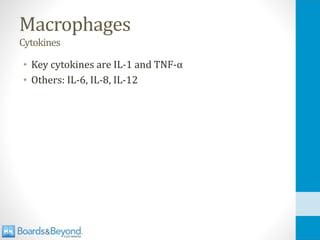
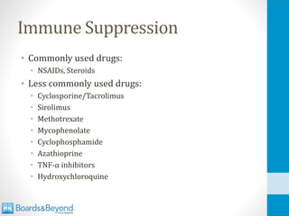
Cytokines
• Cell signalingproteins
• Often released by immune cells
• Stimulate inflammatory response
• Various subsets
• Chemokine: Attracts immune cells (chemotaxis)
• Interleukins: IL-1, IL,2, etc
• Tumor necrosis factor (TNF): Can cause tumor death
• Transforming growth factor (TGF)
• Interferons: Named for interfering with viral replication
6.
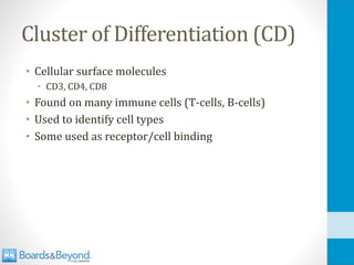
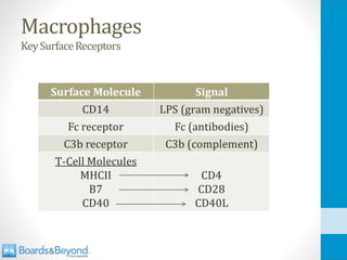
Cluster of Differentiation(CD)
• Cellular surface molecules
• CD3, CD4, CD8
• Found on many immune cells (T-cells, B-cells)
• Used to identify cell types
• Some used as receptor/cell binding
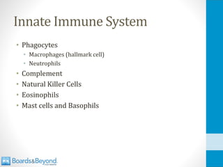
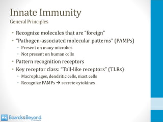
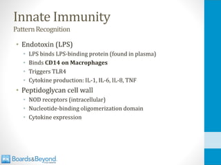
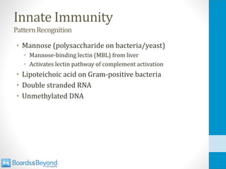
Innate Immunity
PatternRecognition
• Mannose(polysaccharide on bacteria/yeast)
• Mannose-binding lectin (MBL) from liver
• Activates lectin pathway of complement activation
• Lipoteichoic acid on Gram-positive bacteria
• Double stranded RNA
• Unmethylated DNA
11.
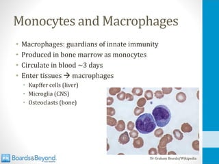
Monocytes and Macrophages
•Macrophages: guardians of innate immunity
• Produced in bone marrow as monocytes
• Circulate in blood ~3 days
• Enter tissues → macrophages
• Kupffer cells (liver)
• Microglia (CNS)
• Osteoclasts (bone)
Dr Graham Beards/Wikipedia
12.
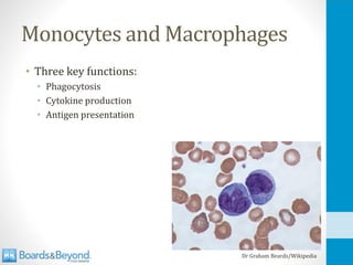
Monocytes and Macrophages
•Three key functions:
• Phagocytosis
• Cytokine production
• Antigen presentation
Dr Graham Beards/Wikipedia
13.
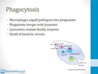
Phagocytosis
• Macrophages engulfpathogens into phagosome
• Phagosome merges with lysosome
• Lysosomes contain deadly enzymes
• Death of bacteria, viruses
Graham Colm/Wikipedia
Phagocytosis
• Some pathogensblock this process
• Tuberculosis modifies phagosome
• Unable to fuse with lysosome
• Proliferation inside macrophages
• Protection from antibodies
• Chediak-Higashi Syndrome
• Immune deficiency syndrome
• Failure of lysosomes to fuse with phagosomes
• Recurrent bacterial infections
CDC/Public Domain/Wikipedia
17.
Macrophages
• Macrophages canexist in several “states”
• Resting: Debris removal
• Activated (“primed”): more effective
• Major activators (via surface TLRs):
• LPS from bacteria
• Peptidoglycan
• Bacterial DNA (no methylation)
• Also, IFN-γ from T-cells, NKC
• Attracted by C5a (complement)
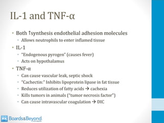
IL-1 and TNF-α
•Both ↑synthesis endothelial adhesion molecules
• Allows neutrophils to enter inflamed tissue
• IL-1
• “Endogenous pyrogen” (causes fever)
• Acts on hypothalamus
• TNF-α
• Can cause vascular leak, septic shock
• “Cachectin:” Inhibits lipoprotein lipase in fat tissue
• Reduces utilization of fatty acids → cachexia
• Kills tumors in animals (“tumor necrosis factor”)
• Can cause intravascular coagulation → DIC
21.
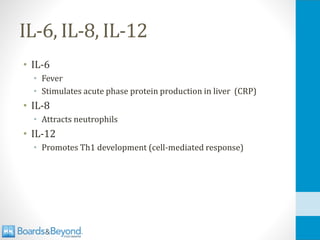
IL-6, IL-8, IL-12
•IL-6
• Fever
• Stimulates acute phase protein production in liver (CRP)
• IL-8
• Attracts neutrophils
• IL-12
• Promotes Th1 development (cell-mediated response)
22.
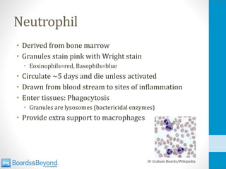
Neutrophil
• Derived frombone marrow
• Granules stain pink with Wright stain
• Eosinophils=red, Basophils=blue
• Circulate ~5 days and die unless activated
• Drawn from blood stream to sites of inflammation
• Enter tissues: Phagocytosis
• Granules are lysosomes (bactericidal enzymes)
• Provide extra support to macrophages
Dr Graham Beards/Wikipedia
23.
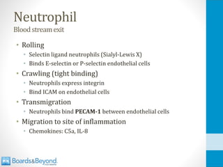
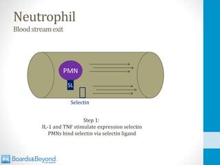
Neutrophil
Bloodstreamexit
• Rolling
• Selectinligand neutrophils (Sialyl-Lewis X)
• Binds E-selectin or P-selectin endothelial cells
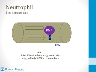
• Crawling (tight binding)
• Neutrophils express integrin
• Bind ICAM on endothelial cells
• Transmigration
• Neutrophils bind PECAM-1 between endothelial cells
• Migration to site of inflammation
• Chemokines: C5a, IL-8
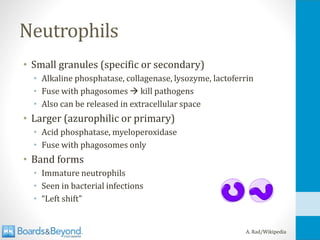
Neutrophils
• Small granules(specific or secondary)
• Alkaline phosphatase, collagenase, lysozyme, lactoferrin
• Fuse with phagosomes → kill pathogens
• Also can be released in extracellular space
• Larger (azurophilic or primary)
• Acid phosphatase, myeloperoxidase
• Fuse with phagosomes only
• Band forms
• Immature neutrophils
• Seen in bacterial infections
• “Left shift”
A. Rad/Wikipedia
27.
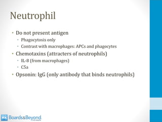
Neutrophil
• Do notpresent antigen
• Phagocytosis only
• Contrast with macrophages: APCs and phagocytes
• Chemotaxins (attracters of neutrophils)
• IL-8 (from macrophages)
• C5a
• Opsonin: IgG (only antibody that binds neutrophils)
28.
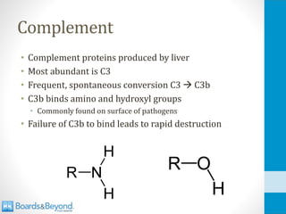
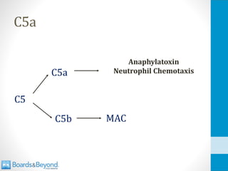
Complement
• Complement proteinsproduced by liver
• Most abundant is C3
• Frequent, spontaneous conversion C3 → C3b
• C3b binds amino and hydroxyl groups
• Commonly found on surface of pathogens
• Failure of C3b to bind leads to rapid destruction
29.
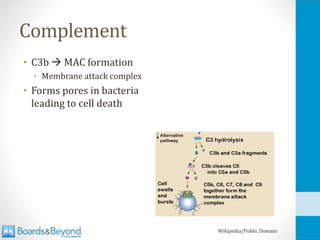
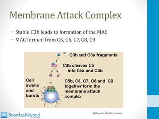
Complement
• C3b →MAC formation
• Membrane attack complex
• Forms pores in bacteria
leading to cell death
Wikipedia/Public Domain
30.
Natural Killer Cells
•Two key roles:
• Kill human cells infected by viruses
• Produce IFN-γ to activates macrophages
31.
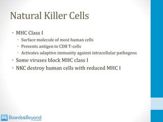
Natural Killer Cells
•MHC Class I
• Surface molecule of most human cells
• Presents antigen to CD8 T-cells
• Activates adaptive immunity against intracellular pathogens
• Some viruses block MHC class I
• NKC destroy human cells with reduced MHC I
32.
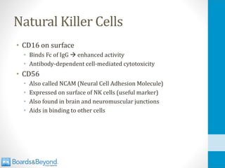
Natural Killer Cells
•CD16 on surface
• Binds Fc of IgG → enhanced activity
• Antibody-dependent cell-mediated cytotoxicity
• CD56
• Also called NCAM (Neural Cell Adhesion Molecule)
• Expressed on surface of NK cells (useful marker)
• Also found in brain and neuromuscular junctions
• Aids in binding to other cells
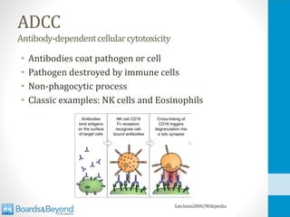
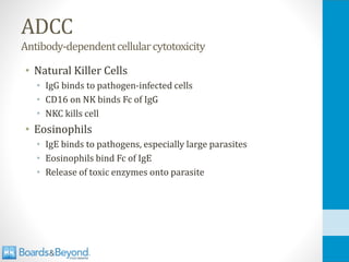
ADCC
Antibody-dependentcellularcytotoxicity
• Natural KillerCells
• IgG binds to pathogen-infected cells
• CD16 on NK binds Fc of IgG
• NKC kills cell
• Eosinophils
• IgE binds to pathogens, especially large parasites
• Eosinophils bind Fc of IgE
• Release of toxic enzymes onto parasite
35.
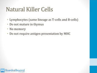
Natural Killer Cells
•Lymphocytes (same lineage as T-cells and B-cells)
• Do not mature in thymus
• No memory
• Do not require antigen presentation by MHC
36.
Eosinophils, Mast Cells,Basophils
• All contain granules with destructive enzymes
• All can be activated/triggered by IgE antibodies
• Important for defense against parasites (helminths)
• Too large for phagocytosis
• Release of toxic substances kills parasite
• Main medical relevance is in allergic disease
37.
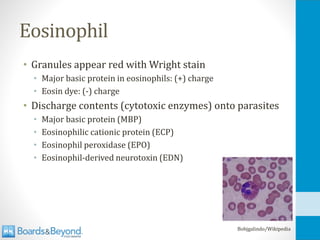
Eosinophil
• Granules appearred with Wright stain
• Major basic protein in eosinophils: (+) charge
• Eosin dye: (-) charge
• Discharge contents (cytotoxic enzymes) onto parasites
• Major basic protein (MBP)
• Eosinophilic cationic protein (ECP)
• Eosinophil peroxidase (EPO)
• Eosinophil-derived neurotoxin (EDN)
Bobjgalindo/Wikipedia
38.
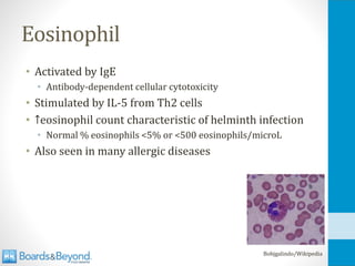
Eosinophil
• Activated byIgE
• Antibody-dependent cellular cytotoxicity
• Stimulated by IL-5 from Th2 cells
• ↑eosinophil count characteristic of helminth infection
• Normal % eosinophils <5% or <500 eosinophils/microL
• Also seen in many allergic diseases
Bobjgalindo/Wikipedia
39.
Mast cells andBasophils
• Granules appear blue with Wright stain
• Basophils: blood stream
• Mast cells: Tissue
• Bind Fc portion of IgE antibodies
• IgE molecules crosslink → degranulation
• Histamine (vasodilation)
• Enzymes (peroxidases, hydrolases)
Wikipedia/Public Domain
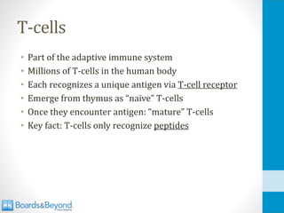
T-cells
• Part ofthe adaptive immune system
• Millions of T-cells in the human body
• Each recognizes a unique antigen via T-cell receptor
• Emerge from thymus as “naïve” T-cells
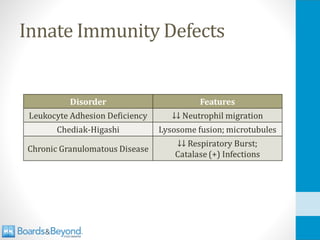
• Once they encounter antigen: “mature” T-cells
• Key fact: T-cells only recognize peptides
47.
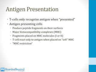
Antigen Presentation
• T-cellsonly recognize antigen when “presented”
• Antigen presenting cells
• Produce peptide fragments on their surfaces
• Major histocompatibility complexes (MHC)
• Fragments placed on MHC molecules (I or II)
• T-cell react only to antigen when placed on “self” MHC
• “MHC restriction”
48.
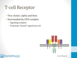
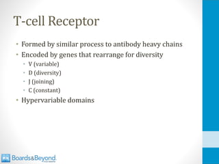
T-cell Receptor
• Twochains: alpha and beta
• Surrounded by CD3 complex
• Signaling complex
• Transmits “bound” signal into cell
Anriar/Wikipedia
49.
T-cell Receptor
• Formedby similar process to antibody heavy chains
• Encoded by genes that rearrange for diversity
• V (variable)
• D (diversity)
• J (joining)
• C (constant)
• Hypervariable domains
50.
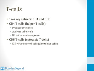
T-cells
• Two keysubsets: CD4 and CD8
• CD4 T-cells (helper T-cells)
• Produce cytokines
• Activate other cells
• Direct immune response
• CD8 T-cells (cytotoxic T-cells)
• Kill virus-infected cells (also tumor cells)
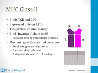
MHC Class II
•Binds TCR and CD4
• Expressed only on APCs
• Two protein chains: α and β
• Bind “invariant” chain in ER
• Prevents binding intracellular proteins
• Must merge with acidified lysosome
• Peptide fragments in lysosome
• Invariant chain released
• Antigen binds to MHC II → surface
atropos235 /Wikipedia
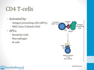
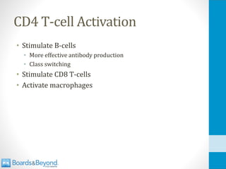
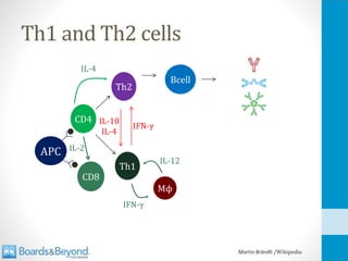
CD4 T-cell Activation
•Stimulate B-cells
• More effective antibody production
• Class switching
• Stimulate CD8 T-cells
• Activate macrophages
55.
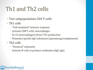
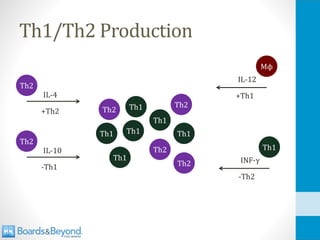
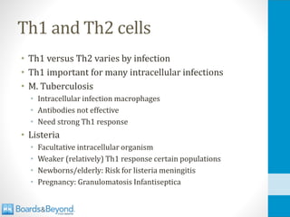
Th1 and Th2cells
• Two subpopulations CD4 T-cells
• Th1 cells
• “Cell-mediated” immune response
• Activate CD8 T-cells, macrophages
• IL-12 (macrophages) drives Th1 production
• Promotes specific IgG subclasses (opsonizing/complement)
• Th2 cells
• “Humoral” immunity
• Activate B-cells to produce antibodies (IgE, IgA)
56.
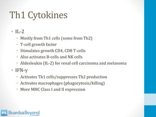
Th1 Cytokines
• IL-2
•Mostly from Th1 cells (some from Th2)
• T-cell growth factor
• Stimulates growth CD4, CD8 T-cells
• Also activates B-cells and NK cells
• Aldesleukin (IL-2) for renal cell carcinoma and melanoma
• IFN-γ
• Activates Th1 cells/suppresses Th2 production
• Activates macrophages (phagocytosis/killing)
• More MHC Class I and II expression
57.
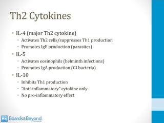
Th2 Cytokines
• IL-4(major Th2 cytokine)
• Activates Th2 cells/suppresses Th1 production
• Promotes IgE production (parasites)
• IL-5
• Activates eosinophils (helminth infections)
• Promotes IgA production (GI bacteria)
• IL-10
• Inhibits Th1 production
• “Anti-inflammatory” cytokine only
• No pro-inflammatory effect
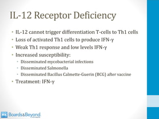
IL-12 Receptor Deficiency
•IL-12 cannot trigger differentiation T-cells to Th1 cells
• Loss of activated Th1 cells to produce IFN-γ
• Weak Th1 response and low levels IFN-γ
• Increased susceptibility:
• Disseminated mycobacterial infections
• Disseminated Salmonella
• Disseminated Bacillus Calmette-Guerin (BCG) after vaccine
• Treatment: IFN-γ
66.
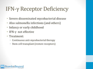
IFN-γ Receptor Deficiency
•Severe disseminated mycobacterial disease
• Also salmonella infections (and others)
• Infancy or early childhood
• IFN-γ not effective
• Treatment:
• Continuous anti-mycobacterial therapy
• Stem cell transplant (restore receptors)
67.
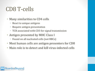
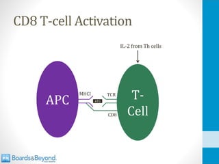
CD8 T-cells
• Manysimilarities to CD4 cells
• React to unique antigens
• Require antigen presentation
• TCR associated with CD3 for signal transmission
• Antigen presented by MHC Class I
• Found on all nucleated cells (not RBCs)
• Most human cells are antigen presenters for CD8
• Main role is to detect and kill virus-infected cells
68.
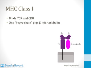
MHC Class I
•Binds TCR and CD8
• One “heavy chain” plus β-microglobulin
atropos235 /Wikipedia
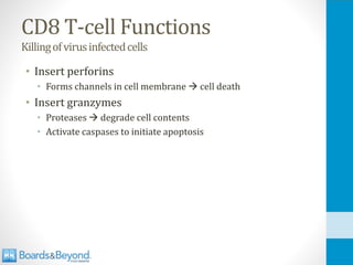
CD8 T-cell Functions
Killingofvirusinfectedcells
•Insert perforins
• Forms channels in cell membrane → cell death
• Insert granzymes
• Proteases → degrade cell contents
• Activate caspases to initiate apoptosis
71.
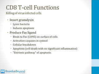
CD8 T-cell Functions
Killingofvirusinfectedcells
•Insert granulysin
• Lyses bacteria
• Induces apoptosis
• Produce Fas ligand
• Binds to Fas (CD95) on surface of cells
• Activation caspases in cytosol
• Cellular breakdown
• Apoptosis (cell death with no significant inflammation)
• “Extrinsic pathway” of apoptosis
72.
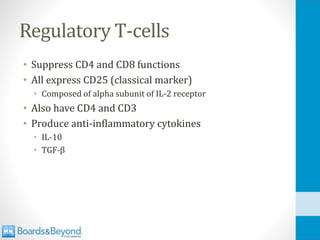
Regulatory T-cells
• SuppressCD4 and CD8 functions
• All express CD25 (classical marker)
• Composed of alpha subunit of IL-2 receptor
• Also have CD4 and CD3
• Produce anti-inflammatory cytokines
• IL-10
• TGF-β
73.
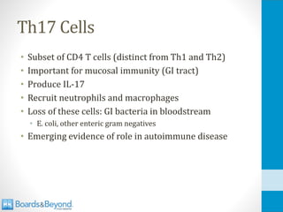
Th17 Cells
• Subsetof CD4 T cells (distinct from Th1 and Th2)
• Important for mucosal immunity (GI tract)
• Produce IL-17
• Recruit neutrophils and macrophages
• Loss of these cells: GI bacteria in bloodstream
• E. coli, other enteric gram negatives
• Emerging evidence of role in autoimmune disease
74.
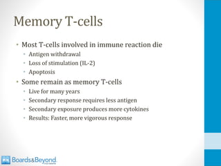
Memory T-cells
• MostT-cells involved in immune reaction die
• Antigen withdrawal
• Loss of stimulation (IL-2)
• Apoptosis
• Some remain as memory T-cells
• Live for many years
• Secondary response requires less antigen
• Secondary exposure produces more cytokines
• Results: Faster, more vigorous response
75.
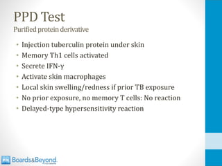
PPD Test
Purifiedproteinderivative
• Injectiontuberculin protein under skin
• Memory Th1 cells activated
• Secrete IFN-γ
• Activate skin macrophages
• Local skin swelling/redness if prior TB exposure
• No prior exposure, no memory T cells: No reaction
• Delayed-type hypersensitivity reaction
76.
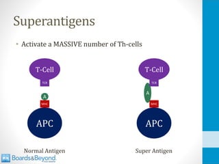
Superantigens
• Activate aMASSIVE number of Th-cells
APC
MHC
A
T-Cell
TCR
Normal Antigen
APC
MHC
A
T-Cell
TCR
Super Antigen
77.
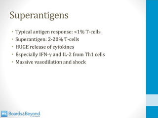
Superantigens
• Typical antigenresponse: <1% T-cells
• Superantigen: 2-20% T-cells
• HUGE release of cytokines
• Especially IFN-γ and IL-2 from Th1 cells
• Massive vasodilation and shock
78.
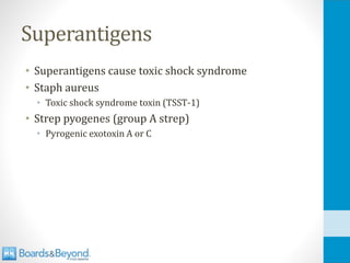
Superantigens
• Superantigens causetoxic shock syndrome
• Staph aureus
• Toxic shock syndrome toxin (TSST-1)
• Strep pyogenes (group A strep)
• Pyrogenic exotoxin A or C
79.
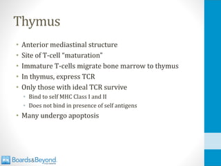
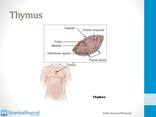
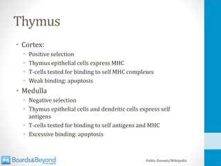
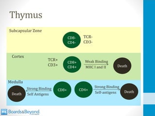
Thymus
• Anterior mediastinalstructure
• Site of T-cell “maturation”
• Immature T-cells migrate bone marrow to thymus
• In thymus, express TCR
• Only those with ideal TCR survive
• Bind to self MHC Class I and II
• Does not bind in presence of self antigens
• Many undergo apoptosis
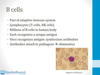
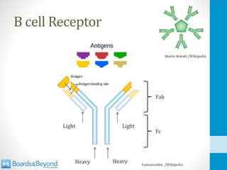
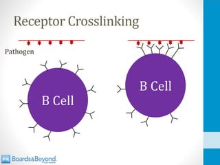
B cells
• Partof adaptive immune system
• Lymphocytes (T-cells, NK cells)
• Millions of B cells in human body
• Each recognizes a unique antigen
• Once recognizes antigen: synthesizes antibodies
• Antibodies attach to pathogens → elimination
Mgiganteus/Wikipedia
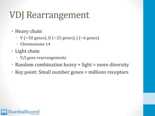
B cell Diversity
•Millions of B cells with unique antigen receptors
• More unique receptors than genes
• If one gene = one receptor, how can this be?
• Answer: Rearrangements of genetic building blocks
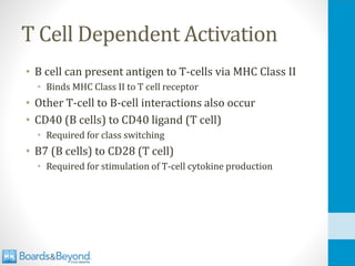
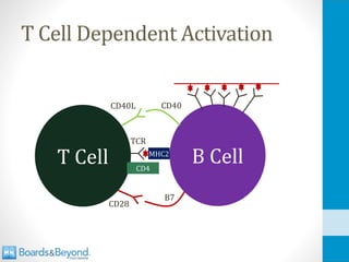
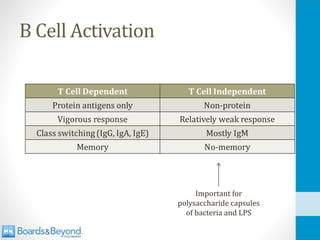
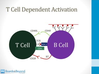
T Cell DependentActivation
• B cell can present antigen to T-cells via MHC Class II
• Binds MHC Class II to T cell receptor
• Other T-cell to B-cell interactions also occur
• CD40 (B cells) to CD40 ligand (T cell)
• Required for class switching
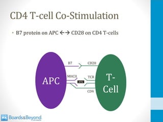
• B7 (B cells) to CD28 (T cell)
• Required for stimulation of T-cell cytokine production
96.
T Cell DependentActivation
B Cell
T Cell MHC2
TCR
CD4
CD40L CD40
CD28
B7
97.
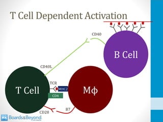
T Cell DependentActivation
Mϕ
T Cell MHC2
TCR
CD4
CD40L
CD28
B7
B Cell
CD40
98.
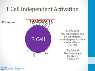
T Cell IndependentActivation
Pathogen
B Cell
Key Point #1
Very important for non-
protein antigens,
especially polysaccharide
capsules of bacteria
and LPS
Key Point #2
Weaker response
Mostly IgM
No memory
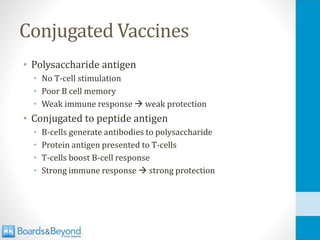
Conjugated Vaccines
• Polysaccharideantigen
• No T-cell stimulation
• Poor B cell memory
• Weak immune response → weak protection
• Conjugated to peptide antigen
• B-cells generate antibodies to polysaccharide
• Protein antigen presented to T-cells
• T-cells boost B-cell response
• Strong immune response → strong protection
101.
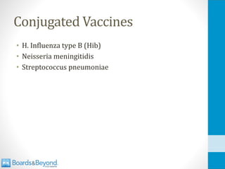
Conjugated Vaccines
• H.Influenza type B (Hib)
• Neisseria meningitidis
• Streptococcus pneumoniae
102.
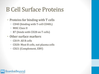
B Cell SurfaceProteins
• Proteins for binding with T cells
• CD40 (binding with T-cell CD40L)
• MHC Class II
• B7 (binds with CD28 on T cells)
• Other surface markers
• CD19: All B cells
• CD20: Most B cells, not plasma cells
• CD21 (Complement, EBV)
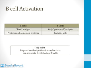
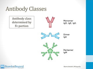
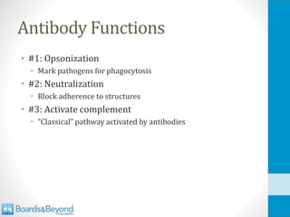
Antibody Functions
• #1:Opsonization
• Mark pathogens for phagocytosis
• #2: Neutralization
• Block adherence to structures
• #3: Activate complement
• “Classical” pathway activated by antibodies
105.
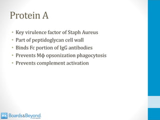
Protein A
• Keyvirulence factor of Staph Aureus
• Part of peptidoglycan cell wall
• Binds Fc portion of IgG antibodies
• Prevents Mϕ opsonization phagocytosis
• Prevents complement activation
106.
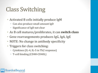
Class Switching
• ActivatedB cells initially produce IgM
• Can also produce small amount IgD
• Significance of IgD not clear
• As B cell matures/proliferates, it can switch class
• Gene rearrangements produces IgG, IgA, IgE
• NOTE: No change in antibody specificity
• Triggers for class switching:
• Cytokines (IL-4, IL-5 in Th2 response)
• T-cell binding (CD40-CD40L)
107.
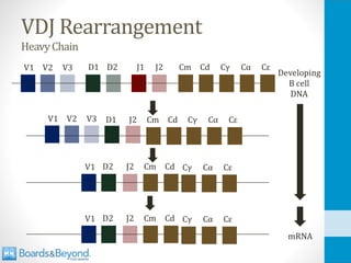
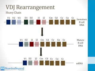
VDJ Rearrangement
HeavyChain
V1 V2V3 D1 D2 J1 J2 Cm Cd
V1 D2 J2 Cm Cd
Immature
B cell
DNA
Mature
B cell
DNA
J1
V1 D2 J2 Cm Cd
mRNA
Cγ Cα Cε
Cγ Cα Cε
Cγ Cα Cε
108.
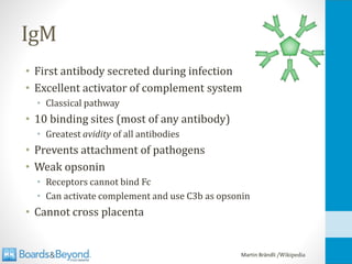
IgM
• First antibodysecreted during infection
• Excellent activator of complement system
• Classical pathway
• 10 binding sites (most of any antibody)
• Greatest avidity of all antibodies
• Prevents attachment of pathogens
• Weak opsonin
• Receptors cannot bind Fc
• Can activate complement and use C3b as opsonin
• Cannot cross placenta
Martin Brändli /Wikipedia
109.
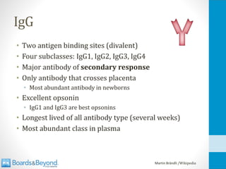
IgG
• Two antigenbinding sites (divalent)
• Four subclasses: IgG1, IgG2, IgG3, IgG4
• Major antibody of secondary response
• Only antibody that crosses placenta
• Most abundant antibody in newborns
• Excellent opsonin
• IgG1 and IgG3 are best opsonins
• Longest lived of all antibody type (several weeks)
• Most abundant class in plasma
Martin Brändli /Wikipedia
110.
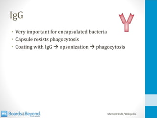
IgG
• Very importantfor encapsulated bacteria
• Capsule resists phagocytosis
• Coating with IgG → opsonization → phagocytosis
Martin Brändli /Wikipedia
111.
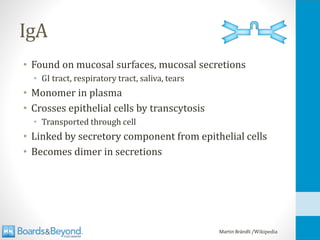
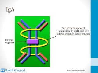
IgA
• Found onmucosal surfaces, mucosal secretions
• GI tract, respiratory tract, saliva, tears
• Monomer in plasma
• Crosses epithelial cells by transcytosis
• Transported through cell
• Linked by secretory component from epithelial cells
• Becomes dimer in secretions
Martin Brändli /Wikipedia
112.
IgA
• Does notfix complement
• Excellent at coating mucosal pathogens
• Ideal for mucosal surfaces
• Coat pathogens so they cannot invade
• Pathogens swept away with mucosal secretions
• No complement = no inflammation
• Secreted into milk to protect baby’s GI tract
Martin Brändli /Wikipedia
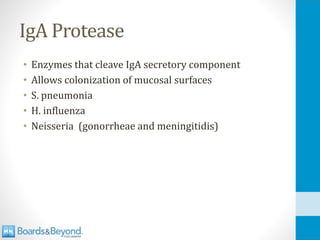
IgA Protease
• Enzymesthat cleave IgA secretory component
• Allows colonization of mucosal surfaces
• S. pneumonia
• H. influenza
• Neisseria (gonorrheae and meningitidis)
115.
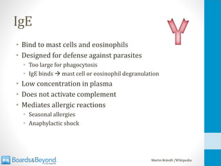
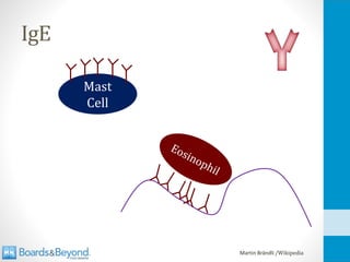
IgE
• Bind tomast cells and eosinophils
• Designed for defense against parasites
• Too large for phagocytosis
• IgE binds → mast cell or eosinophil degranulation
• Low concentration in plasma
• Does not activate complement
• Mediates allergic reactions
• Seasonal allergies
• Anaphylactic shock
Martin Brändli /Wikipedia
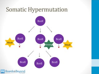
Somatic Hypermutation
• Lateevent during inflammation/infection
• Often after class switching
• High mutation rate in portions of V, D, J genes
• Re-stimulation required for ongoing proliferation
• Strongest binding BCR proliferate the most
• “Affinity maturation”
• Receptors mutate: Stronger antigen binding over time
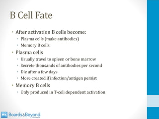
B Cell Fate
•After activation B cells become:
• Plasma cells (make antibodies)
• Memory B cells
• Plasma cells
• Usually travel to spleen or bone marrow
• Secrete thousands of antibodies per second
• Die after a few days
• More created if infection/antigen persist
• Memory B cells
• Only produced in T-cell dependent activation
120.
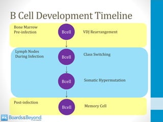
B Cell DevelopmentTimeline
Bcell
Bcell
Bcell
Bcell
VDJ Rearrangement
Class Switching
Somatic Hypermutation
Memory Cell
Bone Marrow
Pre-infection
Lymph Nodes
During Infection
Post-infection
121.
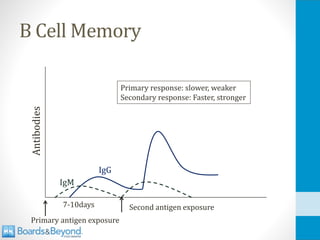
B Cell Memory
Antibodies
Primaryantigen exposure
Second antigen exposure
Primary response: slower, weaker
Secondary response: Faster, stronger
7-10days
IgM
IgG
122.
Vaccines
• B celland T cell response without overt infection
• Protection via immune memory
• Various types:
• Live attenuated
• Killed
• Oral/intramuscular
123.
Vaccines
• Live attenuated
•Pathogens modified to be less virulent
• Can induce a strong, cell-mediated response
• Some risk of infection (especially immunocompromised)
• If given <1yo, maternal antibodies may kill pathogen
• MMR
• Killed
• Pathogen killed but antigens remain intact
• Strong humoral response (antibodies)
• Weaker immune response than live, attenuated
• No risk of infection
Vaccines
• Passive Immunization
•Administration of antibodies
• Short term protection (weeks)
• No memory or long term protection
• Used for dangerous, imminent infections
• Rabies, Tetanus
• Also maternal antibodies → fetus
• Sometimes passive and active done simultaneously
• Rabies immune globulin plus rabies vaccine
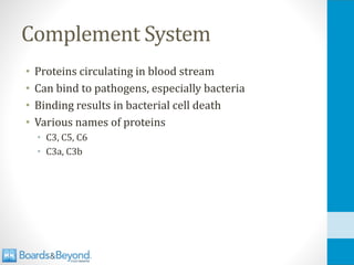
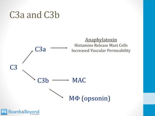
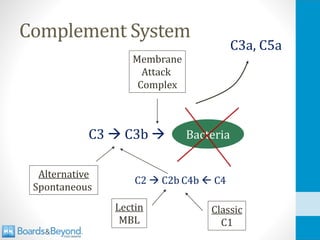
Complement System
• Proteinscirculating in blood stream
• Can bind to pathogens, especially bacteria
• Binding results in bacterial cell death
• Various names of proteins
• C3, C5, C6
• C3a, C3b
128.
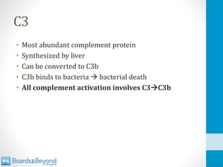
C3
• Most abundantcomplement protein
• Synthesized by liver
• Can be converted to C3b
• C3b binds to bacteria → bacterial death
• All complement activation involves C3→C3b
129.
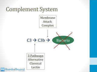
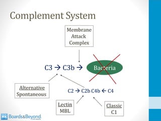
Complement System
C3 →C3b → Bacteria
3 Pathways
Alternative
Classical
Lectin
Membrane
Attack
Complex
130.
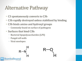
Alternative Pathway
• C3spontaneously converts to C3b
• C3b rapidly destroyed unless stabilized by binding
• C3b binds amino and hydroxyl groups
• Commonly found on surface of pathogens
• Surfaces that bind C3b:
• Bacterial lipopolysaccharides (LPS)
• Fungal cell walls
• Viral envelopes
131.
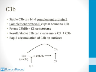
C3b
• Stable C3bcan bind complement protein B
• Complement protein D clips B bound to C3b
• Forms C3bBb = C3 convertase
• Result: Stable C3b can cleave more C3 → C3b
• Rapid accumulation of C3b on surfaces
C3b
(stable)
C3bBb
C3
C3b
B, D
132.
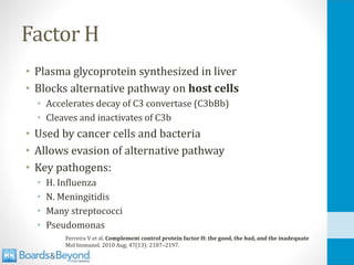
Factor H
• Plasmaglycoprotein synthesized in liver
• Blocks alternative pathway on host cells
• Accelerates decay of C3 convertase (C3bBb)
• Cleaves and inactivates of C3b
• Used by cancer cells and bacteria
• Allows evasion of alternative pathway
• Key pathogens:
• H. Influenza
• N. Meningitidis
• Many streptococci
• Pseudomonas
Ferreira V et al. Complement control protein factor H: the good, the bad, and the inadequate
Mol Immunol. 2010 Aug; 47(13): 2187–2197.
133.
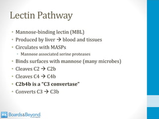
Lectin Pathway
• Mannose-bindinglectin (MBL)
• Produced by liver → blood and tissues
• Circulates with MASPs
• Mannose associated serine proteases
• Binds surfaces with mannose (many microbes)
• Cleaves C2 → C2b
• Cleaves C4 → C4b
• C2b4b is a “C3 convertase”
• Converts C3 → C3b
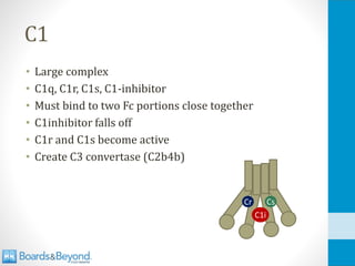
C1
• Large complex
•C1q, C1r, C1s, C1-inhibitor
• Must bind to two Fc portions close together
• C1inhibitor falls off
• C1r and C1s become active
• Create C3 convertase (C2b4b)
Cr Cs
C1i
136.
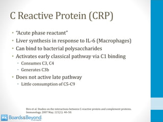
C Reactive Protein(CRP)
• “Acute phase reactant”
• Liver synthesis in response to IL-6 (Macrophages)
• Can bind to bacterial polysaccharides
• Activates early classical pathway via C1 binding
• Consumes C3, C4
• Generates C3b
• Does not active late pathway
• Little consumption of C5-C9
Biro et al. Studies on the interactions between C-reactive protein and complement proteins.
Immunology. 2007 May; 121(1): 40–50.
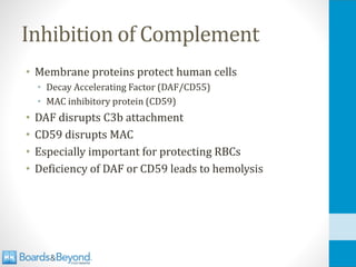
Inhibition of Complement
•Membrane proteins protect human cells
• Decay Accelerating Factor (DAF/CD55)
• MAC inhibitory protein (CD59)
• DAF disrupts C3b attachment
• CD59 disrupts MAC
• Especially important for protecting RBCs
• Deficiency of DAF or CD59 leads to hemolysis
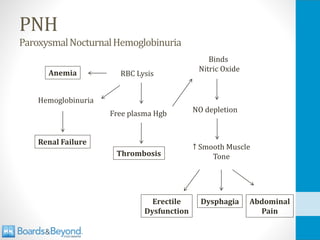
PNH
ParoxysmalNocturnalHemoglobinuria
• Classically causessudden hemolysis at night
• Fatigue, dyspnea (anemia)
• Abdominal pain (smooth muscle tension)
• Thrombosis
• Leading cause of death
• Usually venous clots
• Unusual locations: portal, mesenteric, cerebral veins
145.
Inherited C3 Deficiency
•Recurrent infections encapsulated bacteria
• Pneumococcal and H. flu pneumonia
• Begins in infancy
• Immune complex (IC) deposition
• IC cleared when they bind complement
• Macrophages have complement receptors
• C3 deficiency: glomerulonephritis from IC deposition
• Other type III hypersensitivity syndromes can occur
146.
C5-C9 Deficiency
Terminalcomplementpathwaydeficiency
• LikeC3, impaired defense against encapsulated bugs
• Still have C3a (anaphylatoxin)
• Also have C3b (opsonin for macrophages)
• Recurrent Neisseria infections
• Most often meningitis
147.
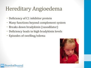
Hereditary Angioedema
• Deficiencyof C1 inhibitor protein
• Many functions beyond complement system
• Breaks down bradykinin (vasodilator)
• Deficiency leads to high bradykinin levels
• Episodes of swelling/edema
148.
Hereditary Angioedema
• Recurrentepisodes swelling without urticaria
• Begins in childhood
• Swelling of skin, GI tract, upper airway
• Airway swelling can be fatal
• Diagnosis: Low C4 level
• Lack of C1 inhibitor
• Consumption of C4
• Can treat with C1 inhibitor concentrate
149.
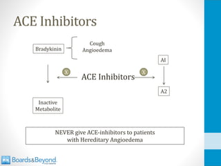
ACE Inhibitors
NEVER giveACE-inhibitors to patients
with Hereditary Angioedema
ACE Inhibitors
X
X
A2
AI
Inactive
Metabolite
Bradykinin
Cough
Angioedema
150.
C3 Nephritic Factor
•Autoantibody
• Stabilizes C3 convertase
• Overactivity of classical pathway
• Found in >80% patients with MPGN II
• Leads to inflammation, hypocomplementemia
151.
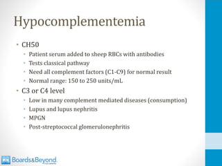
Hypocomplementemia
• CH50
• Patientserum added to sheep RBCs with antibodies
• Tests classical pathway
• Need all complement factors (C1-C9) for normal result
• Normal range: 150 to 250 units/mL
• C3 or C4 level
• Low in many complement mediated diseases (consumption)
• Lupus and lupus nephritis
• MPGN
• Post-streptococcal glomerulonephritis
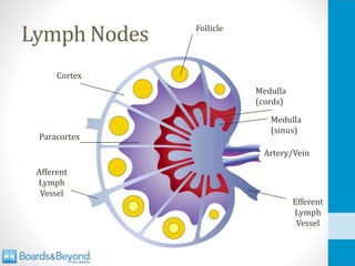
Lymph Nodes
• Lymphfluid drains from site of infection
• Dendritic cells activated
• Express MHC I, MHC II, B7
• Enter lymph carrying processed antigens
• Free antigens also carried with lymph
• Lymph enters nodes
• Many B and T cells waiting for matching antigen
• Dendritic cells present to T cells
• APCs in lymph nodes to process antigen
• B cells react to antigen
• Result: Generation of adaptive immune response
157.
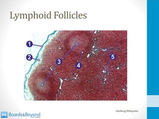
Lymphoid Follicles
• Foundin cortex of lymph nodes
• Site of B-cell activation
• Contain follicular dendritic cells
• Different from tissue dendritic cells
• Permanent cells of lymph nodes
• Surface receptors bind complement-antigen complexes
• Allows easy crosslinking of B cell receptors
• Special note: FDCs important reservoir for HIV
• Early after infection large amounts HIV particles in FDCs
158.
Lymphoid Follicles
• Primaryfollicles
• Inactive follicles
• Follicular dendritic cells and B cells
• Secondary follicles
• “Germinal center”
• B cell growth and differentiation, class switching
• Nearby helper T cells can bind → more growth
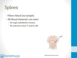
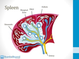
Spleen
• Filters blood(no lymph)
• All blood elements can enter
• No high endothelial venules
• No selective entry T and B cells
Wikipedia/Public Domain
Spleen
• White pulp
•Exposure to B and T cells
• Exposure to macrophages
• Red pulp
• Filters blood in sinusoids
• Removes old RBCs (red)
• Stores many platelets
165.
White Pulp
• Marginalzone
• Macrophages
• Remove debris
• Dendritic cells process antigen
• Follicles
• B cells
• Periarteriolar lymphocyte sheath (PALS)
• T cells
166.
Sinusoids of Spleen
•Red pulp lined by vascular “sinusoids”
• Open endothelium → cells pass in/out
• Capillaries → cords → sinusoids
• Cords contain macrophages (filtration)
167.
Splenic Dysfunction
• Increasedrisk from encapsulated organisms
• Loss of marginal zone macrophages → ↓ phagocytosis
• Also loss of opsonization:
• ↓ IgM and IgG against capsules (splenic B cells)
• Loss of IgG opsonization
• ↓ complement against encapsulated bacteria
• ↓ C3b opsonization
168.
Splenic Dysfunction
• Streppneumo is predominant pathogen for sepsis
• Death in > 50% of patients
• Others: H. flu (Hib), Neisseria meningitidis
• Less common: Strep pyogenes, E coli, Salmonella
• Also malaria and babesia (RBC infections)
Ram e al; Infections of People with Complement Deficiencies and Patients Who Have Undergone Splenectomy
Clin Microbiol Rev. 2010 Oct; 23(4): 740–780.
169.
Splenic Dysfunction
• Splenectomy
•Trauma
• ITP (spleen site of phagocytosis of platelets)
• Hereditary spherocytosis (minimizes anemia)
• Functional asplenia
• Sickle cell anemia
170.
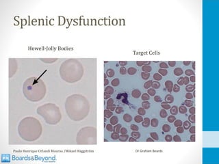
Splenic Dysfunction
• HowellJolly Bodies
• Some RBCs leave marrow with nuclear remnants
• Normally cleared by spleen
• Presence in peripheral blood indicates splenic dysfunction
• Target cells
• Also seen in liver disease, hemoglobin disorders
• From too much surface area (membrane) or too little volume
• Too much surface area: liver disease
• Too little volume: hemoglobin disorders
• Thrombocytosis
• Failure of spleen to remove platelets
Hypersensitivity
• First contactwith antigen “sensitizes”
• Generation of immune response
• Antibodies, Memory cells
• Second contact → hypersensitivity
• Symptoms from overreaction of immune system
• Four patterns of underlying immune response
• Type I, II, III, IV
175.
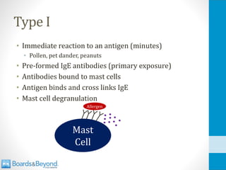
Type I
• Immediatereaction to an antigen (minutes)
• Pollen, pet dander, peanuts
• Pre-formed IgE antibodies (primary exposure)
• Antibodies bound to mast cells
• Antigen binds and cross links IgE
• Mast cell degranulation
Mast
Cell
Allergen
176.
Type I Immunology
•Susceptible individuals make IgE to antigens
• Majority of people make IgG
• IgG does not trigger hypersensitivity response
• IgE results from:
• B cell class switching
• Driven by Th2 cells (humoral response)
• IL-4 is key cytokine for IgE production
• No complement
• IgE does not activate complement
177.
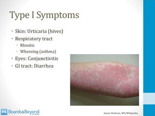
Type I Symptoms
•Skin: Urticaria (hives)
• Respiratory tract
• Rhinitis
• Wheezing (asthma)
• Eyes: Conjunctivitis
• GI tract: Diarrhea
James Heilman, MD/Wikipedia
178.
Anaphylaxis
• Systemic typeI hypersensitivity reaction
• Itching, diffuse hives/erythema
• Respiratory distress from bronchoconstriction
• Hoarseness (laryngeal swelling/edema)
• Vomiting, cramps, diarrhea
• Shock and death
• Treatment: Epinephrine
179.
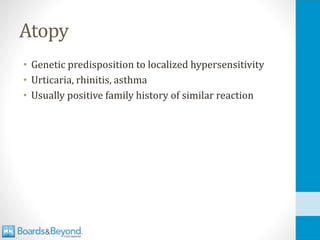
Atopy
• Genetic predispositionto localized hypersensitivity
• Urticaria, rhinitis, asthma
• Usually positive family history of similar reaction
180.
Type I Examples
•Asthma
• Penicillin drug allergy
• Seasonal allergies (allergic rhinitis)
• Allergic conjunctivitis
• Peanut allergy (children)
• Shellfish (food allergy)
181.
Type I
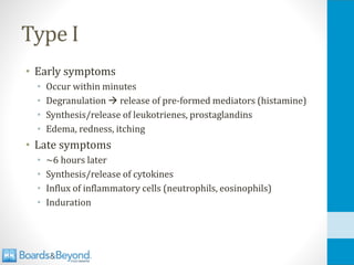
• Earlysymptoms
• Occur within minutes
• Degranulation → release of pre-formed mediators (histamine)
• Synthesis/release of leukotrienes, prostaglandins
• Edema, redness, itching
• Late symptoms
• ~6 hours later
• Synthesis/release of cytokines
• Influx of inflammatory cells (neutrophils, eosinophils)
• Induration
182.
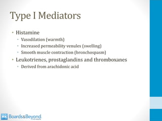
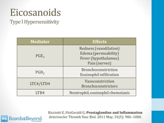
Type I Mediators
•Histamine
• Vasodilation (warmth)
• Increased permeability venules (swelling)
• Smooth muscle contraction (bronchospasm)
• Leukotrienes, prostaglandins and thromboxanes
• Derived from arachidonic acid
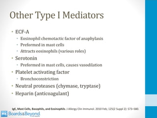
Other Type IMediators
• ECF-A
• Eosinophil chemotactic factor of anaphylaxis
• Preformed in mast cells
• Attracts eosinophils (various roles)
• Serotonin
• Preformed in mast cells, causes vasodilation
• Platelet activating factor
• Bronchoconstriction
• Neutral proteases (chymase, tryptase)
• Heparin (anticoagulant)
IgE, Mast Cells, Basophils, and Eosinophils. J Allergy Clin Immunol. 2010 Feb; 125(2 Suppl 2): S73–S80.
186.
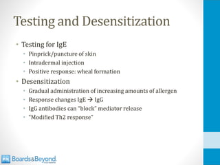
Testing and Desensitization
•Testing for IgE
• Pinprick/puncture of skin
• Intradermal injection
• Positive response: wheal formation
• Desensitization
• Gradual administration of increasing amounts of allergen
• Response changes IgE → IgG
• IgG antibodies can “block” mediator release
• “Modified Th2 response”
187.
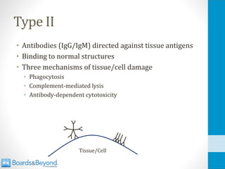
Type II
• Antibodies(IgG/IgM) directed against tissue antigens
• Binding to normal structures
• Three mechanisms of tissue/cell damage
• Phagocytosis
• Complement-mediated lysis
• Antibody-dependent cytotoxicity
Tissue/Cell
188.
Type II
• Phagocytosis
•Fc receptors or C3b receptors on phagocytes
• Complement
• IgM or IgG → classical complement cascade
• Formation of MAC → cell death
• ADCC
• Antibody-dependent cell-mediated cytotoxicity
• Natural killer cells bind Fc portion IgG
189.
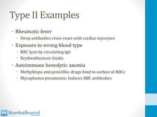
Type II Examples
•Rheumatic fever
• Strep antibodies cross-react with cardiac myocytes
• Exposure to wrong blood type
• RBC lysis by circulating IgG
• Erythroblastosis fetalis
• Autoimmune hemolytic anemia
• Methyldopa and penicillin: drugs bind to surface of RBCs
• Mycoplasma pneumonia: Induces RBC antibodies
190.
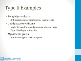
Type II Examples
•Pemphigus vulgaris
• Antibodies against desmosomes in epidermis
• Goodpasture syndrome
• Nephritic syndrome and pulmonary hemorrhage
• Type IV collagen antibodies
• Myasthenia gravis
• Antibodies against Ach receptors
191.
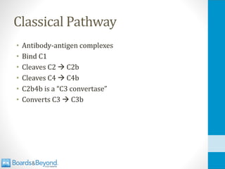
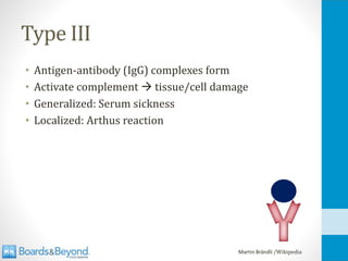
Type III
• Antigen-antibody(IgG) complexes form
• Activate complement → tissue/cell damage
• Generalized: Serum sickness
• Localized: Arthus reaction
Martin Brändli /Wikipedia
192.
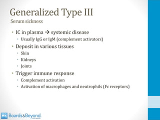
Generalized Type III
Serumsickness
•IC in plasma → systemic disease
• Usually IgG or IgM (complement activators)
• Deposit in various tissues
• Skin
• Kidneys
• Joints
• Trigger immune response
• Complement activation
• Activation of macrophages and neutrophils (Fc receptors)
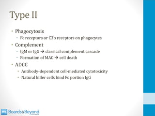
193.
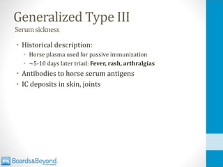
Generalized Type III
Serumsickness
•Historical description:
• Horse plasma used for passive immunization
• ~5-10 days later triad: Fever, rash, arthralgias
• Antibodies to horse serum antigens
• IC deposits in skin, joints
194.
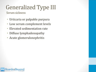
Generalized Type III
Serumsickness
•Urticaria or palpable purpura
• Low serum complement levels
• Elevated sedimentation rate
• Diffuse lymphadenopathy
• Acute glomerulonephritis
195.
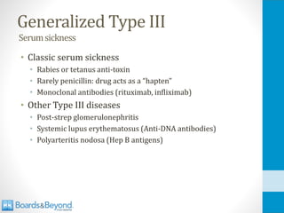
Generalized Type III
Serumsickness
•Classic serum sickness
• Rabies or tetanus anti-toxin
• Rarely penicillin: drug acts as a “hapten”
• Monoclonal antibodies (rituximab, infliximab)
• Other Type III diseases
• Post-strep glomerulonephritis
• Systemic lupus erythematosus (Anti-DNA antibodies)
• Polyarteritis nodosa (Hep B antigens)
196.
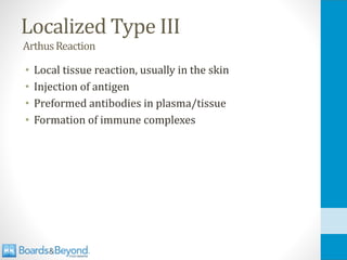
Localized Type III
ArthusReaction
•Local tissue reaction, usually in the skin
• Injection of antigen
• Preformed antibodies in plasma/tissue
• Formation of immune complexes
197.
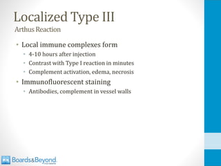
Localized Type III
ArthusReaction
•Local immune complexes form
• 4-10 hours after injection
• Contrast with Type I reaction in minutes
• Complement activation, edema, necrosis
• Immunofluorescent staining
• Antibodies, complement in vessel walls
198.
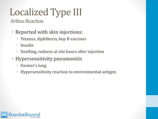
Localized Type III
ArthusReaction
•Reported with skin injections:
• Tetanus, diphtheria, hep B vaccines
• Insulin
• Swelling, redness at site hours after injection
• Hypersensitivity pneumonitis
• Farmer’s lung
• Hypersensitivity reaction to environmental antigen
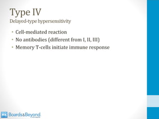
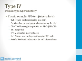
Type IV
Delayed-typehypersensitivity
• Classicexample: PPD test (tuberculosis)
• Tuberculin protein injected into skin
• Previously exposed person has memory T-cells
• CD4 T-cells recognize protein on APCs (MHC II)
• Th1 response
• IFN-γ activates macrophages
• IL-12 from macrophages stimulates Th1 cells
• Result: Redness, induration 24 to 72 hours later
201.
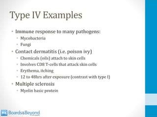
Type IV Examples
•Immune response to many pathogens:
• Mycobacteria
• Fungi
• Contact dermatitis (i.e. poison ivy)
• Chemicals (oils) attach to skin cells
• Involves CD8 T-cells that attack skin cells
• Erythema, itching
• 12 to 48hrs after exposure (contrast with type I)
• Multiple sclerosis
• Myelin basic protein
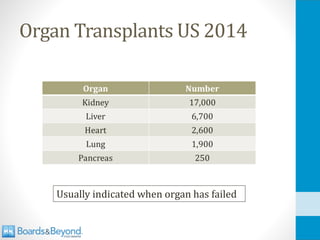
Bone Marrow Transplants
•About 17,000 per year in united states
• Abolish bone marrow with chemotherapy
• Reconstitute all cell lines with donor marrow
• Sometimes autotransplant
• Blood type can change!
205.
Bone Marrow Transplants
•Malignancy (leukemia/lymphoma)
• Inherited red cell disorders
• Pure red cell aplasia, sickle cell disease, beta-thalassemia
• Marrow failure (aplastic anemia, Fanconi anemia)
• Metabolic disorders
• Adrenoleukodystrophy, Gaucher’s disease
• Inherited immune disorders
• Severe combined immunodeficiency, Wiskott-Aldrich
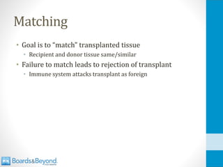
Matching
• Goal isto “match” transplanted tissue
• Recipient and donor tissue same/similar
• Failure to match leads to rejection of transplant
• Immune system attacks transplant as foreign
208.
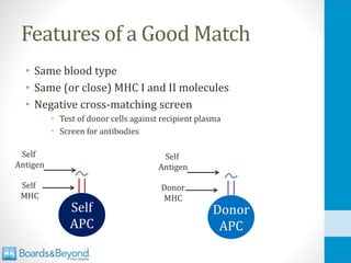
Features of aGood Match
• Same blood type
• Same (or close) MHC I and II molecules
• Negative cross-matching screen
• Test of donor cells against recipient plasma
• Screen for antibodies
Self
APC
Donor
APC
Self
MHC
Self
Antigen
Donor
MHC
Self
Antigen
209.
MHC Matching
• Donorcells express MHC I
• If different from recipient, CD8 cells will react
• MHC Class II also expressed
• Donor APCs may be carried along
• Vascular endothelial cells may express MHC II
210.
Human Leukocyte Antigens
HLAs
•Antigens that make up MHC class I and II molecules
• If different between donor-recipient, immune system
will classify donor tissue as foreign
211.
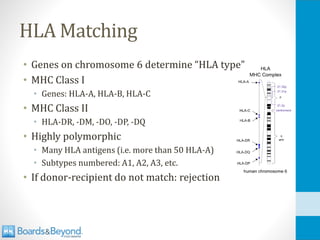
HLA Matching
• Geneson chromosome 6 determine “HLA type”
• MHC Class I
• Genes: HLA-A, HLA-B, HLA-C
• MHC Class II
• HLA-DR, -DM, -DO, -DP, -DQ
• Highly polymorphic
• Many HLA antigens (i.e. more than 50 HLA-A)
• Subtypes numbered: A1, A2, A3, etc.
• If donor-recipient do not match: rejection
212.
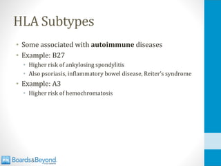
HLA Subtypes
• Someassociated with autoimmune diseases
• Example: B27
• Higher risk of ankylosing spondylitis
• Also psoriasis, inflammatory bowel disease, Reiter’s syndrome
• Example: A3
• Higher risk of hemochromatosis
213.
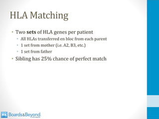
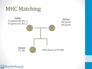
HLA Matching
• Twosets of HLA genes per patient
• All HLAs transferred en bloc from each parent
• 1 set from mother (i.e. A2, B3, etc.)
• 1 set from father
• Sibling has 25% chance of perfect match
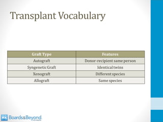
The “Perfect” Match
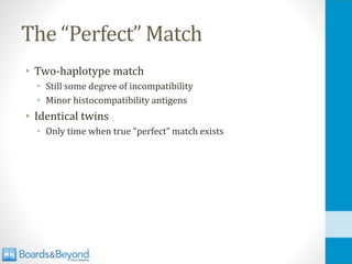
•Two-haplotype match
• Still some degree of incompatibility
• Minor histocompatibility antigens
• Identical twins
• Only time when true “perfect” match exists
216.
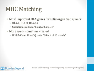
MHC Matching
• Mostimportant HLA genes for solid organ transplants:
• HLA-A, HLA-B, HLA-DR
• Sometimes called a “6 out of 6 match”
• More genes sometimes tested
• If HLA-C and HLA-DQ tests, “10 out of 10 match”
Source: American Society for Histocompatibility and Immunogenetics (ASHI)
217.
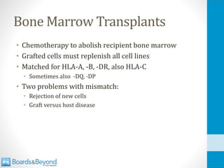
Bone Marrow Transplants
•Chemotherapy to abolish recipient bone marrow
• Grafted cells must replenish all cell lines
• Matched for HLA-A, -B, -DR, also HLA-C
• Sometimes also -DQ, -DP
• Two problems with mismatch:
• Rejection of new cells
• Graft versus host disease
218.
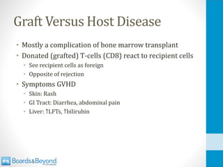
Graft Versus HostDisease
• Mostly a complication of bone marrow transplant
• Donated (grafted) T-cells (CD8) react to recipient cells
• See recipient cells as foreign
• Opposite of rejection
• Symptoms GVHD
• Skin: Rash
• GI Tract: Diarrhea, abdominal pain
• Liver: ↑LFTs, ↑bilirubin
219.
Graft Versus HostDisease
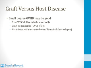
• Small degree GVHD may be good
• New WBCs kill residual cancer cells
• Graft-vs-leukemia (GVL) effect
• Associated with increased overall survival (less relapse)
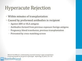
Hyperacute Rejection
• Withinminutes of transplantation
• Caused by preformed antibodies in recipient
• Against ABO or HLA antigens
• Antibodies formed from previous exposure foreign antigens
• Pregnancy, blood transfusion, previous transplantation
• Prevented by cross-matching screen
MULLEY W, KANELLIS J. Understanding crossmatch testing in organ transplantation:
A case-based guide for the general nephrologist. Nephrology 16 (2011) 125–133
222.
Hyperacute Rejection
• Bloodvessels spasm
• Intravascular coagulation
• Ischemia (“white rejection”)
• Rare, usually not treatable
223.
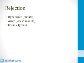
Acute Rejection
• Weeks/monthsafter transplant
• Recipients T cells react to graft (via HLA)
• Cell-mediated immune response
• CD8 T-cells very important
• Biopsy: Infiltrates of lymphocytes/mononuclear cells
• Treatable with immunosuppression
224.
Chronic Rejection
• Monthsor years after transplant
• Inflammation and fibrosis, especially vessels
• Kidneys: fibrosis of capillaries, glomeruli
• Heart: Narrowing coronary arteries
• Lungs: bronchiolitis obliterans
• Complex, incompletely understood process
• Involves cell-mediated and humoral systems
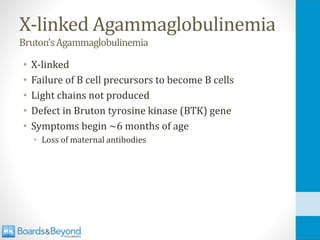
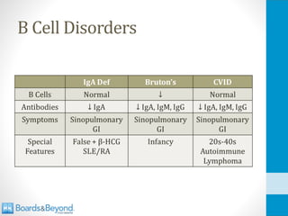
X-linked Agammaglobulinemia
Bruton’sAgammaglobulinemia
• Recurrentrespiratory bacterial infections
• Loss of opsonization by antibodies
• H. Flu, Strep pneumo are common
• Classic presentation: Recurrent otitis media +/- sinusitis/PNA
• GI pathogen infections (loss of IgA)
• Enteroviruses (echo, polio, coxsackie)
• Giardia (GI parasite)
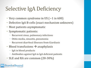
Selective IgA Deficiency
•Very common syndrome in US (~1 in 600)
• Defective IgA B-cells (exact mechanism unknown)
• Most patients asymptomatic
• Symptomatic patients:
• Recurrent sinus, pulmonary infections
• Otitis media, sinusitis, pneumonia
• Recurrent diarrheal illnesses from Giardiasis
• Blood transfusions → anaphylaxis
• IgA in blood products
• Antibodies against IgA in IgA deficient patients
• SLE and RA are common (20-30%)
231.
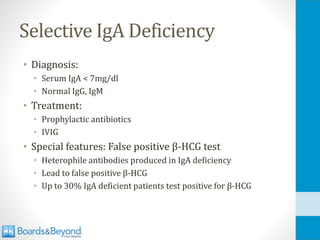
Selective IgA Deficiency
•Diagnosis:
• Serum IgA < 7mg/dl
• Normal IgG, IgM
• Treatment:
• Prophylactic antibiotics
• IVIG
• Special features: False positive β-HCG test
• Heterophile antibodies produced in IgA deficiency
• Lead to false positive β-HCG
• Up to 30% IgA deficient patients test positive for β-HCG
232.
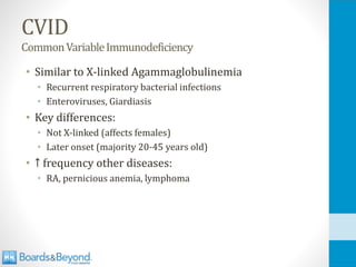
CVID
CommonVariableImmunodeficiency
• Defective Bcell maturation
• Loss of plasma cells and antibodies
• Many underlying genetic causes
• Most cases due to unknown cause
• 10+ genes mutations associated with CVID
• Often sporadic – no family history
• Normal B cell count, absence of antibodies
• Usually IgG
• Sometimes IgA and IgM (variable)
233.
CVID
CommonVariableImmunodeficiency
• Similar toX-linked Agammaglobulinemia
• Recurrent respiratory bacterial infections
• Enteroviruses, Giardiasis
• Key differences:
• Not X-linked (affects females)
• Later onset (majority 20-45 years old)
• ↑ frequency other diseases:
• RA, pernicious anemia, lymphoma
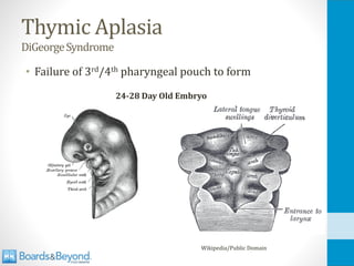
Thymic Aplasia
DiGeorgeSyndrome
• Mostcases: 22q11 chromosomal deletion
• Key point: Not familial
• Classic triad:
• Loss of thymus (Loss of T-cells, recurrent infections)
• Loss of parathyroid glands (hypocalcemia, tetany)
• Congenital heart defects (“conotruncal”)
• Heart Defects:
• Abnormal aortic arch
• Truncus arteriosus
• Tetralogy of Fallot
• ASDs/VSDs
237.
Thymic Aplasia
DiGeorgeSyndrome
• Immunesymptoms
• Recurrent infections
• Viral, fungal, protozoal, intracellular bacteria
• Immune symptoms sometimes improve
• Cleft palate, mandible problems also common
238.
Thymic Aplasia
KeyFindings
• Nothymus shadow on CXR
• Thymus large in newborns
• Faint white shadow on chest x-ray
• Also seen in SCID (without ↓Ca, facial/heart abnormalities)
• Low T-cell count
• Underdeveloped T-cell structures
• Paracortex in lymph nodes
• Peri-arteriolar sheaths in spleen
• Treatment:
• Thymic transplantation
• Hematopoietic cell transplantation
239.
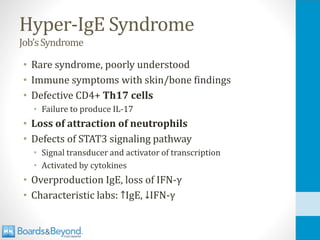
Hyper-IgE Syndrome
Job’sSyndrome
• Raresyndrome, poorly understood
• Immune symptoms with skin/bone findings
• Defective CD4+ Th17 cells
• Failure to produce IL-17
• Loss of attraction of neutrophils
• Defects of STAT3 signaling pathway
• Signal transducer and activator of transcription
• Activated by cytokines
• Overproduction IgE, loss of IFN-γ
• Characteristic labs: ↑IgE, ↓IFN-γ
240.
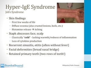
Hyper-IgE Syndrome
Job’sSyndrome
• Skinfindings
• First few weeks of life
• Diffuse eczema (also crusted lesions, boils, etc.)
• Histamine release → itching
• Staph abscesses face, scalp
• Classically “cold” - lacking warmth/redness of inflammation
• Loss of cytokine production
• Recurrent sinusitis, otitis (often without fever)
• Facial deformities (broad nasal bridge)
• Retained primary teeth (two rows of teeth!)
Wikipedia/Public Domain
241.
Hyper-IgE Syndrome
Job’sSyndrome
• Classiccase:
• Newborn baby
• Deformed face/teeth
• Diffuse rash
• Skin abscesses that are “cold”
• Recurrent infections without fever
• Labs: Elevated IgE
242.
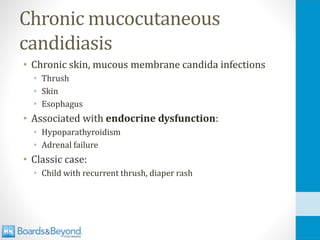
Chronic mucocutaneous
candidiasis
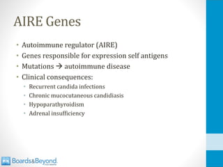
• Defectin autoimmune regulator (AIRE) genes
• AIRE Function #1:
• Associates with Dectin-1 receptor
• Dectin-1 responds to Candida antigens
• Result of defect: Recurrent candida infections
• AIRE Function #2:
• Promotes self antigens production in thymus
• Self antigens presented to T-cells (negative selection)
• Result of defect: Autoimmune T-cells
• Endocrine dysfunction (parathyroid/adrenal)
243.
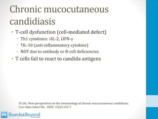
Chronic mucocutaneous
candidiasis
• T-celldysfunction (cell-mediated defect)
• Th1 cytokines: ↓IL-2, ↓IFN-γ
• ↑IL-10 (anti-inflammatory cytokine)
• NOT due to antibody or B-cell deficiencies
• T cells fail to react to candida antigens
D Lilic. New perspectives on the immunology of chronic mucocutaneous candidiasis.
Curr Opin Infect Dis. 2002; 15(2):143-7
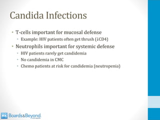
Candida Infections
• T-cellsimportant for mucosal defense
• Example: HIV patients often get thrush (↓CD4)
• Neutrophils important for systemic defense
• HIV patients rarely get candidemia
• No candidemia in CMC
• Chemo patients at risk for candidemia (neutropenia)
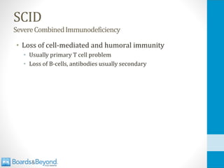
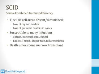
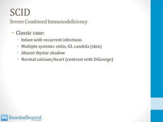
SCID
SevereCombinedImmunodeficiency
• T-cell/B cellareas absent/diminished:
• Loss of thymic shadow
• Loss of germinal centers in nodes
• Susceptible to many infections
• Thrush, bacterial, viral, fungal
• Babies: Thrush, diaper rash, failure to thrive
• Death unless bone marrow transplant
248.
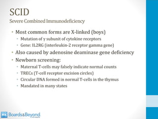
SCID
SevereCombinedImmunodeficiency
• Most commonforms are X-linked (boys)
• Mutation of γ subunit of cytokine receptors
• Gene: IL2RG (interleukin-2 receptor gamma gene)
• Also caused by adenosine deaminase gene deficiency
• Newborn screening:
• Maternal T-cells may falsely indicate normal counts
• TRECs (T-cell receptor excision circles)
• Circular DNA formed in normal T-cells in the thymus
• Mandated in many states
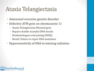
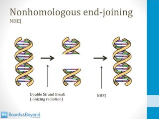
Ataxia Telangiectasia
• Mixof systems involved with varying findings
• CNS (ataxia)
• Skin (telangiectasias)
• Immune system (infections, malignancies)
• Presents in childhood with progressive symptoms
• Usually begins with gait and balance problems
254.
Ataxia Telangiectasia
• Cerebellaratrophy
• Ataxia in 1st year of life
• Telangiectasias
• Dilation of capillary vessels on skin
• Repeated sinus/respiratory infections
• Low levels immunoglobulins, especially IgA and IgG
• High risk of cancer (lymphomas)
• Commonly identified lab abnormalities:
• Most consistent lab finding: ↑AFP
• Low IgA level
255.
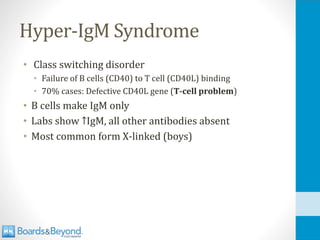
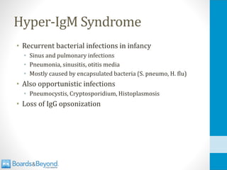
Hyper-IgM Syndrome
• Classswitching disorder
• Failure of B cells (CD40) to T cell (CD40L) binding
• 70% cases: Defective CD40L gene (T-cell problem)
• B cells make IgM only
• Labs show ↑IgM, all other antibodies absent
• Most common form X-linked (boys)
256.
T Cell DependentActivation
B Cell
T Cell MHC2
TCR
CD4
CD40L CD40
CD28
B7
257.
Hyper-IgM Syndrome
• Recurrentbacterial infections in infancy
• Sinus and pulmonary infections
• Pneumonia, sinusitis, otitis media
• Mostly caused by encapsulated bacteria (S. pneumo, H. flu)
• Also opportunistic infections
• Pneumocystis, Cryptosporidium, Histoplasmosis
• Loss of IgG opsonization
258.
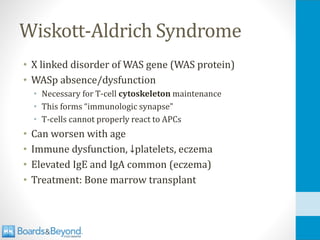
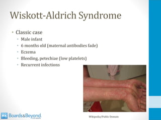
Wiskott-Aldrich Syndrome
• Xlinked disorder of WAS gene (WAS protein)
• WASp absence/dysfunction
• Necessary for T-cell cytoskeleton maintenance
• This forms “immunologic synapse”
• T-cells cannot properly react to APCs
• Can worsen with age
• Immune dysfunction, ↓platelets, eczema
• Elevated IgE and IgA common (eczema)
• Treatment: Bone marrow transplant
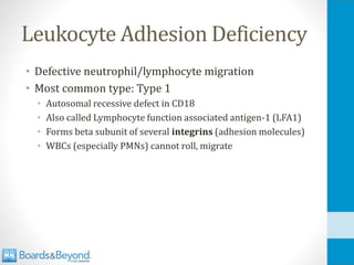
Leukocyte Adhesion Deficiency
•Defective neutrophil/lymphocyte migration
• Most common type: Type 1
• Autosomal recessive defect in CD18
• Also called Lymphocyte function associated antigen-1 (LFA1)
• Forms beta subunit of several integrins (adhesion molecules)
• WBCs (especially PMNs) cannot roll, migrate
261.
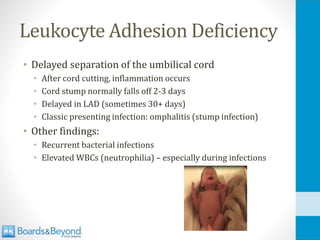
Leukocyte Adhesion Deficiency
•Delayed separation of the umbilical cord
• After cord cutting, inflammation occurs
• Cord stump normally falls off 2-3 days
• Delayed in LAD (sometimes 30+ days)
• Classic presenting infection: omphalitis (stump infection)
• Other findings:
• Recurrent bacterial infections
• Elevated WBCs (neutrophilia) – especially during infections
262.
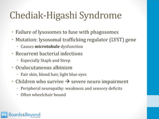
Chediak-Higashi Syndrome
• Failureof lysosomes to fuse with phagosomes
• Mutation: lysosomal trafficking regulator (LYST) gene
• Causes microtubule dysfunction
• Recurrent bacterial infections
• Especially Staph and Strep
• Oculocutaneous albinism
• Fair skin, blond hair, light blue eyes
• Children who survive → severe neuro impairment
• Peripheral neuropathy: weakness and sensory deficits
• Often wheelchair bound
263.
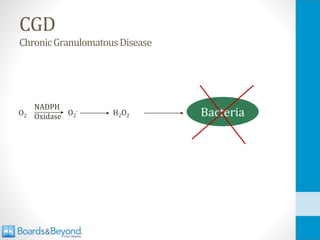
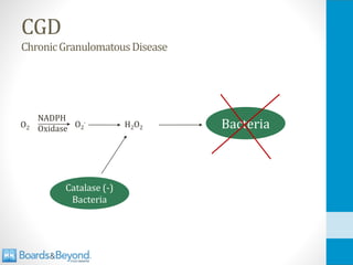
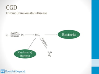
CGD
ChronicGranulomatousDisease
• Loss offunction of NADPH oxidase
• Phagocytes use NADPH oxidase to generate H2O2 from
oxygen (respiratory burst)
• Catalase (-) bacteria generate their own H2O2 which
phagocytes use despite enzyme deficiency
• Catalase (+) bacteria breakdown H2O2
• Host cells have no H2O2 to use → recurrent infections
• Five organisms cause almost all CGD infections:
• Staph aureus, Pseudomonas, Serratia, Nocardia, Aspergillus
Source: UpToDate
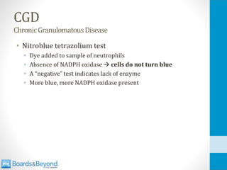
CGD
ChronicGranulomatousDisease
• Nitroblue tetrazoliumtest
• Dye added to sample of neutrophils
• Absence of NADPH oxidase → cells do not turn blue
• A “negative” test indicates lack of enzyme
• More blue, more NADPH oxidase present
Acute Interstitial Nephritis
•Inflammation of “interstitium”
• Space between cells
• Not disease of nephron itself
• Hypersensitivity (allergic) reaction
• Usually triggered by drugs
• Sometimes infections or autoimmune disease
• Classic finding: Urine eosinophils
277.
Acute Interstitial Nephritis
•Classic presentation
• Days to weeks after exposure to typical drug
• Fever, rash
• Oliguria
• Increased BUN/Cr
• Eosinophils in urine
278.
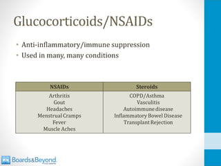
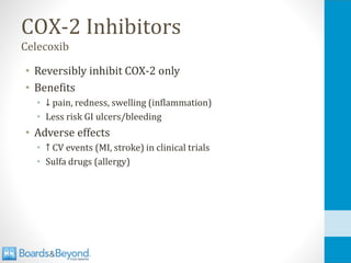
COX-2 Inhibitors
Celecoxib
• Reversiblyinhibit COX-2 only
• Benefits
• ↓ pain, redness, swelling (inflammation)
• Less risk GI ulcers/bleeding
• Adverse effects
• ↑ CV events (MI, stroke) in clinical trials
• Sulfa drugs (allergy)
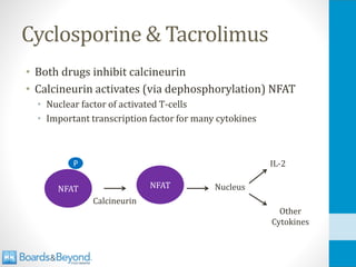
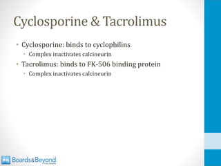
Cyclosporine & Tacrolimus
•Both drugs inhibit calcineurin
• Calcineurin activates (via dephosphorylation) NFAT
• Nuclear factor of activated T-cells
• Important transcription factor for many cytokines
NFAT
P
NFAT
Calcineurin
Nucleus
IL-2
Other
Cytokines
290.
Cyclosporine & Tacrolimus
•Cyclosporine: binds to cyclophilins
• Complex inactivates calcineurin
• Tacrolimus: binds to FK-506 binding protein
• Complex inactivates calcineurin
291.
Cyclosporine & Tacrolimus
•Autoimmune diseases, organ transplants
• Similar side effects
• Both drugs metabolized P450 system
• Many drug-drug interactions
• Can raise/lower levels/effects
292.
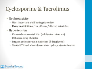
Cyclosporine & Tacrolimus
•Nephrotoxicity
• Most important and limiting side effect
• Vasoconstriction of the afferent/efferent arterioles
• Hypertension
• Via renal vasoconstriction (salt/water retention)
• Diltiazem drug of choice
• Impairs cyclosporine metabolism (↑ drug levels)
• Treats HTN and allows lower dose cyclosporine to be used
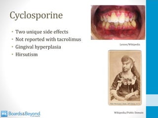
Cyclosporine
• Two uniqueside effects
• Not reported with tacrolimus
• Gingival hyperplasia
• Hirsutism
Lesion/Wikipedia
Wikipedia/Public Domain
295.
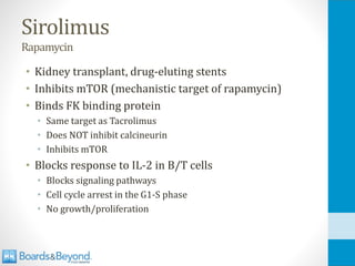
Sirolimus
Rapamycin
• Kidney transplant,drug-eluting stents
• Inhibits mTOR (mechanistic target of rapamycin)
• Binds FK binding protein
• Same target as Tacrolimus
• Does NOT inhibit calcineurin
• Inhibits mTOR
• Blocks response to IL-2 in B/T cells
• Blocks signaling pathways
• Cell cycle arrest in the G1-S phase
• No growth/proliferation
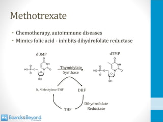
Methotrexate
SideEffects
• Myelosuppression
• Reversiblewith leucovorin (folinic acid )
• Converted to THF
• Does not require dihydrofolate reductase
• “Leucovorin rescue”
• Stomatitis/Mucositis (mouth soreness)
• Occurs with many chemo agents
• DNA damage → cytokine release
• Cytokines damage epithelium
• Loss of mucosal integrity → pain, bacterial growth
• Abnormal LFTs, GI upset
The pathobiology of mucositis. Sonis ST. Nat Rev Cancer. 2004;4(4):277
300.
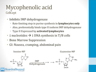
Mycophenolic acid
CellCept
• InhibitsIMP dehydrogenase
• Rate-limiting step in purine synthesis in lymphocytes only
• Also, preferentially binds type II isoform IMP dehydrogenase
• Type II Expressed by activated lymphocytes
• ↓ nucleotides → ↓ DNA synthesis in T/B cells
• Bone Marrow Suppression
• GI: Nausea, cramping, abdominal pain
Inosine-MP Guanosine-MP
IMP
Dehydrogenase
301.
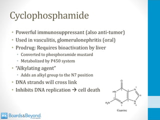
Cyclophosphamide
• Powerful immunosuppressant(also anti-tumor)
• Used in vasculitis, glomerulonephritis (oral)
• Prodrug: Requires bioactivation by liver
• Converted to phosphoramide mustard
• Metabolized by P450 system
• “Alkylating agent”
• Adds an alkyl group to the N7 position
• DNA strands will cross link
• Inhibits DNA replication → cell death
302.
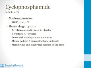
Cyclophosphamide
SideEffects
• Myelosuppression
• ↓WBC,↓Hct, ↓Plt
• Hemorrhagic cystitis
• Acrolein metabolite toxic to bladder
• Hematuria +/- dysuria
• Lower risk with hydration and mesna
• Mesna: sodium 2-mercaptoethane sulfonate
• Mesna binds and inactivates acrolein in the urine
303.
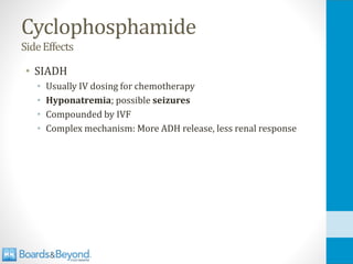
Cyclophosphamide
SideEffects
• SIADH
• UsuallyIV dosing for chemotherapy
• Hyponatremia; possible seizures
• Compounded by IVF
• Complex mechanism: More ADH release, less renal response
304.
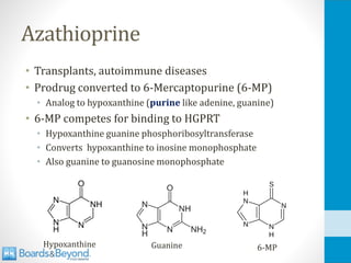
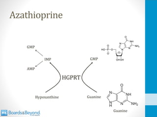
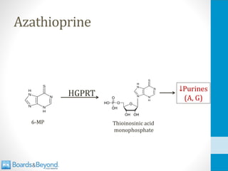
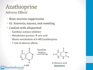
Azathioprine
• Transplants, autoimmunediseases
• Prodrug converted to 6-Mercaptopurine (6-MP)
• Analog to hypoxanthine (purine like adenine, guanine)
• 6-MP competes for binding to HGPRT
• Hypoxanthine guanine phosphoribosyltransferase
• Converts hypoxanthine to inosine monophosphate
• Also guanine to guanosine monophosphate
Hypoxanthine 6-MP
Guanine
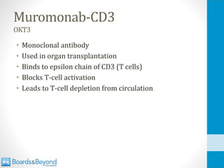
Muromonab-CD3
OKT3
• Monoclonal antibody
•Used in organ transplantation
• Binds to epsilon chain of CD3 (T cells)
• Blocks T-cell activation
• Leads to T-cell depletion from circulation
309.
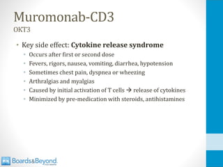
Muromonab-CD3
OKT3
• Key sideeffect: Cytokine release syndrome
• Occurs after first or second dose
• Fevers, rigors, nausea, vomiting, diarrhea, hypotension
• Sometimes chest pain, dyspnea or wheezing
• Arthralgias and myalgias
• Caused by initial activation of T cells → release of cytokines
• Minimized by pre-medication with steroids, antihistamines
310.
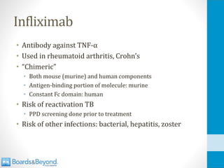
Infliximab
• Antibody againstTNF-α
• Used in rheumatoid arthritis, Crohn’s
• “Chimeric”
• Both mouse (murine) and human components
• Antigen-binding portion of molecule: murine
• Constant Fc domain: human
• Risk of reactivation TB
• PPD screening done prior to treatment
• Risk of other infections: bacterial, hepatitis, zoster
311.
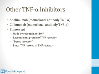
Other TNF-α Inhibitors
•Adalimumab (monoclonal antibody TNF-α)
• Golimumab (monoclonal antibody TNF-α)
• Etanercept
• Made by recombinant DNA
• Recombinant protein of TNF receptor
• “Decoy receptor”
• Binds TNF instead of TNF receptor
312.
Malaria Drugs
• Chloroquineand hydroxychloroquine
• Malaria drugs with immunosuppressive actions
• Block TLRs in B-cells (↓activation)
• Weak bases: ↑pH in immune cells → ↓ activity
• Other actions
• Used in rheumatoid arthritis, SLE
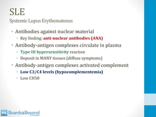
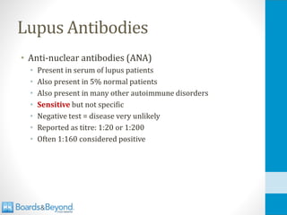
Lupus Antibodies
• Anti-nuclearantibodies (ANA)
• Present in serum of lupus patients
• Also present in 5% normal patients
• Also present in many other autoimmune disorders
• Sensitive but not specific
• Negative test = disease very unlikely
• Reported as titre: 1:20 or 1:200
• Often 1:160 considered positive
318.
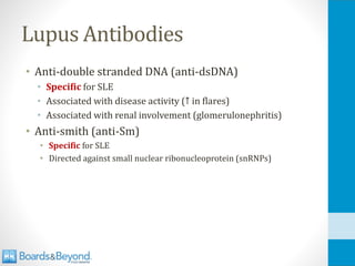
Lupus Antibodies
• Anti-doublestranded DNA (anti-dsDNA)
• Specific for SLE
• Associated with disease activity (↑ in flares)
• Associated with renal involvement (glomerulonephritis)
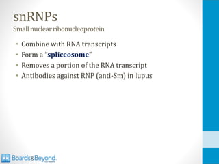
• Anti-smith (anti-Sm)
• Specific for SLE
• Directed against small nuclear ribonucleoprotein (snRNPs)
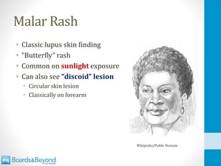
Malar Rash
• Classiclupus skin finding
• “Butterfly” rash
• Common on sunlight exposure
• Can also see “discoid” lesion
• Circular skin lesion
• Classically on forearm
Wikipedia/Public Domain
323.
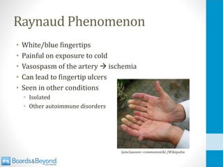
Raynaud Phenomenon
• White/bluefingertips
• Painful on exposure to cold
• Vasospasm of the artery → ischemia
• Can lead to fingertip ulcers
• Seen in other conditions
• Isolated
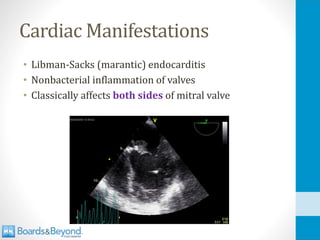
• Other autoimmune disorders
Jamclaassen~commonswiki /Wikipedia
324.
SLE
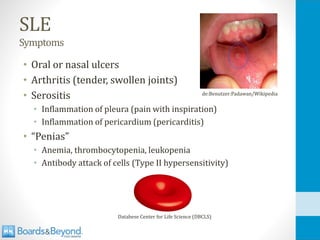
Symptoms
• Oral ornasal ulcers
• Arthritis (tender, swollen joints)
• Serositis
• Inflammation of pleura (pain with inspiration)
• Inflammation of pericardium (pericarditis)
• “Penias”
• Anemia, thrombocytopenia, leukopenia
• Antibody attack of cells (Type II hypersensitivity)
de:Benutzer:Padawan/Wikipedia
Databese Center for Life Science (DBCLS)
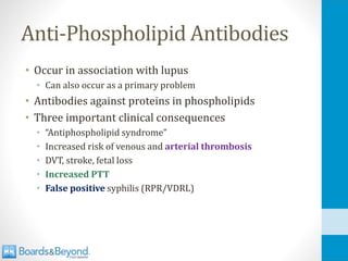
Anti-Phospholipid Antibodies
• Occurin association with lupus
• Can also occur as a primary problem
• Antibodies against proteins in phospholipids
• Three important clinical consequences
• “Antiphospholipid syndrome”
• Increased risk of venous and arterial thrombosis
• DVT, stroke, fetal loss
• Increased PTT
• False positive syphilis (RPR/VDRL)
329.
Anti-Phospholipid Antibodies
• Anti-cardiolipin
•False positive RPR/VDRL
• Syphilis also produces these antibodies
• “Lupus anticoagulant”
• Interferes with PTT test
• False elevation
• Anti-β2 glycoprotein
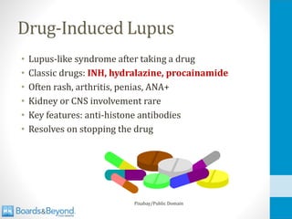
Drug-Induced Lupus
• Lupus-likesyndrome after taking a drug
• Classic drugs: INH, hydralazine, procainamide
• Often rash, arthritis, penias, ANA+
• Kidney or CNS involvement rare
• Key features: anti-histone antibodies
• Resolves on stopping the drug
Pixabay/Public Domain
332.
SLE
Treatment
• Steroids
• Otherimmunosuppressants
• Avoid sunlight
• Many patients photosensitive
• Can trigger flares
• Causes of death
• Renal failure
• Infection (immunosuppression drugs)
• Coronary disease (SLE → increased risk)
Pixabay/Public Domain
333.
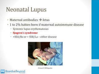
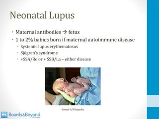
Neonatal Lupus
• Maternalantibodies → fetus
• 1 to 2% babies born if maternal autoimmune disease
• Systemic lupus erythematosus
• Sjogren's syndrome
• +SSA/Ro or + SSB/La – either disease
Ernest F/Wikipedia
334.
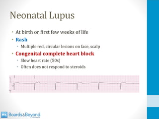
Neonatal Lupus
• Atbirth or first few weeks of life
• Rash
• Multiple red, circular lesions on face, scalp
• Congenital complete heart block
• Slow heart rate (50s)
• Often does not respond to steroids
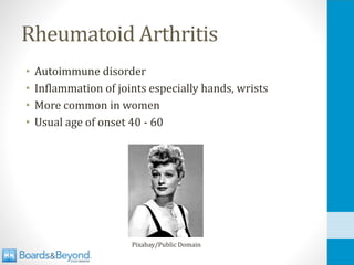
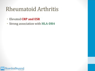
Rheumatoid Arthritis
• Autoimmunedisorder
• Inflammation of joints especially hands, wrists
• More common in women
• Usual age of onset 40 - 60
Pixabay/Public Domain
337.
Rheumatoid Arthritis
• Synovium
•Thin layer of tissue (few cells thick)
• Lines joints and tendon sheaths
• Secretes hyaluronic acid to lubricate joint space
• Inflammation
• Unknown trigger
• Overproduction of TNF and IL-6
338.
Rheumatoid Arthritis
• Synovialhypertrophy
• Thickens into pannus
• Infiltrated with inflammatory cells, granulation tissue
• Increase in synovial fluid
• Erodes into cartilage, bone
• Antibody-mediated
• Type III hypersensitivity
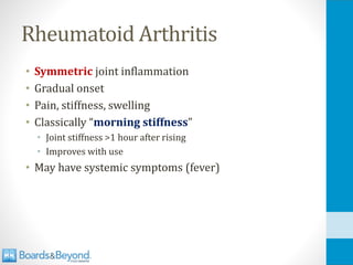
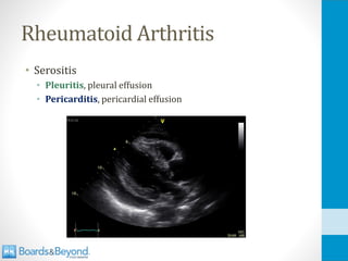
Rheumatoid Arthritis
• Symmetricjoint inflammation
• Gradual onset
• Pain, stiffness, swelling
• Classically “morning stiffness”
• Joint stiffness >1 hour after rising
• Improves with use
• May have systemic symptoms (fever)
341.
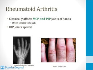
Rheumatoid Arthritis
• Classicallyaffects MCP and PIP joints of hands
• Often tender to touch
• DIP joints spared
James Heilman, MD/Wikipedia
davida__jones/Flikr
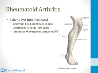
Rheumatoid Arthritis
• Baker'scyst (popliteal cyst)
• Synovium-lined sac at back of knee
• Continuous with the joint space
• If ruptures → symptoms similar to DVT
Wikipedia/Public Domain
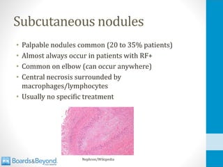
Subcutaneous nodules
• Palpablenodules common (20 to 35% patients)
• Almost always occur in patients with RF+
• Common on elbow (can occur anywhere)
• Central necrosis surrounded by
macrophages/lymphocytes
• Usually no specific treatment
Nephron/Wikipedia
347.
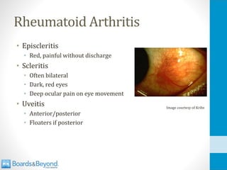
Rheumatoid Arthritis
• Episcleritis
•Red, painful without discharge
• Scleritis
• Often bilateral
• Dark, red eyes
• Deep ocular pain on eye movement
• Uveitis
• Anterior/posterior
• Floaters if posterior
Image courtesy of Kribz
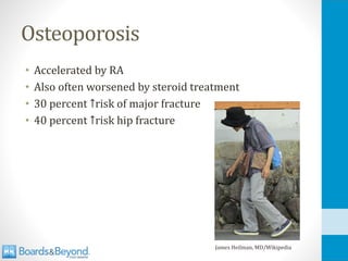
Osteoporosis
• Accelerated byRA
• Also often worsened by steroid treatment
• 30 percent ↑risk of major fracture
• 40 percent ↑risk hip fracture
James Heilman, MD/Wikipedia
350.
Rheumatoid Arthritis
• ~80%positive rheumatoid factor
• Antibodies against Fc portion of IgG antibody
• “Seropositive” rheumatoid arthritis
• Poor specificity
• Positive in endocarditis, Hep B, Hep C
• Positive in Sjogren’s, Lupus
• Antibodies to citrullinated peptides (ACPA)
• Specific marker of RA
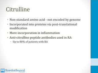
Citrulline
• Non-standard aminoacid - not encoded by genome
• Incorporated into proteins via post-translational
modification
• More incorporation in inflammation
• Anti-citrulline peptide antibodies used in RA
• Up to 80% of patients with RA
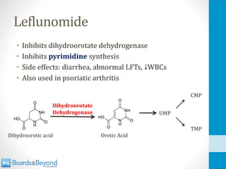
• Inhibits dihydroorotatedehydrogenase
• Inhibits pyrimidine synthesis
• Side effects: diarrhea, abnormal LFTs, ↓WBCs
• Also used in psoriatic arthritis
Dihydroorotate
Dehydrogenase
Leflunomide
UMP
CMP
TMP
Orotic Acid
Dihydroorotic acid
357.
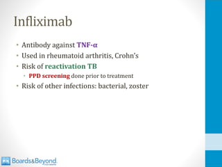
Infliximab
• Antibody againstTNF-α
• Used in rheumatoid arthritis, Crohn’s
• Risk of reactivation TB
• PPD screening done prior to treatment
• Risk of other infections: bacterial, zoster
358.
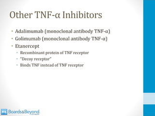
Other TNF-α Inhibitors
•Adalimumab (monoclonal antibody TNF-α)
• Golimumab (monoclonal antibody TNF-α)
• Etanercept
• Recombinant protein of TNF receptor
• “Decoy receptor”
• Binds TNF instead of TNF receptor
359.
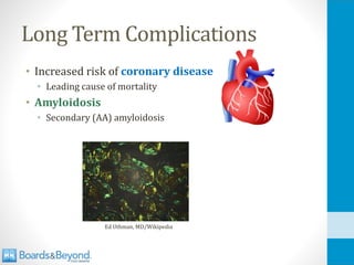
Long Term Complications
•Increased risk of coronary disease
• Leading cause of mortality
• Amyloidosis
• Secondary (AA) amyloidosis
Ed Uthman, MD/Wikipedia
360.
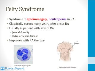
Felty Syndrome
• Syndromeof splenomegaly, neutropenia in RA
• Classically occurs many years after onset RA
• Usually in patient with severe RA
• Joint deformity
• Extra-articular disease
• Improves with RA therapy
Wikipedia/Public Domain
Bob Blaylock/Wikipedia
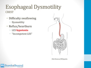
Diffuse Scleroderma
• Diffuseskin thickening
• Raynaud’s phenomenon
• Often initial sign
• Followed ~ 1 year with other signs/symptoms
• Early involvement of visceral organs
• Renal disease – renal failure
• GI tract – dysmotility, heartburn
• Heart: pericarditis, myocarditis, conduction disease
• Joints/muscles: Arthralgia, myalgias
366.
Pulmonary Disease
• Pulmonaryhypertension
• Can progress to right heart failure
• RV heave
• Elevated jugular veins
• Pitting edema
• Routine monitoring: echocardiography
• Interstitial lung disease
367.
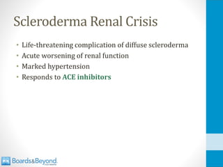
Scleroderma Renal Crisis
•Life-threatening complication of diffuse scleroderma
• Acute worsening of renal function
• Marked hypertension
• Responds to ACE inhibitors
368.
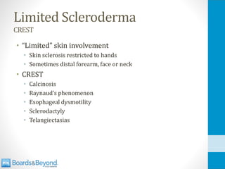
Limited Scleroderma
CREST
• “Limited”skin involvement
• Skin sclerosis restricted to hands
• Sometimes distal forearm, face or neck
• CREST
• Calcinosis
• Raynaud’s phenomenon
• Esophageal dysmotility
• Sclerodactyly
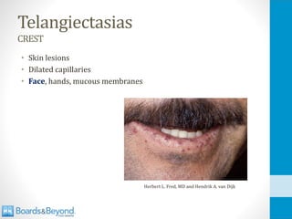
• Telangiectasias
369.
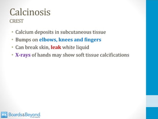
Calcinosis
CREST
• Calcium depositsin subcutaneous tissue
• Bumps on elbows, knees and fingers
• Can break skin, leak white liquid
• X-rays of hands may show soft tissue calcifications
370.
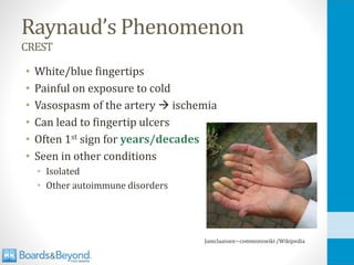
Raynaud’s Phenomenon
CREST
• White/bluefingertips
• Painful on exposure to cold
• Vasospasm of the artery → ischemia
• Can lead to fingertip ulcers
• Often 1st sign for years/decades
• Seen in other conditions
• Isolated
• Other autoimmune disorders
Jamclaassen~commonswiki /Wikipedia
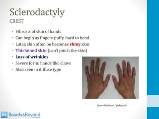
Sclerodactyly
CREST
• Fibrosis ofskin of hands
• Can begin as fingers puffy, hard to bend
• Later, skin often be becomes shiny skin
• Thickened skin (can’t pinch the skin)
• Loss of wrinkles
• Severe form: hands like claws
• Also seen in diffuse type
James Heilman /Wikipedia
Limited Scleroderma
CREST
• Generallymore benign course than diffuse
• Rarely involves heart, kidneys
• Main risk is pulmonary disease
• Leading cause of death
• Pulmonary hypertension
• Interstitial lung disease
• Similar features to diffuse scleroderma
375.
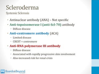
Scleroderma
SystemicSclerosis
• Antinuclear antibody(ANA) – Not specific
• Anti-topoisomerase I (anti-Scl-70) antibody
• Diffuse disease
• Anti-centromere antibody (ACA)
• Limited disease
• CREST = centromere
• Anti-RNA polymerase III antibody
• Diffuse disease
• Associated with rapidly progressive skin involvement
• Also increased risk for renal crisis
376.
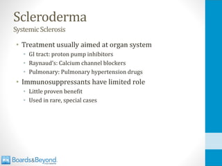
Scleroderma
SystemicSclerosis
• Treatment usuallyaimed at organ system
• GI tract: proton pump inhibitors
• Raynaud’s: Calcium channel blockers
• Pulmonary: Pulmonary hypertension drugs
• Immunosuppressants have limited role
• Little proven benefit
• Used in rare, special cases
377.
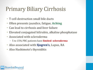
Primary Biliary Cirrhosis
•T-cell destruction small bile ducts
• Often presents jaundice, fatigue, itching
• Can lead to cirrhosis and liver failure
• Elevated conjugated bilirubin, alkaline phosphatase
• Associated with scleroderma
• 5 to 15% PBC patients have limited scleroderma
• Also associated with Sjogren’s, Lupus, RA
• Also Hashimoto’s thyroiditis
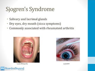
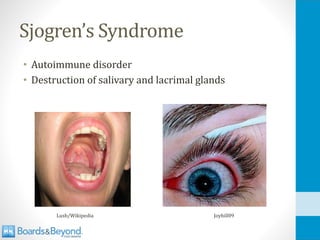
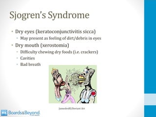
Sjogren’s Syndrome
• Morecommon among women
• Age of onset usually in 40s
• Many elderly patients have “sicca symptoms”
• Dry mouth, dry eyes
• Not due to Sjogren’s
• Antibody tests and/or biopsy = normal
383.
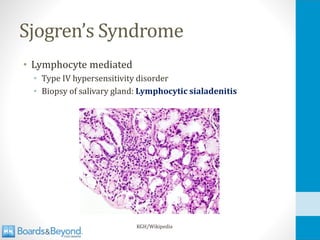
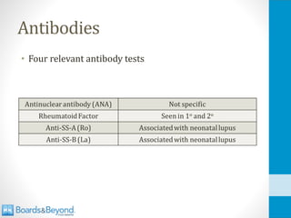
Sjogren’s Syndrome
• Lymphocytemediated
• Type IV hypersensitivity disorder
• Biopsy of salivary gland: Lymphocytic sialadenitis
KGH/Wikipedia
384.
Sjogren’s Syndrome
• Primaryor secondary
• Often associated with rheumatoid arthritis and lupus
• 40-65% of primary biliary cirrhosis patients have Sjögren's
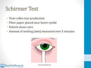
Schirmer Test
• Testsreflex tear production
• Filter paper placed near lower eyelid
• Patient closes eyes
• Amount of wetting (mm) measured over 5 minutes
Jmarchn/Wikipedia
387.
Salivary Testing
• Salivarygland scintigraphy
• Nuclear test
• Low uptake of radionuclide in patients with SS
• Whole sialometry
• Measurement of saliva production
• Patient collects all saliva over 15 minutes
• Sample weighed
388.
Diagnosis
• Any 4of 6 criteria
• Must include either histopathology or autoantibodies
389.
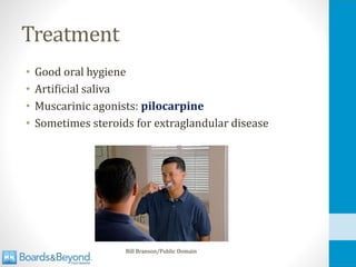
Treatment
• Good oralhygiene
• Artificial saliva
• Muscarinic agonists: pilocarpine
• Sometimes steroids for extraglandular disease
Bill Branson/Public Domain
390.
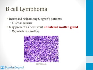
B cell Lymphoma
•Increased risk among Sjogren’s patients
• 5-10% of patients
• May present as persistent unilateral swollen gland
• May mimic past swelling
KGH/Wikipedia
391.
Neonatal Lupus
• Maternalantibodies → fetus
• 1 to 2% babies born if maternal autoimmune disease
• Systemic lupus erythematosus
• Sjögren's syndrome
• +SSA/Ro or + SSB/La – either disease
Ernest F/Wikipedia
392.
Neonatal Lupus
• Atbirth or first few weeks of life
• Rash
• Multiple red, circular lesions on face, scalp
• Congenital complete heart block
• Slow heart rate (50s)
• Often does not respond to steroids
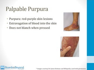
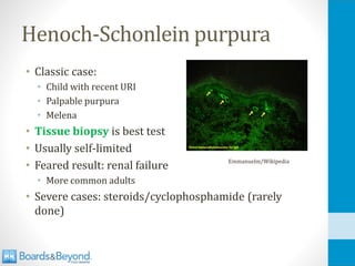
Palpable Purpura
• Purpura:red-purple skin lesions
• Extravagation of blood into the skin
• Does not blanch when pressed
* Images courtesy Dr. James Heilman and Wikipedia; used with permission
397.
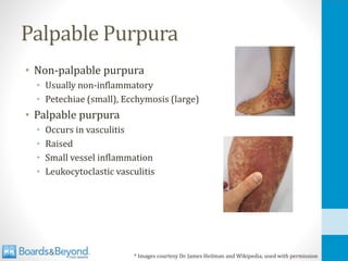
Palpable Purpura
• Non-palpablepurpura
• Usually non-inflammatory
• Petechiae (small), Ecchymosis (large)
• Palpable purpura
• Occurs in vasculitis
• Raised
• Small vessel inflammation
• Leukocytoclastic vasculitis
* Images courtesy Dr. James Heilman and Wikipedia; used with permission
Large Vessel Vasculitis
•Temporal arteritis
• Takayasu’s arteritis
• Granulomatous inflammation
• Narrowing of large arteries
401.
Temporal Arteritis
GiantCellArteritis
• Narrowingof temporal artery system
• Headache, jaw claudication (pain on chewing)
• If not treated → blindness
• Ophthalmic artery occlusion
Wikipedia/Public Domain
402.
Temporal Arteritis
GiantCellArteritis
• HighESR
• Diagnosis: Biopsy temporal artery (granulomas)
• Treat with high dose steroids (don’t wait for biopsy)
• Classic case:
• Elderly female with headache
• Pain on chewing
• High ESR
Dr. Ryan’s Grandmother
403.
Takayasu’s Arteritis
• Granulomatousthickening of aortic arch and branches
• Up to 90% of cases occur in women
• Greatest prevalence in Asia
Public Domain
404.
Takayasu’s Arteritis
• Classicsymptoms: Weak pulses one arm
• "Pulseless disease"
• Proximal great vessels
• BP difference between arms/legs
• Bruits over arteries
• ↑ESR
• Treat with steroids
Wikipedia/Public Domain
405.
Kawasaki Disease
• Autoimmuneattack of medium vessels
• Most cases occur in children
• Classic involvement: skin, lips, tongue
• Diffuse, red rash
• Palms, soles → later desquamates
• Changes in lips/oral mucosa: "strawberry tongue”
• Feared complication: coronary aneurysms
• Rupture → myocardial infarction
• Treatment: IV immunoglobulin and aspirin
AHA Scientific Statement: Diagnosis, Treatment, and Long-Term Management of Kawasaki Disease.
Circulation 2004:110:2747-2771
Scarlet Fever
• Fever,sore throat, diffuse red rash
• Many small papules ("sandpaper" skin)
• Classic finding: Strawberry tongue
• Eventually skin desquamates
Image courtesy of Wikipedia/Public Domain
Image courtesy of Afag Azizova/Wikipedia
409.
Reye’s Syndrome
• Encephalopathy,liver failure, fatty infiltration
• Symptoms: vomiting, confusion, seizures, coma
• Often follows viral illness
• Influenza, varicella
• Caused by diffuse mitochondrial insult
• Associated with aspirin use in children
• Generally, aspirin not used for kids
• Only exception is Kawasaki
410.
Buerger’s Disease
thromboangiitisobliterans
• Malesmokers
• Poor blood flow to hands/feet
• Gangrene
• Autoamputation of digits
• Superficial nodular phlebitis
• Tender nodules over course of a vein
• Raynaud's phenomenon
• Segmental thrombosing vasculitis
• Treatment: Smoking cessation
Dr. James Heilman/Wikipedia
411.
Polyarteritis Nodosa
• Immunecomplex mediated disease: medium vessels
• Type III hypersensitivity reactions
• Classic demographic: Hep B+
• Nerves: Motor/sensory deficits
• Skin: Nodules, purpura
• Kidneys: Renal failure
James Heilman, MD/Wikipedia
James Heilman, MD/Wikipedia
412.
Polyarteritis Nodosa
• Manyaneurysms and constrictions on arteriogram
• Kidney, liver, and mesenteric arteries
• Rosary sign
• Transmural inflammation of medium vessel wall
• Fibrinoid necrosis
413.
Polyarteritis Nodosa
• Classiccase:
• Hep B+, nerve defects, skin nodules, purpura, renal failure
• Diagnosis:
• Angiogram (aneurysms)
• Tissue biopsy of affected system
• Treatment: Corticosteroids, cyclophosphamide
Wikipedia/mprice18
414.
Henoch-Schonlein purpura
• Mostcommon childhood systemic vasculitis
• Often follows URI
• Associated with IgA
• Vasculitis from IgA complex deposition
• IgA nephropathy
• Also C3 deposition
Martin Brändli /Wikipedia
IgA Antibody
415.
Henoch-Schonlein purpura
• Skin:palpable purpura on buttocks/legs
• GI: abdominal pain, melena
• Kidney: Nephritis
Public Domain/Wikipedia
416.
Henoch-Schonlein purpura
• Classiccase:
• Child with recent URI
• Palpable purpura
• Melena
• Tissue biopsy is best test
• Usually self-limited
• Feared result: renal failure
• More common adults
• Severe cases: steroids/cyclophosphamide (rarely
done)
Emmanuelm/Wikipedia
417.
ANCA Diseases
Anti-neutrophilcytoplasmicantibodies
• Churg-Strausssyndrome
• Wegener's Granulomatosis
• Microscopic Polyangiitis
• All have pulmonary involvement
• All have renal involvement
• Crescentic RPGN
• “Pauci-immune”
• Paucity of Ig (negative IF)
• Nephritic syndrome
• Proteinuria, hematuria
Images courtesy of bilalbanday
Daisuke Koya, Kazuyuki Shibuya,
Ryuichi Kikkawa and Masakazu Haneda.
418.
ANCA Diseases
Anti-neutrophilcytoplasmicantibodies
• ANCA
•Autoantibodies
• Attack neutrophil proteins
• Two patterns distinguish diseases
• c-ANCA (cytoplasmic)
• Usually proteinase 3 (PR3) antibodies
• Wegener’s only
• p-ANCA (perinuclear)
• Usually myeloperoxidase (MPO) antibodies
• Churg-Strauss and Microscopic Polyangiitis
Dr Graham Beards/Wikipedia
Microscopic Polyangiitis
• Hemoptysis,kidney failure, purpura
• Just like Wegner’s except
• No upper airway disease (sinusitis)
• p-ANCA not c-ANCA
• No granulomas on biopsy
• Treatment: steroids and cyclophosphamide
422.
Goodpasture’s Syndrome
• Antibodyto collagen (type II hypersensitivity)
• Antibodies to alpha-3 chain of type IV collagen
• Anti-GBM
• Anti-alveoli
• Hemoptysis and nephritic syndrome
• Linear IF (IgG, C3)
• Classic case
• Young adult
• Male
• Hemoptysis
• Hematuria
Images courtesy of bilalbanday