This document describes a case of granuloma annulare developing within the red dye of a tattoo. A 36-year-old man presented with an itchy, inflamed reaction restricted to the red part of a leg tattoo that he had received 10 years prior. A biopsy showed features consistent with granuloma annulare. The reaction was confined to the red areas of the multi-colored tattoo, sparing the black areas and other prior tattoos. This represents a rare case of granuloma annulare developing specifically within the red pigment of an older tattoo.
The DUBLiN Lift: To establish the clinical effectiveness of combining five treatments in the rejuvenation of the ageing face in an effort to increase aesthetic effect, patient safety, and reduce laser downtime.
Minimal Invasive treatment for Aged EyelidsYong Hyun KWON
This is about minimal invasive procedure to lift drooped eyelids.
This presentation was spoken in 3rd Int`l congress of Korea-Japan-China Aesthetic Surgery & Medicine on 25, Oct, 2015
You can watch procedure video on YouTube.
https://youtu.be/lOITt9fk5Bw
Pathological evaluation of melanocytic lesionsHisashi Uhara
In this lecture, the following basic steps by which I routinely scan specimens in our hospital will be presented with examples.
1. Evaluate the specimen preparation.
1) Is the incision for the specimen made perpendicular to the skin surface?
2) Is the slice of tissue from volar skin made perpendicular to the furrows of skin?
2. Estimate the specimen size and location.
1) Estimate the size of the lesion from the magnification of the objective lens.
2) Estimate the specimen location.
3. Precaution before evaluation
1) Observe the specimens without clinical information as much as possible.
2) Obtain as much information as possible at low magnification.
4. The steps for observation
1) At low magnification: Check the symmetric properties and circumscription of the lesion based on the following points.
a. Distance from the densest area of the lesion to both ends.
b. Variation of the thickness of epidermis from the center to both ends.
c. Distribution of melanin in the coronoid layer, epidermis, and dermis.
d. Distribution of nests and distance between each nest.
e. Density of solitary distributed melanocytes.
f. Existence of inflammatory infiltration in the dermis and its distribution.
g. Continuity of the spread of nests and tumor cells in both ends.
h. Is the bottom of the lesion smooth or not?
2) At high magnification: Check the details of tumor cells.
a. Tumor cells in the epidermis: Existence of necrosis, atypia (large nucleolus), or mitosis.
b. Other findings in the epidermis: Distribution of melanin in the cornified layer, the existence of tumor cells in the upper epidermis, the polymorphism of tumor cells, the relationship between tumor cells and keratinocytes.
c. In the dermis: An overlapping, crowded, or sheet-like gathering of tumor cells, maturation of tumor cells, mitotic figures, or melanin of tumor cells at the bottom of the lesion.
d. In the adnexal area: The existence of tumor cells in adnexal walls.
5. After provisionally giving a pathological diagnosis, check discrepancies between the pathological diagnosis and clinical findings. Return to the pathological evaluation if necessary.
Photodynamic therapy in treatment of oral lichen planus: Dr AparnaAparna Srivastava
PHOTODYNAMIC THERAPY is also known as Photoradiation therapy,
Phototherapy,
Photochemotherapy.
Photodynamic therapy (PDT) is a treatment that uses a drug, called a photosensitizer or photosensitizing agent.
Photosensitizers are exposed to a specific wavelength of light, photoactivation causes the formation of singlet oxygen, which produces peroxidative reactions that can cause cell damage and death.
Know more about Psoriasis ,Types and TreatmentsiCliniq
Psoriasis is a prototypic papulosquamous skin
diseases characterised by erythematous papules. It is a chronic inflammatory skin disease with increased epidermal proliferation related to dysregulation of the immune system.
It needs long time medication to get it control, the permanent is not found yet.
To Get guidance to treat Psoriasis from a doctor --> https://www.icliniq.com/ask-a-doctor-online/dermatologist/psoriasis
Atypical Presentation of Orbital Natural Killer cell Lymphoma | Crimson Publi...CrimsonpublishersMSOR
Natural killer (NK)/T-cell lymphoma is a rare malignancy accounting for less than 1 percent of all lymphomas in North America and Europe. Ocular manifestations frequently presented as orbital cellulites that does not improve with adequate antibiotics. We report a case of NK/T cell lymphoma with rare ocular manifestation that was initially confused with orbital cellulites and chronic sinusitis due to multiple negative biopsies. 92-year-old female presented with right eye vision loss, and ipsi lateral orbital swelling for four weeks. MRI demonstrated right intra-orbital extension to the orbital apex. Workups for infection and vasculitis were negative. Multiple biopsies with histopathology and flow cytology were unrevealing for malignancy. Patient failed to improve on antibiotics and steroids. Five months after the initial presentation, patient presented with the same complaint; however, the orbital mass had grown in size and involved the maxillary sinus and contra lateral side. Re-biopsy revealed positive CD 56 for NK lymphoma.
Lichen planus (LP) is a chronic mucocutaneous disorder
of the stratified squamous epithelium that affects oral
and genital mucous membranes, skin, nails, and scalp
Pilomatrixoma of the Arm After IPL Photodepilation Treat-ments Complication(B...semualkaira
Pilomatrixoma calcifying epithelioma of Malherbe is a rare, benign, limited, calcifying epithelial neoplasm that arises from the hair pluripotent precursor matrix cells. Standard treatment for pilomatrixoma is surgical excision. We report a case of a 38-year-old female patient with a rare localisation of pilomatrixoma on the upper extremities following IPL hair removal treatment?s complication
Pilomatrixoma of The Arm After IPL Photodepilation Treatment’s Complication(B...semualkaira
Pilomatrixoma calcifying epithelioma of Malherbe is a rare,
benign, limited, calcifying epithelial neoplasm that arises from
the hair pluripotent precursor matrix cells. It occurs with a rate
of 0.1% among skin tumors. It is observed in the head-neck
region and less frequently in the trunk and extremities
Pilomatrixoma of the Arm After IPL Photodepilation Treat-ments Complication(B...semualkaira
Pilomatrixoma calcifying epithelioma of Malherbe is a rare, benign, limited, calcifying epithelial neoplasm that arises from the hair pluripotent precursor matrix cells. Standard treatment for pilomatrixoma is surgical excision. We report a case of a 38-year-old female patient with a rare localisation of pilomatrixoma on the upper extremities following IPL hair removal treatment?s complication
These lecture slides, by Dr Sidra Arshad, offer a quick overview of physiological basis of a normal electrocardiogram.
Learning objectives:
1. Define an electrocardiogram (ECG) and electrocardiography
2. Describe how dipoles generated by the heart produce the waveforms of the ECG
3. Describe the components of a normal electrocardiogram of a typical bipolar leads (limb II)
4. Differentiate between intervals and segments
5. Enlist some common indications for obtaining an ECG
Study Resources:
1. Chapter 11, Guyton and Hall Textbook of Medical Physiology, 14th edition
2. Chapter 9, Human Physiology - From Cells to Systems, Lauralee Sherwood, 9th edition
3. Chapter 29, Ganong’s Review of Medical Physiology, 26th edition
4. Electrocardiogram, StatPearls - https://www.ncbi.nlm.nih.gov/books/NBK549803/
5. ECG in Medical Practice by ABM Abdullah, 4th edition
6. ECG Basics, http://www.nataliescasebook.com/tag/e-c-g-basics
Explore natural remedies for syphilis treatment in Singapore. Discover alternative therapies, herbal remedies, and lifestyle changes that may complement conventional treatments. Learn about holistic approaches to managing syphilis symptoms and supporting overall health.
Pulmonary Thromboembolism - etilogy, types, medical- Surgical and nursing man...VarunMahajani
Disruption of blood supply to lung alveoli due to blockage of one or more pulmonary blood vessels is called as Pulmonary thromboembolism. In this presentation we will discuss its causes, types and its management in depth.
Title: Sense of Smell
Presenter: Dr. Faiza, Assistant Professor of Physiology
Qualifications:
MBBS (Best Graduate, AIMC Lahore)
FCPS Physiology
ICMT, CHPE, DHPE (STMU)
MPH (GC University, Faisalabad)
MBA (Virtual University of Pakistan)
Learning Objectives:
Describe the primary categories of smells and the concept of odor blindness.
Explain the structure and location of the olfactory membrane and mucosa, including the types and roles of cells involved in olfaction.
Describe the pathway and mechanisms of olfactory signal transmission from the olfactory receptors to the brain.
Illustrate the biochemical cascade triggered by odorant binding to olfactory receptors, including the role of G-proteins and second messengers in generating an action potential.
Identify different types of olfactory disorders such as anosmia, hyposmia, hyperosmia, and dysosmia, including their potential causes.
Key Topics:
Olfactory Genes:
3% of the human genome accounts for olfactory genes.
400 genes for odorant receptors.
Olfactory Membrane:
Located in the superior part of the nasal cavity.
Medially: Folds downward along the superior septum.
Laterally: Folds over the superior turbinate and upper surface of the middle turbinate.
Total surface area: 5-10 square centimeters.
Olfactory Mucosa:
Olfactory Cells: Bipolar nerve cells derived from the CNS (100 million), with 4-25 olfactory cilia per cell.
Sustentacular Cells: Produce mucus and maintain ionic and molecular environment.
Basal Cells: Replace worn-out olfactory cells with an average lifespan of 1-2 months.
Bowman’s Gland: Secretes mucus.
Stimulation of Olfactory Cells:
Odorant dissolves in mucus and attaches to receptors on olfactory cilia.
Involves a cascade effect through G-proteins and second messengers, leading to depolarization and action potential generation in the olfactory nerve.
Quality of a Good Odorant:
Small (3-20 Carbon atoms), volatile, water-soluble, and lipid-soluble.
Facilitated by odorant-binding proteins in mucus.
Membrane Potential and Action Potential:
Resting membrane potential: -55mV.
Action potential frequency in the olfactory nerve increases with odorant strength.
Adaptation Towards the Sense of Smell:
Rapid adaptation within the first second, with further slow adaptation.
Psychological adaptation greater than receptor adaptation, involving feedback inhibition from the central nervous system.
Primary Sensations of Smell:
Camphoraceous, Musky, Floral, Pepperminty, Ethereal, Pungent, Putrid.
Odor Detection Threshold:
Examples: Hydrogen sulfide (0.0005 ppm), Methyl-mercaptan (0.002 ppm).
Some toxic substances are odorless at lethal concentrations.
Characteristics of Smell:
Odor blindness for single substances due to lack of appropriate receptor protein.
Behavioral and emotional influences of smell.
Transmission of Olfactory Signals:
From olfactory cells to glomeruli in the olfactory bulb, involving lateral inhibition.
Primitive, less old, and new olfactory systems with different path
Ethanol (CH3CH2OH), or beverage alcohol, is a two-carbon alcohol
that is rapidly distributed in the body and brain. Ethanol alters many
neurochemical systems and has rewarding and addictive properties. It
is the oldest recreational drug and likely contributes to more morbidity,
mortality, and public health costs than all illicit drugs combined. The
5th edition of the Diagnostic and Statistical Manual of Mental Disorders
(DSM-5) integrates alcohol abuse and alcohol dependence into a single
disorder called alcohol use disorder (AUD), with mild, moderate,
and severe subclassifications (American Psychiatric Association, 2013).
In the DSM-5, all types of substance abuse and dependence have been
combined into a single substance use disorder (SUD) on a continuum
from mild to severe. A diagnosis of AUD requires that at least two of
the 11 DSM-5 behaviors be present within a 12-month period (mild
AUD: 2–3 criteria; moderate AUD: 4–5 criteria; severe AUD: 6–11 criteria).
The four main behavioral effects of AUD are impaired control over
drinking, negative social consequences, risky use, and altered physiological
effects (tolerance, withdrawal). This chapter presents an overview
of the prevalence and harmful consequences of AUD in the U.S.,
the systemic nature of the disease, neurocircuitry and stages of AUD,
comorbidities, fetal alcohol spectrum disorders, genetic risk factors, and
pharmacotherapies for AUD.
ARTIFICIAL INTELLIGENCE IN HEALTHCARE.pdfAnujkumaranit
Artificial intelligence (AI) refers to the simulation of human intelligence processes by machines, especially computer systems. It encompasses tasks such as learning, reasoning, problem-solving, perception, and language understanding. AI technologies are revolutionizing various fields, from healthcare to finance, by enabling machines to perform tasks that typically require human intelligence.
New Directions in Targeted Therapeutic Approaches for Older Adults With Mantl...i3 Health
i3 Health is pleased to make the speaker slides from this activity available for use as a non-accredited self-study or teaching resource.
This slide deck presented by Dr. Kami Maddocks, Professor-Clinical in the Division of Hematology and
Associate Division Director for Ambulatory Operations
The Ohio State University Comprehensive Cancer Center, will provide insight into new directions in targeted therapeutic approaches for older adults with mantle cell lymphoma.
STATEMENT OF NEED
Mantle cell lymphoma (MCL) is a rare, aggressive B-cell non-Hodgkin lymphoma (NHL) accounting for 5% to 7% of all lymphomas. Its prognosis ranges from indolent disease that does not require treatment for years to very aggressive disease, which is associated with poor survival (Silkenstedt et al, 2021). Typically, MCL is diagnosed at advanced stage and in older patients who cannot tolerate intensive therapy (NCCN, 2022). Although recent advances have slightly increased remission rates, recurrence and relapse remain very common, leading to a median overall survival between 3 and 6 years (LLS, 2021). Though there are several effective options, progress is still needed towards establishing an accepted frontline approach for MCL (Castellino et al, 2022). Treatment selection and management of MCL are complicated by the heterogeneity of prognosis, advanced age and comorbidities of patients, and lack of an established standard approach for treatment, making it vital that clinicians be familiar with the latest research and advances in this area. In this activity chaired by Michael Wang, MD, Professor in the Department of Lymphoma & Myeloma at MD Anderson Cancer Center, expert faculty will discuss prognostic factors informing treatment, the promising results of recent trials in new therapeutic approaches, and the implications of treatment resistance in therapeutic selection for MCL.
Target Audience
Hematology/oncology fellows, attending faculty, and other health care professionals involved in the treatment of patients with mantle cell lymphoma (MCL).
Learning Objectives
1.) Identify clinical and biological prognostic factors that can guide treatment decision making for older adults with MCL
2.) Evaluate emerging data on targeted therapeutic approaches for treatment-naive and relapsed/refractory MCL and their applicability to older adults
3.) Assess mechanisms of resistance to targeted therapies for MCL and their implications for treatment selection
HOT NEW PRODUCT! BIG SALES FAST SHIPPING NOW FROM CHINA!! EU KU DB BK substit...GL Anaacs
Contact us if you are interested:
Email / Skype : kefaya1771@gmail.com
Threema: PXHY5PDH
New BATCH Ku !!! MUCH IN DEMAND FAST SALE EVERY BATCH HAPPY GOOD EFFECT BIG BATCH !
Contact me on Threema or skype to start big business!!
Hot-sale products:
NEW HOT EUTYLONE WHITE CRYSTAL!!
5cl-adba precursor (semi finished )
5cl-adba raw materials
ADBB precursor (semi finished )
ADBB raw materials
APVP powder
5fadb/4f-adb
Jwh018 / Jwh210
Eutylone crystal
Protonitazene (hydrochloride) CAS: 119276-01-6
Flubrotizolam CAS: 57801-95-3
Metonitazene CAS: 14680-51-4
Payment terms: Western Union,MoneyGram,Bitcoin or USDT.
Deliver Time: Usually 7-15days
Shipping method: FedEx, TNT, DHL,UPS etc.Our deliveries are 100% safe, fast, reliable and discreet.
Samples will be sent for your evaluation!If you are interested in, please contact me, let's talk details.
We specializes in exporting high quality Research chemical, medical intermediate, Pharmaceutical chemicals and so on. Products are exported to USA, Canada, France, Korea, Japan,Russia, Southeast Asia and other countries.
Couples presenting to the infertility clinic- Do they really have infertility...Sujoy Dasgupta
Dr Sujoy Dasgupta presented the study on "Couples presenting to the infertility clinic- Do they really have infertility? – The unexplored stories of non-consummation" in the 13th Congress of the Asia Pacific Initiative on Reproduction (ASPIRE 2024) at Manila on 24 May, 2024.
- Video recording of this lecture in English language: https://youtu.be/lK81BzxMqdo
- Video recording of this lecture in Arabic language: https://youtu.be/Ve4P0COk9OI
- Link to download the book free: https://nephrotube.blogspot.com/p/nephrotube-nephrology-books.html
- Link to NephroTube website: www.NephroTube.com
- Link to NephroTube social media accounts: https://nephrotube.blogspot.com/p/join-nephrotube-on-social-media.html
Hemodialysis: Chapter 3, Dialysis Water Unit - Dr.Gawad
medical exam
1. Lichen planus can affect any part of the body surface, but is most
often seen on the volar aspect of the wrists, the lumbar region and
around the ankles. Flexural sites like axillae, groins and inframam-
mary regions may be rarely involved in typical lichen planus. Reports
on flexural LP in the published work are either associated with LP
pigmentosus or erosive variants.1,2
In most LP cases, the papule
lesions eventually flatten after a few months, often to be replaced by
an area of pigmentation that retains the shape of the papule and
persists for months or years. In this case, we can see three kinds of
different period lesions (violaceous-brown papules, papules with
the pitchy edge and annular dark brownish macules) on the flexural
sites representing the gradual regression.
Follicular lesions usually appear during the course of typical LP,
sometimes as sole manifestation of the disease in the scalp. But
they rarely occur in flexural LP. Gunduz et al.3
reported the first case
of combination of follicular and flexural variants of LP. But there was
a little difference between the two cases because the follicular
lesions localized to the flexures and the waist, respectively.
The infiltrating cells in LP are predominantly T-lymphocytes with
very few B-lymphocytes. The identification of various subtypes of
T-lymphocytes has given contradictory results with regards to the
predominance of CD4+
helper-inducer T-lymphocytes and CD8+
suppressor-cytotoxic T-lymphocytes in the infiltrate. It is likely that
both subsets participate in the immunological reaction.4
Our
immunohistochemical study demonstrated the same result and it
was easy to distinguish with LP-like keratosis because CD4+
lymphocytes were abundant in the dermis as Jang et al.5
observed. Contrasted to the flexural lesion, the follicular lesion
of the waist was characterized by a higher CD4 ⁄ CD8 ratio of
T-lymphocytes.
Our patient did not use any special treatment in the 6-month
course of disease, but we can see the submammary and groin
lesions are undergoing progressive spontaneous regression.
We conclude that the process is benign and tends to resolve
spontaneously. We are now following up the patient without admin-
istering any special treatment.
Han MA, Lei GUAN, Xiang-yang SU, Wei LAI,
Chun LU
Department of Dermatology, The Third Affiliated Hospital of Sun Yat-sen
University, Guangzhou, Guangdong, China
REFERENCES
1 Pock L, Jelinkova L, Drlik L et al. Lichen planus pigmentosus-inversus.
J Eur Acad Dermatol Venereol 2001; 15: 452–454.
2 Eisman S, Orteu CH. Recalcitrant erosive flexural lichen planus, successful
treatment with a combination of thalidomide and 0.1% tacrolimus ointment.
Clin Exp Dermatol 2004; 29: 268–270.
3 Gunduz K, Sacar T, Inanir I et al. Flexural follicular lichen planus. Clin Exp
Dermatol 2009; 34: 297–298.
4 Elder DE, Elenitsas R, Johnson BL et al. Lever’s Histopathology of the Skin.
In: Narciss M, Sonia T, Hideko K, eds. Noninfectious Erythematous, Papu-
lar, and Squamous Diseases, 9nd edn. Philadelphia: Lippincott Williams &
Wilkins Press, 2005; 198–199.
5 Jang KA, Kim SH, Choi JH et al. Lichenoid keratosis: a clinicopathologic
study of 17 patients. J Am Acad Dermatol 2000; 43: 511–516.
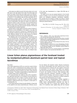
Linear lichen planus pigmentosus of the forehead treated
by neodymium:yttrium–aluminum–garnet laser and topical
tacrolimus
Dear Editor,
Lichen planus pigmentosus (LPP) is an uncommon variant of lichen
planus (LP), which is characterized by the insidious onset of dark-
brown macules in sun-exposed areas and flexural folds. It was origi-
nally reported from India but it tends to occur also in other racial and
ethnic groups.1,2
The pattern of pigmentation is mostly diffuse and it
can present rarely in linear or follicular pattern.3
A 61-year-old Korean man visited our clinic with an asymptom-
atic, pigmented linear lesion on his forehead. He was working as a
security guard and had no history of trauma and there was no
preceding erythema or scaly skin eruption. Examination revealed
ill-defined, mottled brownish array of macules in a linear patch on
his forehead (Fig. 1a).
A skin biopsy specimen showed vacuolar alteration of basal cells
of the epidermis and dense lymphohistiocytic infiltrates around
the hair follicles with apoptotic bodies. Pigment incontinence
and dermal melanophages were also observed (Fig. 2). Direct
immunofluorescence of lesional skin was negative. At the time
of examination, he had chronic renal failure and diabetes,
with abnormal laboratory findings including blood urea nitrogen
37 mg ⁄ dL and creatinine 2.3 mg ⁄ dL. Other laboratory tests,
including antinuclear antibodies, antibodies to dsDNA, and
hepatitis B and C serology, were normal or negative. For treat-
ment, he had been taking an angiotensin II receptor antagonist,
beta-blocker and sulfonylurea for over 10 years.
These clinical and histopathological findings suggested a
diagnosis of LPP with overlapping features of follicular LP.
Initially, topical methylprednisolone aceponate was used for
3 months twice daily but it showed poor response. After a
while, 0.1% tacrolimus cream applied along with low fluenced
Correspondence: Mi-Woo Lee, M.D., Ph.D., Department of Dermatology, Asan Medical Center, University of Ulsan College of Medicine, 388-1
Pungnapdong, Songpagu, 138-736 Seoul, Korea. Email: dermakim@gmail.com
Conflict of interest: None.
Letters to the Editor
Ó 2011 Japanese Dermatological Association 189
2. (1.8 J ⁄ cm2
) 1064-nm Q-switched neodymium:yttrium–aluminum–
garnet laser (QSNY) (Spectra VRM; Lutronic, Gyeonggi, South
Korea) every 3 weeks. Six weeks later, the lesions were much
lighter and after 4 months, the lesion had cleared without a scar
and there has been no evidence of recurrence for over 6 months
(Fig. 1b–d).
(a) (b)
(c) (d)
Figure 1. Photographs showing (a) brownish linear patch on the forehead before treatment, (b) partial improvement after 6 weeks of treatment
with a 0.1% topical tacrolimus and 1064-nm Q-switched neodymium:yttrium–aluminum–garnet laser, (c) resolution without a scar after 4 months
of treatment and (d) maintenance without recurrence 6 months after ceasing treatment.
(a) (b)
Figure 2. Histopathology showing epidermal basal cell vacuolar degeneration, lymphohistiocytic infiltration and basal cell liquefaction around
the hair follicle, as well as pigment incontinence (hematoxylin–eosin, original magnifications: [a] ·100, [b] ·200).
Letters to the Editor
190 Ó 2011 Japanese Dermatological Association
3. The histopathological changes of LPP consisted of vacuolar
degeneration of the basal layer in the epidermis. In the dermis, peri-
vascular or lichenoid infiltrate and the presence of melanin inconti-
nence were the predominant changes noted. A recently developed
lesion tends to show more predominant band-like lymphocytic
infiltration and epidermal vacuolization rather than epidermal
atrophy.3,4
Linear lesions can frequently occur at sites of scratching or
trauma in patients with LP as a result of Koebner’s phenomenon, or,
as in our case, they may appear spontaneously within the lines of
Blaschko on the face.5
In acquired Blaschko linear inflammatory
dermatosis, cutaneous antigenic mosaicism could be responsible
for the susceptibility to induce mosaic T-cell responses. Because
drugs had not been changed in type or dosage over several years
of treatment, and underlying medical diseases had been well con-
trolled, the possibility of drug-related reaction was thought to be
low. Considering the clinical features in our patient, and the fact
that exposed sites were frequently the first to be involved, it can be
suggested that exposure to sunlight (even in a casual dose) may be
a kind of stimuli to induce the lesion of LPP in a genetically suscepti-
ble patient.4
Usually the course is chronic and treatments are less effective
for follicular LP or LPP than for classical LP.3–7
Topical tacrolimus,
a member of the immunosuppressive macrolide family that sup-
presses T-cell activation, has been shown to be effective in the
treatment of some mucosal and follicular LP.3,6,7
There is only one
article about the successful treatment of LPP with topical tacroli-
mus.3
Although they showed over 50% improvement in seven of
13 patients after 4 months of treatment, the authors did not men-
tion any case of complete clearance in their article. Moreover,
the other six of the 13 patients did not show improvement in
pigmentation.
Therefore, in the present case, 1064-nm QSNY with low fluence
treatment was chosen for treating pigmentation. The 1064-nm
QSNY in nanosecond (ns) domain is strongly absorbed by the
finely distributed melanin in dermal pigmented lesions. Moreover,
1064-nm QSNY with low fluence, which in a ‘‘top-hat’’ beam
mode can evenly distribute energy density throughout the whole
spot, is now widely used when treating darker skin types, because
it greatly reduces the risk of epidermal injury and post-therapy
dyschromia.8,9
In our patient, because of poor response to topical
steroid, we started tacrolimus ointment for mainly targeting T cells,
and for the treatment of pigmentation, we added QSNY treatment.
It suggests that the combination treatment of 1064-nm low flu-
enced QSNY with topical tacrolimus may be a good therapeutic
option for patients with recalcitrant facial LPP in dark-skinned
individuals.
Jeong-Eun KIM, Chong-Hyun WON,
Sungeun CHANG, Mi-Woo LEE, Jee-Ho CHOI,
Kee-Chan MOON
Department of Dermatology, Asan Medical Center,
University of Ulsan College of Medicine, Seoul, Korea
REFERENCES
1 Bhutani LK, Bedi TR, Pandhi RK, Nayak NC. Lichen planus pigmentosus.
Dermatologica 1974; 149: 43–50.
2 Kanwar AJ, Kaur S. Lichen planus pigmentosus. J Am Acad Dermatol
1989; 21: 815.
3 Al-Mutairi N, El-Khalawany M. Clinicopathological characteristics of lichen
planus pigmentosus and its response to tacrolimus ointment: an open
label, non-randomized, prospective study. J Eur Acad Dermatol Venereol
2010; 24: 535–540.
4 Kanwar AJ, Dogra S, Handa S et al. A study of 124 Indian patients with
lichen planus pigmentosus. Clin Exp Dermatol 2003; 28: 481–485.
5 Ezzedine K, Simonart T, Vereecken P et al. Facial actinic lichen planus
following the Blaschko’s line: successful treatment with topical 0.1%
pimecrolimus cream. J Eur Acad Dermatol Venereol 2009; 23: 458–
459.
6 Volz T, Caroli U, Ludtke H et al. Pimecrolimus cream 1% in erosive oral
lichen planus – a prospective randomized double-blind vehicle-controlled
study. Br J Dermatol 2008; 159: 936–941.
7 Blazek C, Megahed M. Lichen planopilaris. Successful treatment with
tacrolimus. Hautarzt 2008; 59: 874–877.
8 Cho SB, Park SJ, Kim SJ et al. Treatment of post-inflammatory hyperpig-
mentation using 1064-nm Q-switched Nd:YAG laser with low fluence:
report of three cases. J Eur Acad Dermatol Venereol 2009; 23: 1206–
1207.
9 Kim JH, Kim H, Park HC et al. Subcellular selective photothermolysis of
melanosomes in adult zebrafish skin following 1064-nm Q-switched
Nd:YAG laser irradiation. J Invest Dermatol 2010; 130: 2333–2335.
Granuloma annulare within the red dye of a tattoo
Dear Editor,
Eczematous, lymphohistiocytic, lichenoid, granulomatous, sarcoid-
osis-like or pseudolymphomatous reactions may occur in tattoos
with highly variable delays.1,2
Granuloma annulare (GA) is a com-
mon dermatosis that has seldom been reported in tattoos.3–5
We
report a new case within the red dye of a tattoo.
A 36-year-old otherwise healthy Caucasian man was referred for
an inflamed and infiltrated itchy reaction restricted to the red part of
a leg tattoo that had developed 6 months earlier. He had been tat-
tooed on three different occasions without any complication. One
tattoo depicting a dragon was performed on the left lower leg in
2000 in a tattoo parlor. The healing phase had been unremarkable.
Ten years later, he developed an itchy infiltrated reaction restricted
to its red part (Fig. 1). The rest of the tattoo was spared, as were
two prior black-colored tattoos. Examination was otherwise
unremarkable with no sign of systemic disease. A 3-mm punch
biopsy of the inflamed tattoo revealed large areas of necrobiosis
surrounded by a heavy interstitial and perivascular inflammatory
Correspondence: Nicolas Kluger, M.D., Departments of Dermatology, Allergology and Venereology, Institute of Clinical Medicine, University of
Helsinki, Helsinki University Central Hospital, Meilahdentie 2, PO Box 160, 00029 HUS, Finland. Email: nicolaskluger@yahoo.fr
Letters to the Editor
Ó 2011 Japanese Dermatological Association 191
4. Copyright of Journal of Dermatology is the property of Wiley-Blackwell and its content may not be copied or
emailed to multiple sites or posted to a listserv without the copyright holder's express written permission.
However, users may print, download, or email articles for individual use.
![(1.8 J ⁄ cm2
) 1064-nm Q-switched neodymium:yttrium–aluminum–
garnet laser (QSNY) (Spectra VRM; Lutronic, Gyeonggi, South
Korea) every 3 weeks. Six weeks later, the lesions were much
lighter and after 4 months, the lesion had cleared without a scar
and there has been no evidence of recurrence for over 6 months
(Fig. 1b–d).
(a) (b)
(c) (d)
Figure 1. Photographs showing (a) brownish linear patch on the forehead before treatment, (b) partial improvement after 6 weeks of treatment
with a 0.1% topical tacrolimus and 1064-nm Q-switched neodymium:yttrium–aluminum–garnet laser, (c) resolution without a scar after 4 months
of treatment and (d) maintenance without recurrence 6 months after ceasing treatment.
(a) (b)
Figure 2. Histopathology showing epidermal basal cell vacuolar degeneration, lymphohistiocytic infiltration and basal cell liquefaction around
the hair follicle, as well as pigment incontinence (hematoxylin–eosin, original magnifications: [a] ·100, [b] ·200).
Letters to the Editor
190 Ó 2011 Japanese Dermatological Association](data:image/gif;base64,R0lGODlhAQABAIAAAAAAAP///yH5BAEAAAAALAAAAAABAAEAAAIBRAA7)