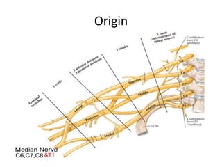
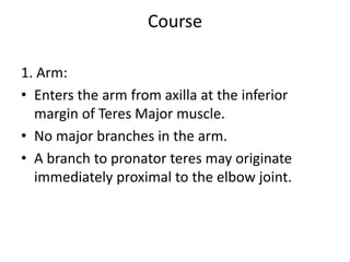
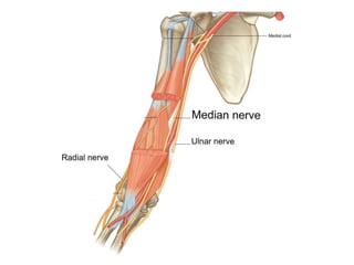
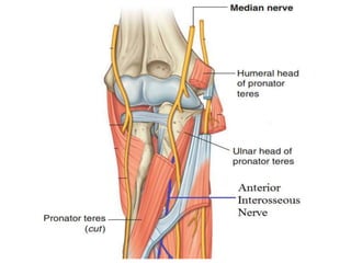
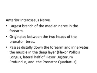
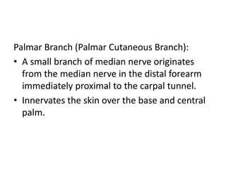
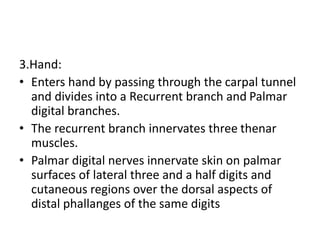
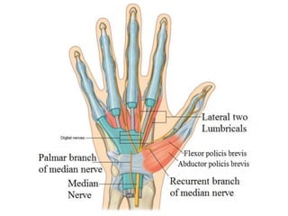
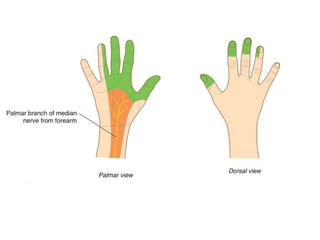
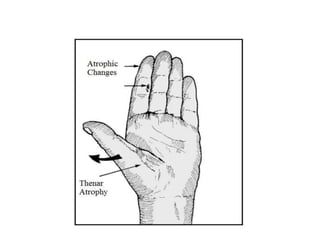
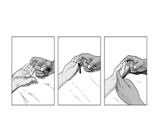
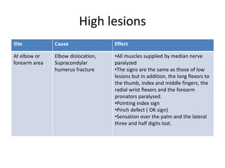
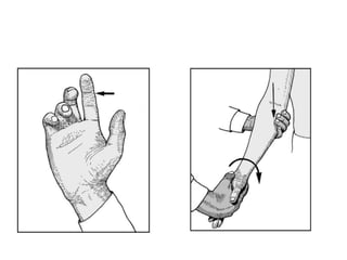
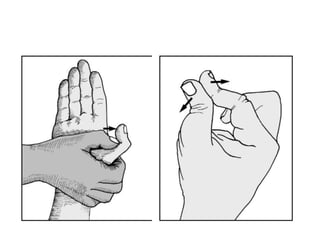
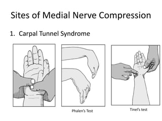
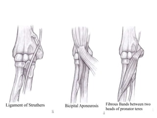
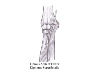
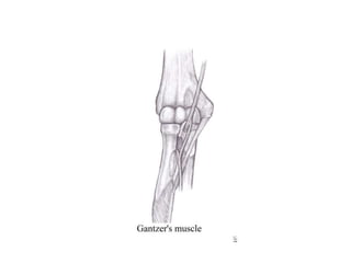
Median Nerve
Deep Medicine
Origin
Course
1. Arm:
• Enters the arm from axilla at the inferior
margin of Teres Major muscle.
• No major branches in the arm.
• A branch to pronator teres may originate
immediately proximal to the elbow joint.
2. Forearm:
• Exits cubital fossa between the humeral and
ulnar heads of Pronator Teres
• Innervates all the muscles of anterior
compartment except Flexor Carpi Ulnaris and
the medial part of the Flexor Digitorum
Profundus.
Anterior Interosseus Nerve
• Largest branch of the median nerve in the
forearm
• Originates between the two heads of the
pronator teres.
• Passes distally down the forearm and innervates
the muscle in the deep layer (Flexor Pollicis
Longus, lateral half of Flexor Digitorum
Profundus, and the Pronator Quadratus).
Palmar Branch (Palmar Cutaneous Branch):
• A small branch of median nerve originates
from the median nerve in the distal forearm
immediately proximal to the carpal tunnel.
• Innervates the skin over the base and central
palm.
3.Hand:
• Enters hand by passing through the carpal tunnel
and divides into a Recurrent branch and Palmar
digital branches.
• The recurrent branch innervates three thenar
muscles.
• Palmar digital nerves innervate skin on palmar
surfaces of lateral three and a half digits and
cutaneous regions over the dorsal aspects of
distal phallanges of the same digits
• In addition to skin, the digital nerves supply
the lateral two lumbrical muscles.
Median Nerve Injury
• Low lesions
• High lesions
Low lesions
Site Cause Effect
At the level of Wrist
Joint
Carpal Tunnel Syndrome
Carpal Dislocations
•Paralysis of the Three thenar
muscles and the lateral two
lumbricals
•Patient unable to abduct the
thumb.
•Sensation over the lateral three
and half digits lost.
•In long standing cases thenar
eminence is wasted, thumb may
come to lie in the plane of palm
(Ape thumb Deformity)
High lesions
Site Cause Effect
At elbow or
forearm area
Elbow dislocation,
Supracondylar
humerus fracture
•All muscles supplied by median nerve
paralyzed
•The signs are the same as those of low
lesions but in addition, the long flexors to
the thumb, index and middle fingers, the
radial wrist flexors and the forearm
pronators paralysed.
•Pointing index sign
•Pinch defect ( OK sign)
•Sensation over the palm and the lateral
three and half digits lost.
Sites of Medial Nerve Compression
1. Carpal Tunnel Syndrome
Phalen’s Test Tinel’s test
2. Pronator syndrome:
i. Ligament of Struthers
ii. Bicipital Aponeurosis
iii. Fibrous bands between the deep and
superficial heads of the Pronator Teres.
iv. Fibrous Arch of Flexor Digitorum Superficialis
3. Anterior Interosseous Nerve Syndrome:
• Spontaneously (Parsonage-Turner Syndrome)
or fracture, fibrous bands, tumours
• Gantzer’s muscle
Thank You

Median Nerve.pptx

  • 1.
  • 2.
  • 3.
    Course 1. Arm: • Entersthe arm from axilla at the inferior margin of Teres Major muscle. • No major branches in the arm. • A branch to pronator teres may originate immediately proximal to the elbow joint.
  • 5.
    2. Forearm: • Exitscubital fossa between the humeral and ulnar heads of Pronator Teres • Innervates all the muscles of anterior compartment except Flexor Carpi Ulnaris and the medial part of the Flexor Digitorum Profundus.
  • 7.
    Anterior Interosseus Nerve •Largest branch of the median nerve in the forearm • Originates between the two heads of the pronator teres. • Passes distally down the forearm and innervates the muscle in the deep layer (Flexor Pollicis Longus, lateral half of Flexor Digitorum Profundus, and the Pronator Quadratus).
  • 8.
    Palmar Branch (PalmarCutaneous Branch): • A small branch of median nerve originates from the median nerve in the distal forearm immediately proximal to the carpal tunnel. • Innervates the skin over the base and central palm.
  • 9.
    3.Hand: • Enters handby passing through the carpal tunnel and divides into a Recurrent branch and Palmar digital branches. • The recurrent branch innervates three thenar muscles. • Palmar digital nerves innervate skin on palmar surfaces of lateral three and a half digits and cutaneous regions over the dorsal aspects of distal phallanges of the same digits
  • 10.
    • In additionto skin, the digital nerves supply the lateral two lumbrical muscles.
  • 13.
    Median Nerve Injury •Low lesions • High lesions
  • 14.
    Low lesions Site CauseEffect At the level of Wrist Joint Carpal Tunnel Syndrome Carpal Dislocations •Paralysis of the Three thenar muscles and the lateral two lumbricals •Patient unable to abduct the thumb. •Sensation over the lateral three and half digits lost. •In long standing cases thenar eminence is wasted, thumb may come to lie in the plane of palm (Ape thumb Deformity)
  • 17.
    High lesions Site CauseEffect At elbow or forearm area Elbow dislocation, Supracondylar humerus fracture •All muscles supplied by median nerve paralyzed •The signs are the same as those of low lesions but in addition, the long flexors to the thumb, index and middle fingers, the radial wrist flexors and the forearm pronators paralysed. •Pointing index sign •Pinch defect ( OK sign) •Sensation over the palm and the lateral three and half digits lost.
  • 20.
    Sites of MedialNerve Compression 1. Carpal Tunnel Syndrome Phalen’s Test Tinel’s test
  • 21.
    2. Pronator syndrome: i.Ligament of Struthers ii. Bicipital Aponeurosis iii. Fibrous bands between the deep and superficial heads of the Pronator Teres. iv. Fibrous Arch of Flexor Digitorum Superficialis
  • 24.
    3. Anterior InterosseousNerve Syndrome: • Spontaneously (Parsonage-Turner Syndrome) or fracture, fibrous bands, tumours • Gantzer’s muscle
  • 26.

Editor's Notes

  • #3 Formed Anterior to the third part of the axillary artery by the union of lateral and medial roots originating from lateral and medial cords of brachial plexus
  • #6 Median: Flexor Carpi Radialis, Palmaris Longus, Pronator Teres, Flexor Digitorum Superficialis (Superficial Compartment) AIN: Flexor Digitorum Profundus- Lateral Part, Pronator Quadratus, Flexor Policis Longus (Deep Compartment)
  • #8 Solely Motor Nerve
  • #9 Palmar cutaneous branch is spared in Carpal Tunnel Syndrome
  • #10 Recurrent Branch: Thenar Muscles (Flexor pollicis brevis, Opponens Policis, Abductor pollicis Brevis) Palmar digital branch: The lateral Two lumbricals and sensory supply
  • #18 Typically the hand is held with the ulnar fingers flexed and the index straight (the ‘pointing index sign’)
  • #21 Compression: First the median nerve is identified between flexor carpi radialis and palmaris longus, the nerve is compressed with both the thumbs with firm pressure for 30 seconds, intervel between pain, paresthesia or numbness is noted usually about 16 seconds in carpal tunnel syndrome. Phalen’s test:Both wrists in a fully flexed position for 1–2 minutes. The appearance or exacerbation of paraesthesia in the median distribution is suggestive of the carpal tunnel syndrome, and is positive in 70% of those suffering from this condition Tinel’s test:the test is positive if gentle finger percussion over the median nerve produces paraesthesia in its distribution. This test is said to be positive in 56% of cases of carpal tunnel syndrome.
  • #23 Ligament of Struthers: The ligament of Struthers connects the supracondylar process to the medial epicondyle, encasing the median nerve and brachial artery. It is seen in approximately 13% of the general population and rarely causes median nerve entrapment.
  • #25 Isolated AIN injury is rare. Spontaneous (and usually temporary) Physiological failure (Parsonage–Turner syndrome) is a more likely cause. There is motor weakness without sensory symptoms. Gantzer’s muscle: This is the accessory head of the FPL and has been postulated to be a cause of AINS ; in an anatomic study, the muscle was found in 52% of limbs and was supplied by the AIN, and it was found to be posterior to both the median nerve and the AIN in all cases