SlideShare a Scribd company logo
1 of 3
Download to read offline
Fig. 1: Components of MEASSuRE
MicroElectrode Array Stretching Stimulating und Recording Equipment
(MEASSuRE)
Cells in the body are constantly subject to mechanical forces, with yet poorly understood effects on
cell division, gene expression, cell migration, morpho-genesis, ion channel gating, and others. In a
physiological stretch, the cells are stretched within their healthy limits. In a pathological stretch, the
cells are stretched beyond the healthy limits and normal cell function is disrupted, causing a trauma. It
is currently difficult to investigate reproducibly and reliably the effects of physiological and
pathological mechanical stretch on the electrophysiology of biological tissue, both in vitro and in vivo,
because current electrodes are hard and stiff, thus break or damage the tissue upon stretching.
Moreover, existing commercial technologies only allow to either stretch cells mechanically or to
stimulate them electrically, but not both. MEASSuRE solves this problem. It provides the capability to
reproduce the mechanical and electrical in vivo environment in vitro by allowing the application of a
controlled stretch (strain, strain rate) to a cell or tissue culture while simultaneously stimulating
electrophysiolgical activity to assess the health of the cells. This capability is enabled by incorporating
BMSEED’s proprietary elastically stretchable microelectrodes. In addition, MEASSuRE will be the
only commercial system that allows optical imaging of the cells while they are being stretched
because the tissue remains in the focal plane of the lens throughout the stretching process. The
microelectrodes on the sMEA consist of a proprietary microcracked gold film, which is critical for the
stretchability of the electrodes (for further information, see: http://www.bmseed.com/References.html).
The development work is funded by an SBIR from the National Institute of Neurological Disorders and
Stroke (NINDS).
MEASSuRE is a complete solution for researchers that require to stretch, record/stimulate, and
image cells in vitro, separately or concurrently. It consists of six modules.
1. Stretchable microelectrode array (sMEA): The sMEA
interfaces with the cell or tissue culture at its center, and the
data acquisition system at its perimeter, and contains the
stretchable microelectrodes.
2. Stretcher: The stretcher applies the strain to the cell or
tissue culture at a chosen strain rate. It consists of a voice
coil actuator (VCA) with PID controller from BEIKimco, a
power supply, and the custom BMSEED hardware.
3. Software: The motion of the stretcher, the camera, and the
data acquisition system are software controlled.
4. High frame rate video camera: The camera (DELimaging)
and lens (Zoom 6000, Navitar) allow capturing images of
the cells before, during and after stretching to verify how the
applied strain is translated to the tissue.
5. Plexon board: The interface board electrically connects the
microelectrodes on the sMEA to the data acquisition system
6. Data acquisition system: The data acquisition system (using electrical components from Intan
Technologies) allows amplification, storage, and display of the recorded data; it is interfaced with
the sMEA via the Plexon interface board (see E). Other data acquisition systems, e.g., from
MultiChannel Systems (MCS), are directly interfaced with the sMEAs via the headstage.
Capabilities of MEASSuRE:
A) Mechanical and electrical interface with a cell culture: MEASSuRE is the first research tool that
combines the capability to electrically and mechanically stimulate cell or tissue cultures, either
individually or concurrently.
B) Comparison of pre and post stretch: MEASSuRE allows the normalization of post-stretch cell
response to pre-stretch level because the microelectrodes remain in contact with the same
location on the tissue before, during, and after stretching.
C) High reproducibility: The strain on the tissue and the strain rate can be precisely controlled.
D) Versatility: MEASSuRE provides high versatility with respect to the stretch pattern that can be
programmed with the Ingenia MotionLab PID controller and software. The acceleration, velocity,
and strain (vertical position of the sMEA) can be controlled using macros.
E) Maintenance of sterility: The sMEAs can be sterilized and are biocompatible, thus allowing long-
term studies because sterility does not need to be broken to insert electrodes or transfer the
tissue to an MEA after stretching.
F) Repeated stretch and relaxation: The microelectrodes that are embedded in the sMEA elastically
stretch and relax with the tissue, allowing for cyclic or repeated stretching.
G) Optical imaging: The high speed camera and lens capture sharp images of the stretched cells at
up to 1700 frames per second. This capability is important because it allows the verification that
the strain applied to the sMEA is properly translated to the cells.
Applications for MEASSuRE
The applications for MEASSuRE can be grouped in the following segments:
Physiological stretching of cells
• tissue engineering: Stem cells that differentiate into a specific tissue have properties that
resemble adult tissue more closely when the cells are under mechanical and electrical
stimulation during the differentiation process. MEASSuRE has the capability to provide both,
electrical and mechanical stimulation.
• toxicity testing of drugs: Stem cells that differentiate into a specific tissue have properties that
resemble adult tissue more closely when the cells are under mechanical and electrical
stimulation during the differentiation process. Tissue grown from stem cells in this manner are
therefore more representative of the respective organ in an adult human, thus, increasing the
validity of drug toxicity testing by pharmaceutical companies. MEASSuRE has the capability to
provide both, electrical and mechanical stimulation.
• mechano-biology: There are a variety of mechanisms for transducing and sensing mechanical
forces in neurons and other cell types. MEASSuRE provides capabilities to fundamentally
understand the effect of mechanical forces.
Pathological stretching of cells
• neurotrauma treatments: MEASSuRE allows to reproduce reliably and repeatedly the
biomechanics of traumatic brain injury and spinal cord injury in a controlled environment.
Changes in the electrophysiology of the injured neurons can be assessed with in a
straightforward manner with the embedded microelectrodes by comparing the post-injury
electrophysiology to pre-injury level. The effectiveness of drugs or other treatment strategies
to minimize the damage after injury can therefore be readily assessed.
• concussion protocols: MEASSuRE will allow researchers and physicians to develop improved
concussion protocols that are based on the electrophysiology of the underlying injury rather
than cognitive tests.
• muscle injuries: MEASSuRE will allow the investigation of the mechanism of those muscle
injuries that are caused by excessive tension or compression, and to evaluate the efficacy of
drugs to speed up recovery.
• stem cell repair: Stem cells are involved in repair processes after injury in different parts of the
body, e.g., in the brain after a traumatic brain injury. The mechanism of the activation of the
mechanoreceptors is not understood. MEASSuRE will be a useful tool to elucidate and study
this mechanism.
• Other - Neurodegenerative diseases: Neurodegenerative diseases such as Alzheimer’s
disease have common pathological pathways with traumatic brain injury, e.g., the build up of
amyloid-β plaques. Therefore, MEASSuRE might be a valuable tool for the early evaluation of
the efficacy of drug candidates against Alzheimer’s disease.
MEASSuRE is well-suited to both physiological and pathological stretching of cells. The main
differences between these two applications are (a) the level of strain, (b) the strain rate, and (c) the
number of stretch and relaxation cycles. In neurotrauma applications, tissue cultures are stretched
once or a few times at high strain rates (up to 50 s-1
), up to maximal strains of 5% to >30%. In tissue
engineering applications, the cells are stretched elastically for many cycles at strain rates of less than
0.5 s-1
, at physiological strain of typically not more than 5%.
Product Specifications
Parameter Values
Strain rate up to 20s-1
Maximum strain 50%
2,000 cycles at to 10%,
20%, and 30% strain
No change in number of
functional electrodesSlow dynamic
cyclic strain (1Hz) 150,000 cycles to 10%
strain
No change in number of
functional electrodes
Fast dynamic
stretch and
relaxation
40 times fast stretch
(100ms) to 10%, 20%,
30%, 40%, 50% strain
>90% of electrodes remain
functional
Stretch and hold (static strain)
Up to 15% strain; all electrodes
recover upon relaxation
Substrate and encapsulation material PDMS
Electrode Material Gold
Well for medium
Polycarbonate; diameter 1”;
height 0.5”, 0.375”, and 0.25”
Vpeak-to-peak 10-20 µV
Electrode impedance at 1kHz 5-20 kohm
Stimulation capability Yes: Pt black or IrOx coating
Number of electrodes 28
Total recording area 1.5mm x 1.5mm
Recording area of individual electrodes 100µm x 100µm
Camera and lens Depending on application
Radial Lagrangian strain calculated per equation below:
From:
Journal of Neuroscience Methods 150 (2006) 192–201
An in vitro model of traumatic brain injury utilising two-dimensional stretch of organotypic
hippocampal slice cultures
Barclay Morrison III, Heather L. Cater, Christopher D. Benhamc, Lars E. Sundstrom

More Related Content

What's hot

MURA Dataset: Towards Radiologist-Level Abnormality Detection in Musculoskele...
MURA Dataset: Towards Radiologist-Level Abnormality Detection in Musculoskele...MURA Dataset: Towards Radiologist-Level Abnormality Detection in Musculoskele...
MURA Dataset: Towards Radiologist-Level Abnormality Detection in Musculoskele...
Utkupek
 
Implementation of Medical Image Analysis using Image Processing Techniques
Implementation of Medical Image Analysis using Image Processing TechniquesImplementation of Medical Image Analysis using Image Processing Techniques
Implementation of Medical Image Analysis using Image Processing Techniques
YogeshIJTSRD
 
Final Draft of Research Paper 1
Final Draft of Research Paper 1Final Draft of Research Paper 1
Final Draft of Research Paper 1
Trevor Davis
 

What's hot (20)

Wheeler_Benjamin_MyPURA
Wheeler_Benjamin_MyPURAWheeler_Benjamin_MyPURA
Wheeler_Benjamin_MyPURA
 
Stereotaxy Brain
Stereotaxy BrainStereotaxy Brain
Stereotaxy Brain
 
IMAGE GUIDED SURGERY
IMAGE GUIDED SURGERYIMAGE GUIDED SURGERY
IMAGE GUIDED SURGERY
 
MURA Dataset: Towards Radiologist-Level Abnormality Detection in Musculoskele...
MURA Dataset: Towards Radiologist-Level Abnormality Detection in Musculoskele...MURA Dataset: Towards Radiologist-Level Abnormality Detection in Musculoskele...
MURA Dataset: Towards Radiologist-Level Abnormality Detection in Musculoskele...
 
High Precision And Fast Functional Mapping Of Cortical Circuitry Through A No...
High Precision And Fast Functional Mapping Of Cortical Circuitry Through A No...High Precision And Fast Functional Mapping Of Cortical Circuitry Through A No...
High Precision And Fast Functional Mapping Of Cortical Circuitry Through A No...
 
2009.12.29, Draft of New Chapter
2009.12.29, Draft of New Chapter2009.12.29, Draft of New Chapter
2009.12.29, Draft of New Chapter
 
Improving radiologists’ and orthopedists’ QoE in diagnosing lumbar disk herni...
Improving radiologists’ and orthopedists’ QoE in diagnosing lumbar disk herni...Improving radiologists’ and orthopedists’ QoE in diagnosing lumbar disk herni...
Improving radiologists’ and orthopedists’ QoE in diagnosing lumbar disk herni...
 
Implementation of Medical Image Analysis using Image Processing Techniques
Implementation of Medical Image Analysis using Image Processing TechniquesImplementation of Medical Image Analysis using Image Processing Techniques
Implementation of Medical Image Analysis using Image Processing Techniques
 
Why Outsource To Neurotar
Why Outsource To NeurotarWhy Outsource To Neurotar
Why Outsource To Neurotar
 
Bioplasty
Bioplasty Bioplasty
Bioplasty
 
Multimodality Molecular Imaging – An Overview With Special Focus on PET/CT
Multimodality Molecular Imaging – An Overview With Special Focus on PET/CTMultimodality Molecular Imaging – An Overview With Special Focus on PET/CT
Multimodality Molecular Imaging – An Overview With Special Focus on PET/CT
 
IEEE Medical image Title and Abstract 2016
IEEE Medical image Title and Abstract 2016 IEEE Medical image Title and Abstract 2016
IEEE Medical image Title and Abstract 2016
 
Detecting the basis of cognitive dysfunction in mild Traumatic Brain Injury
Detecting the basis of cognitive dysfunction in mild Traumatic Brain InjuryDetecting the basis of cognitive dysfunction in mild Traumatic Brain Injury
Detecting the basis of cognitive dysfunction in mild Traumatic Brain Injury
 
Design and development of calibration Unit for precision planter
Design and development of calibration Unit for precision planterDesign and development of calibration Unit for precision planter
Design and development of calibration Unit for precision planter
 
Neuronavigation Software to visualize and surgically approach brain structures
Neuronavigation Software to visualize and surgically approach brain structuresNeuronavigation Software to visualize and surgically approach brain structures
Neuronavigation Software to visualize and surgically approach brain structures
 
International Conference On Electrical Engineering
International Conference On Electrical EngineeringInternational Conference On Electrical Engineering
International Conference On Electrical Engineering
 
Final Draft of Research Paper 1
Final Draft of Research Paper 1Final Draft of Research Paper 1
Final Draft of Research Paper 1
 
nature17435
nature17435nature17435
nature17435
 
Gamma knife radiosurgery
Gamma knife radiosurgeryGamma knife radiosurgery
Gamma knife radiosurgery
 
Ucsf 4 4
Ucsf 4 4Ucsf 4 4
Ucsf 4 4
 

Similar to MEASSuRE_product description

Experimental Muscle Mechanics in Aging and Disease
Experimental Muscle Mechanics in Aging and DiseaseExperimental Muscle Mechanics in Aging and Disease
Experimental Muscle Mechanics in Aging and Disease
InsideScientific
 
Abba mems pepar
Abba mems peparAbba mems pepar
Abba mems pepar
Abba_33
 
Saenz Cogollo et al. - 2011 - A new integrated system combining atomic force ...
Saenz Cogollo et al. - 2011 - A new integrated system combining atomic force ...Saenz Cogollo et al. - 2011 - A new integrated system combining atomic force ...
Saenz Cogollo et al. - 2011 - A new integrated system combining atomic force ...
Jose Francisco Saenz Cogollo
 
ZMPCZM017000.11.11 Home based EMG Triggered stimulation in chronic stroke
ZMPCZM017000.11.11 Home based EMG Triggered stimulation in chronic strokeZMPCZM017000.11.11 Home based EMG Triggered stimulation in chronic stroke
ZMPCZM017000.11.11 Home based EMG Triggered stimulation in chronic stroke
painezeeman
 
International Journal of Biometrics and Bioinformatics(IJBB) Volume (3) Issue...
International Journal of Biometrics and Bioinformatics(IJBB) Volume (3) Issue...International Journal of Biometrics and Bioinformatics(IJBB) Volume (3) Issue...
International Journal of Biometrics and Bioinformatics(IJBB) Volume (3) Issue...
CSCJournals
 
In vivo characterization of breast tissue by non-invasive bio-impedance measu...
In vivo characterization of breast tissue by non-invasive bio-impedance measu...In vivo characterization of breast tissue by non-invasive bio-impedance measu...
In vivo characterization of breast tissue by non-invasive bio-impedance measu...
ijbesjournal
 

Similar to MEASSuRE_product description (20)

Experimental Muscle Mechanics in Aging and Disease
Experimental Muscle Mechanics in Aging and DiseaseExperimental Muscle Mechanics in Aging and Disease
Experimental Muscle Mechanics in Aging and Disease
 
Mvit
MvitMvit
Mvit
 
MEMS
MEMSMEMS
MEMS
 
Three Techniques, One System: How to Effectively Characterize Complete Muscle...
Three Techniques, One System: How to Effectively Characterize Complete Muscle...Three Techniques, One System: How to Effectively Characterize Complete Muscle...
Three Techniques, One System: How to Effectively Characterize Complete Muscle...
 
Abba mems pepar
Abba mems peparAbba mems pepar
Abba mems pepar
 
Saenz Cogollo et al. - 2011 - A new integrated system combining atomic force ...
Saenz Cogollo et al. - 2011 - A new integrated system combining atomic force ...Saenz Cogollo et al. - 2011 - A new integrated system combining atomic force ...
Saenz Cogollo et al. - 2011 - A new integrated system combining atomic force ...
 
MEMS BASED OPTICAL COHERENCE TOMOGRAPHY IMAGING SYSTEM AND OPTICAL BIOPSY PRO...
MEMS BASED OPTICAL COHERENCE TOMOGRAPHY IMAGING SYSTEM AND OPTICAL BIOPSY PRO...MEMS BASED OPTICAL COHERENCE TOMOGRAPHY IMAGING SYSTEM AND OPTICAL BIOPSY PRO...
MEMS BASED OPTICAL COHERENCE TOMOGRAPHY IMAGING SYSTEM AND OPTICAL BIOPSY PRO...
 
A MEMS BASED OPTICAL COHERENCE TOMOGRAPHY IMAGING SYSTEM AND OPTICAL BIOPSY P...
A MEMS BASED OPTICAL COHERENCE TOMOGRAPHY IMAGING SYSTEM AND OPTICAL BIOPSY P...A MEMS BASED OPTICAL COHERENCE TOMOGRAPHY IMAGING SYSTEM AND OPTICAL BIOPSY P...
A MEMS BASED OPTICAL COHERENCE TOMOGRAPHY IMAGING SYSTEM AND OPTICAL BIOPSY P...
 
Paper22
Paper22Paper22
Paper22
 
ZMPCZM017000.11.11 Home based EMG Triggered stimulation in chronic stroke
ZMPCZM017000.11.11 Home based EMG Triggered stimulation in chronic strokeZMPCZM017000.11.11 Home based EMG Triggered stimulation in chronic stroke
ZMPCZM017000.11.11 Home based EMG Triggered stimulation in chronic stroke
 
ZMPCZM017000.11.11
ZMPCZM017000.11.11ZMPCZM017000.11.11
ZMPCZM017000.11.11
 
International Journal of Biometrics and Bioinformatics(IJBB) Volume (3) Issue...
International Journal of Biometrics and Bioinformatics(IJBB) Volume (3) Issue...International Journal of Biometrics and Bioinformatics(IJBB) Volume (3) Issue...
International Journal of Biometrics and Bioinformatics(IJBB) Volume (3) Issue...
 
WAVELET DECOMPOSITION METHOD BASED AUTOMATED DIAGNOSIS OF MUSCLE DISEASES
WAVELET DECOMPOSITION METHOD BASED AUTOMATED DIAGNOSIS OF MUSCLE DISEASESWAVELET DECOMPOSITION METHOD BASED AUTOMATED DIAGNOSIS OF MUSCLE DISEASES
WAVELET DECOMPOSITION METHOD BASED AUTOMATED DIAGNOSIS OF MUSCLE DISEASES
 
Neurobionics and robotic neurorehabilitations
Neurobionics and robotic neurorehabilitationsNeurobionics and robotic neurorehabilitations
Neurobionics and robotic neurorehabilitations
 
In vivo characterization of breast tissue by non-invasive bio-impedance measu...
In vivo characterization of breast tissue by non-invasive bio-impedance measu...In vivo characterization of breast tissue by non-invasive bio-impedance measu...
In vivo characterization of breast tissue by non-invasive bio-impedance measu...
 
New Technology for Communication
New Technology for CommunicationNew Technology for Communication
New Technology for Communication
 
Spinal cord Stimulation
Spinal cord StimulationSpinal cord Stimulation
Spinal cord Stimulation
 
Force Quantification of IASTM device
Force Quantification of IASTM deviceForce Quantification of IASTM device
Force Quantification of IASTM device
 
Dr. Frankenstein’s Dream Made Possible: Implanted Electronic Devices
Dr. Frankenstein’s Dream Made Possible:  Implanted Electronic Devices Dr. Frankenstein’s Dream Made Possible:  Implanted Electronic Devices
Dr. Frankenstein’s Dream Made Possible: Implanted Electronic Devices
 
W P Biomechanics
W P  BiomechanicsW P  Biomechanics
W P Biomechanics
 

MEASSuRE_product description

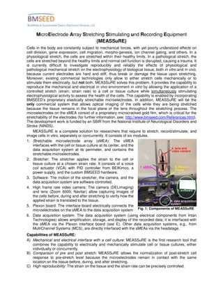
  • 1. Fig. 1: Components of MEASSuRE MicroElectrode Array Stretching Stimulating und Recording Equipment (MEASSuRE) Cells in the body are constantly subject to mechanical forces, with yet poorly understood effects on cell division, gene expression, cell migration, morpho-genesis, ion channel gating, and others. In a physiological stretch, the cells are stretched within their healthy limits. In a pathological stretch, the cells are stretched beyond the healthy limits and normal cell function is disrupted, causing a trauma. It is currently difficult to investigate reproducibly and reliably the effects of physiological and pathological mechanical stretch on the electrophysiology of biological tissue, both in vitro and in vivo, because current electrodes are hard and stiff, thus break or damage the tissue upon stretching. Moreover, existing commercial technologies only allow to either stretch cells mechanically or to stimulate them electrically, but not both. MEASSuRE solves this problem. It provides the capability to reproduce the mechanical and electrical in vivo environment in vitro by allowing the application of a controlled stretch (strain, strain rate) to a cell or tissue culture while simultaneously stimulating electrophysiolgical activity to assess the health of the cells. This capability is enabled by incorporating BMSEED’s proprietary elastically stretchable microelectrodes. In addition, MEASSuRE will be the only commercial system that allows optical imaging of the cells while they are being stretched because the tissue remains in the focal plane of the lens throughout the stretching process. The microelectrodes on the sMEA consist of a proprietary microcracked gold film, which is critical for the stretchability of the electrodes (for further information, see: http://www.bmseed.com/References.html). The development work is funded by an SBIR from the National Institute of Neurological Disorders and Stroke (NINDS). MEASSuRE is a complete solution for researchers that require to stretch, record/stimulate, and image cells in vitro, separately or concurrently. It consists of six modules. 1. Stretchable microelectrode array (sMEA): The sMEA interfaces with the cell or tissue culture at its center, and the data acquisition system at its perimeter, and contains the stretchable microelectrodes. 2. Stretcher: The stretcher applies the strain to the cell or tissue culture at a chosen strain rate. It consists of a voice coil actuator (VCA) with PID controller from BEIKimco, a power supply, and the custom BMSEED hardware. 3. Software: The motion of the stretcher, the camera, and the data acquisition system are software controlled. 4. High frame rate video camera: The camera (DELimaging) and lens (Zoom 6000, Navitar) allow capturing images of the cells before, during and after stretching to verify how the applied strain is translated to the tissue. 5. Plexon board: The interface board electrically connects the microelectrodes on the sMEA to the data acquisition system 6. Data acquisition system: The data acquisition system (using electrical components from Intan Technologies) allows amplification, storage, and display of the recorded data; it is interfaced with the sMEA via the Plexon interface board (see E). Other data acquisition systems, e.g., from MultiChannel Systems (MCS), are directly interfaced with the sMEAs via the headstage. Capabilities of MEASSuRE: A) Mechanical and electrical interface with a cell culture: MEASSuRE is the first research tool that combines the capability to electrically and mechanically stimulate cell or tissue cultures, either individually or concurrently. B) Comparison of pre and post stretch: MEASSuRE allows the normalization of post-stretch cell response to pre-stretch level because the microelectrodes remain in contact with the same location on the tissue before, during, and after stretching. C) High reproducibility: The strain on the tissue and the strain rate can be precisely controlled.
  • 2. D) Versatility: MEASSuRE provides high versatility with respect to the stretch pattern that can be programmed with the Ingenia MotionLab PID controller and software. The acceleration, velocity, and strain (vertical position of the sMEA) can be controlled using macros. E) Maintenance of sterility: The sMEAs can be sterilized and are biocompatible, thus allowing long- term studies because sterility does not need to be broken to insert electrodes or transfer the tissue to an MEA after stretching. F) Repeated stretch and relaxation: The microelectrodes that are embedded in the sMEA elastically stretch and relax with the tissue, allowing for cyclic or repeated stretching. G) Optical imaging: The high speed camera and lens capture sharp images of the stretched cells at up to 1700 frames per second. This capability is important because it allows the verification that the strain applied to the sMEA is properly translated to the cells. Applications for MEASSuRE The applications for MEASSuRE can be grouped in the following segments: Physiological stretching of cells • tissue engineering: Stem cells that differentiate into a specific tissue have properties that resemble adult tissue more closely when the cells are under mechanical and electrical stimulation during the differentiation process. MEASSuRE has the capability to provide both, electrical and mechanical stimulation. • toxicity testing of drugs: Stem cells that differentiate into a specific tissue have properties that resemble adult tissue more closely when the cells are under mechanical and electrical stimulation during the differentiation process. Tissue grown from stem cells in this manner are therefore more representative of the respective organ in an adult human, thus, increasing the validity of drug toxicity testing by pharmaceutical companies. MEASSuRE has the capability to provide both, electrical and mechanical stimulation. • mechano-biology: There are a variety of mechanisms for transducing and sensing mechanical forces in neurons and other cell types. MEASSuRE provides capabilities to fundamentally understand the effect of mechanical forces. Pathological stretching of cells • neurotrauma treatments: MEASSuRE allows to reproduce reliably and repeatedly the biomechanics of traumatic brain injury and spinal cord injury in a controlled environment. Changes in the electrophysiology of the injured neurons can be assessed with in a straightforward manner with the embedded microelectrodes by comparing the post-injury electrophysiology to pre-injury level. The effectiveness of drugs or other treatment strategies to minimize the damage after injury can therefore be readily assessed. • concussion protocols: MEASSuRE will allow researchers and physicians to develop improved concussion protocols that are based on the electrophysiology of the underlying injury rather than cognitive tests. • muscle injuries: MEASSuRE will allow the investigation of the mechanism of those muscle injuries that are caused by excessive tension or compression, and to evaluate the efficacy of drugs to speed up recovery. • stem cell repair: Stem cells are involved in repair processes after injury in different parts of the body, e.g., in the brain after a traumatic brain injury. The mechanism of the activation of the mechanoreceptors is not understood. MEASSuRE will be a useful tool to elucidate and study this mechanism. • Other - Neurodegenerative diseases: Neurodegenerative diseases such as Alzheimer’s disease have common pathological pathways with traumatic brain injury, e.g., the build up of amyloid-β plaques. Therefore, MEASSuRE might be a valuable tool for the early evaluation of the efficacy of drug candidates against Alzheimer’s disease. MEASSuRE is well-suited to both physiological and pathological stretching of cells. The main differences between these two applications are (a) the level of strain, (b) the strain rate, and (c) the number of stretch and relaxation cycles. In neurotrauma applications, tissue cultures are stretched once or a few times at high strain rates (up to 50 s-1 ), up to maximal strains of 5% to >30%. In tissue engineering applications, the cells are stretched elastically for many cycles at strain rates of less than 0.5 s-1 , at physiological strain of typically not more than 5%.
  • 3. Product Specifications Parameter Values Strain rate up to 20s-1 Maximum strain 50% 2,000 cycles at to 10%, 20%, and 30% strain No change in number of functional electrodesSlow dynamic cyclic strain (1Hz) 150,000 cycles to 10% strain No change in number of functional electrodes Fast dynamic stretch and relaxation 40 times fast stretch (100ms) to 10%, 20%, 30%, 40%, 50% strain >90% of electrodes remain functional Stretch and hold (static strain) Up to 15% strain; all electrodes recover upon relaxation Substrate and encapsulation material PDMS Electrode Material Gold Well for medium Polycarbonate; diameter 1”; height 0.5”, 0.375”, and 0.25” Vpeak-to-peak 10-20 µV Electrode impedance at 1kHz 5-20 kohm Stimulation capability Yes: Pt black or IrOx coating Number of electrodes 28 Total recording area 1.5mm x 1.5mm Recording area of individual electrodes 100µm x 100µm Camera and lens Depending on application Radial Lagrangian strain calculated per equation below: From: Journal of Neuroscience Methods 150 (2006) 192–201 An in vitro model of traumatic brain injury utilising two-dimensional stretch of organotypic hippocampal slice cultures Barclay Morrison III, Heather L. Cater, Christopher D. Benhamc, Lars E. Sundstrom