1. Examining the expression of potential cell cycle regulators in the developing
mouse inner ear
Maryam Ebrahimi1, Gillian L. Drury2, Giselle Boukhaled2, Melissa A. Vollrath1, 2
1 Department of Neurology and Neurosurgery and 2 Department of Physiology, McGill University, Montreal, Quebec, Canada!
ABSTRACT!
RESULTS!
RT-‐PCR
confirmed
the
expression
of
10
candidate
genes
in
mouse
utricle
at
postnatal
day
7
BACKGROUND!
Nega&ve
cell-‐cycle
regulators
with
ongoing
expression
in
suppor&ng
cells
prevent
them
from
dividing
and
differen&a&ng
into
hair
cells
HYPOTHESIS !
APPROACH!
DISCUSSION AND FUTURE DIRECTIONS!
Analyzing
SHIELD
data
Selected
10
candidate
cell-‐cycle
genes
with
high
and
persistent
expression
in
utricle
suppor&ng
cells
at
embryonic
day
16
and
postnatal
days
4,7,16
Confirma&on
of
SHIELD
data
by
RT-‐PCR
To
date
we
have
confirmed
expression
of
10
of
these
genes
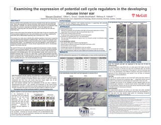
Localiza&on
of
candidate
genes
by
in
situ
hybridiza&on
Here
we
show
RNA
localiza&on
for
genes
Cav2,
Frk,
AnnexA1
and
Igf-‐1
at
embryonic
days
13.5,15.5,18.5
In
situ
hybridizaAon
method:
Gene-‐specific
RNA
probes
labeled
using
Digoxygenin.
Embryos
fixed
at
different
embryonic
&me
points
and
frozen
in
OCT.
16-‐um
sec&ons
cut
on
cryostat.
Candidate
gene
probes
are
hybridized
to
inner
ear
sec&ons.
Control
probes
(Atoh1)
are
hybridized
to
adjacent
sec&ons
to
visualize
hair
cell
layer.
A[er
color
development,
slides
are
cover-‐slipped
and
visualized
under
the
microscope.
REFERENCES!
Cav2,
Frk,
AnnexA1
and
Igf-‐1
are
expressed
in
the
inner
ear
of
mice
at
embryonic
days
Our
primary
analysis
using
SHIELD
led
us
to
select
genes
with
higher
and
more
persistent
expression
in
utricular
SCs
than
HCs
at
postnatal
days.
Our
RT-‐PCR
results
also
confirmed
the
expression
of
these
genes
in
utricle
at
postnatal
day
7.
Our
in
situ
hybridiza&on,
from
different
embryonic
&me
points
localized
the
expression
of
four
selected
genes
more
spread
in
hair
cell
layer,
as
compared
with
posi&ve
control
probe(Atoh1).
Next
Step
We
will
use
the
specific
probes
for
Sox
9
and
P27kip
which
have
been
shown
to
be
expressed
in
utricular
SCs
at
embryonic
ages
7,8
.
Using
these
controls,
we
can
visualize
to
what
extend
our
selected
genes
are
expressed
in
these
two
cells.
To
inves&gate
the
func&on
of
the
candidate
cell-‐cycle
regulator
genes
we
will
knockdown
gene
expression
in
cultured
mouse
utricles
using
siRNA
and
assay
for
cell
prolifera&on.
Reentry
into
the
cell
cycle
will
be
measured
by
BrdU
uptake
(a
standard
assay
for
measuring
cell
division).
Future
direcAons
This
experimental
approach
will
allow
us
to
test
these
and
other
candidate
nega&ve
cell-‐cycle
regulators
for
their
ability
to
produce
hair
cell
prolifera&on.
In
the
long
term
this
may
iden&fy
target
genes
for
developing
therapeu&c
strategies
for
the
treatment
of
hearing
loss.
Igf-‐1
and
AnnexA1
associate
with
cell
prolifera&on
of
CNS
and
PNS
in
different
developmental
&me
point.5,7
Igf-‐1
maintains
cell
prolifera&on
of
O&c
Vesicle
during
development.8
AnnexA1
Is
secreted
from
cochlear
suppor&ng
cells
a[er
hair
cell
loss.9
AnnexA1
associates
with
Cyclin
D
to
control
cell
cycle
progress
in
different
cell
lines.6
1.Groves
A.K.
The
challenge
of
hair
cell
regenera&on.
Exp
Biol
Med.
2010;
235:
434-‐446
2.Lowenheim
H,
Furness
DN,
Kil
J,
Zinn
C,
Gol&g
K,
Fero
ML,
et
al.
Gene
disrup&on
of
p27Kip1
allows
cell
prolifera&on
in
the
postnatal
and
adult
organ
of
Cor&.
Proceedings
of
the
Na&onal
Academy
of
Sciences.
1999
March
30,
1999;96(7):4084-‐8.
3.
Sage
C,
Huang
M,
Karimi
K,
Gu&errez
G,
Vollrath
MA,
Zhang
D-‐S,
et
al.
Prolifera&on
of
Func&onal
Hair
Cells
in
Vivo
in
the
Absence
of
the
Re&noblastoma
Protein.
Science.
2005
February
18,
2005;307(5712):1114-‐8.
4.Xie
L,
Frank
PG,
Lisan&
MP,
Sowa
G.
Endothelial
cells
isolated
from
caveolin-‐2
knockout
mice
display
higher
prolifera&on
rate
and
cell
cycle
progression
rela&ve
to
their
wild-‐type
counterparts.
2010;
298:
C693-‐701
5.Alldrige
LC,
Bryant
CE.
Annexin
1
regulates
cell
prolifera&on
by
disrup&on
of
cell
morphology
and
inhibi&on
of
cyclin
D1
expression
through
sustained
ac&va&on
of
the
ERK1/2
MAPK
signal.
Experimental
cell
research.
2003;290:
93-‐107.
6.Mak
ACY,
Szeto
IYY,
Fritzsch
B,
Cheah
KSE.
Differen&al
and
overlapping
expression
of
SOX2
and
SOX9
in
inner
ear
development.
Gene
Expr
Pajerns.2009;9(6):444-‐453.
7.Varela-‐Nieto
I,
Morales-‐Garcia
JA,
Vigil
P,
Diaz-‐Casares
A,
Gorospe
I,
Sanchez-‐Galiano
S,
Canon
S,
Camarero
G,
Contreras
J,
Cediel
R,
Leon
Y.
Trophic
effects
of
insulin-‐like
growth
factor-‐I
in
the
inner
ear.
Hearing
research.2004;196:
19-‐25.
8.
Leon
Y,
Vazquez
CS,
Vega
JA,
Mato
JM,
Giraldez
F,
Represa
J,
Varela-‐Nieto
I.
Insulin-‐Like
growth
factor_I
regulates
cell
prolifera&on
in
the
developing
Inner
ear.
Endocrinology.
1995;136(8):
3494-‐3503
.
9.
Kalinec
F,
Webster
P,
Maricle
A,
Guerrero
D,
Chakravar&
DN,
Chakravar&
B,
Gellibolian
R,
Kalinec
G.
Glucocor&coid-‐s&mulated,
transcrip&on-‐independent
release
of
Annexin
A1
by
Cochlear
Hensen
cells.
Bri&sh
Journal
of
Phramacology.2009;
158:
1820-‐1834.
10.
Yim
EK,
Siwko
S,
Lin
SY.
Exploring
Rak
tyrosine
kinase
func&on
in
breast
cancer.
Cell
cycle.2009;8:
2360-‐2364.
NegaAve
regulators
of
cell
cycle
prevent
supporAng
cell
division
In
the
development
of
the
mammalian
inner
ear,
progenitor
cells
exit
the
cell
cycle
around
embryonic
day
13
and
differen&ate
into
hair
cells
and
suppor&ng
cells.
Both
cell
types
remain
quiescent
a[er
birth.
The
persistent
expression
of
nega&ve
cell-‐cycle
regulators
is
thought
to
be
responsible
for
the
inability
of
inner
ear
suppor&ng
cells
to
proliferate
a[er
hair
cell
death.
Knocking
out
cell-‐cycle
inhibitors
such
as
Re&noblastoma
(Rb)
and
P27kip1
lead
to
hair
cell
prolifera&on
indica&ng
that
these
genes
ac&vely
prevent
suppor&ng
cell
differen&a&on.
Atoh1
(HC
control)
In
situ
hybridizaAon
shows
Atoh1
and
Cav2
expression
in
utricular
HCs
at:
Frk
Sensory epithelia of the mammalian inner ear are composed of hair cells and supporting
cells. These cells originate from common precursors, which exit the cell cycle during late
embryogenesis. Thereafter, hair cells and supporting cells maintain a non-proliferative state.
As a result, hair cell loss in mammals is irreversible. If inner ear organs are to be
regenerated, supporting cells must reenter the cell cycle to produce new hair cells and
supporting cells.
Little is known about genes that maintain the post-mitotic state of inner ear supporting cells.
Previous studies revealed the role of cell cycle inhibitors such as p27kip in regulating the
post-mitotic fate of differentiated supporting cells (Lowenheim et al., 1999).
The purpose of our study was to identify other potential regulators of cell cycle in supporting
cells and determine their expression at different ages. We used the Shared Harvard Inner-
Ear Laboratory data base (SHIELD; shield.hms.harvard.edu) to select 15 candidate genes
following these criteria: 1) an established role in cell cycle regulation in other cell types, 2)
maintained postnatal expression in mouse utricular supporting cells, and 3) not previously
studied in the mouse inner ear.
We used RT-PCR and in situ hybridization to determine the expression of these candidate
genes in inner ear sensory epithelia. Our RT-PCR results, from mouse utricles at postnatal
day 7, (which contained hair cells, supporting cells, and non-sensory cells) confirmed the
expression of all candidate genes. Our in situ hybridization results, which came from mice
between embryonic days 13.5 and 18.5, were mixed. In some cases expression was seen
mainly in supporting cells, as expected based on the SHIELD data and in other cases in hair
cells or non-sensory cells. Further studies are required to test whether these genes play a
role in regulating cell cycle in the mouse developing inner ear.
In
situ
hybridizaAon
shows
Frk,
AnnexA1
and
Igf-‐1
expression
in
utricular
HCs
at:
E13.5
E15.5
E18.5
Otolithic membrane
Hair cells
Supporting cells
Nerve fiber
AnnexA1
Igf-‐1
Cav2,
Frk,
AnnexA1
and
Igf-‐I
regulate
cell
proliferaAon
during
development
Frk
is
a
nucleated
tyrosine
kinas
with
regulatory
role
in
mammalian
epithelial
tumor
cells.
10
Cave
is
a
member
of
Caveolae
proteins
which
func&on
in
lipid
ra[.
Isolated
endothelial
cells
from
Cav2
null
mouse
lung
have
shown
higher
rate
of
cell
prolifera&on
when
compared
with
their
normal
counterparts.4
These
genes
have
not
been
previously
studied
in
mouse
developing
utricle
E13.5 E15.5
E18.5
Cav2
Inner
ear
development
in
the
mouse
100uM