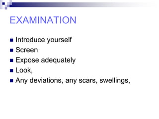
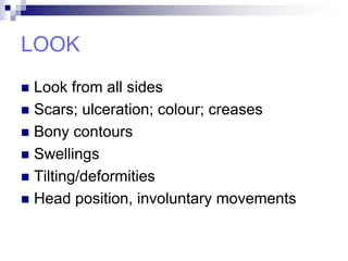
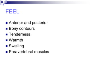
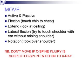
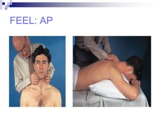
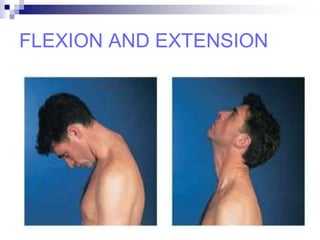
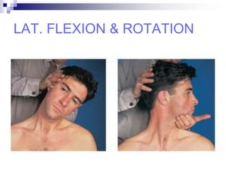
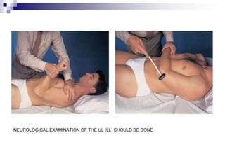
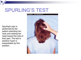
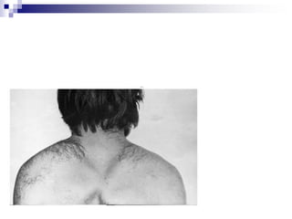
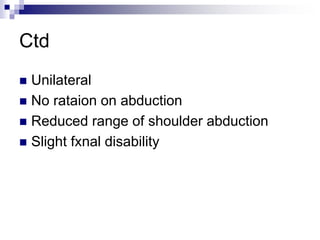
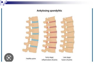
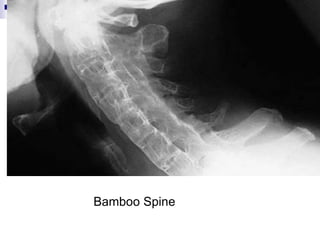
EXAMINATION OF
THE NECK AND
NECK
DEFORMITIES
Olusola Anifowose Richard
OUTLINE
 INTRODUCTION
 BRIEF ANATOMY
 THE STEPS
LOOK
FEEL
MOVE
 CONCLUSION
INTRODUCTION
 The connection between the brain and
body is by the spinal cord, which passes
through spinal canal of cervical vertebrae.
 The cervical spine is the bone of the neck,
its examination is of paramount important
in patient evaluation.
A BRIEF ANATOMY
 Neck supports the head
 Major conduit between head, trunk and
limbs
 Many important structures are crowded
together-muscles, aa, vv, nn, lymphatics,
trachea, esophagus and vertebrae
SKELETON OF THE NECK
 Cervical vertebrae-enclosing the spinal
cord and meninges
 Hyoid bone-mobile bone in the ant part of
the neck at C3 level
 Manubrium of the sternum
 Clavicles
VISCERA OF THE NECK
 Grouped into 3 layers:
1.Endocrine Layer-thyroid and parathyroid
glands
2.Respiratory Layer-larynx and trachea
3.Alimentary Layer-pharynx and
esophagus
NECK ROM
 Flexion
 Extension
 Lateral flexion
 Rotation
SYMPTOMS
 Pain
 Stiffness
 Deformity
 Headache
 Numbness; weakness in the UL and LL
EXAMINATION
 Introduce yourself
 Screen
 Expose adequately
 Look,
 Any deviations, any scars, swellings,
LOOK
 Look from all sides
 Scars; ulceration; colour; creases
 Bony contours
 Swellings
 Tilting/deformities
 Head position, involuntary movements
FEEL
 Anterior and posterior
 Bony contours
 Tenderness
 Warmth
 Swelling
 Paravertebral muscles
MOVE
 Active & Passive
 Flexion (touch chin to chest)
 Extend (look at ceiling)
 Lateral flexion (try to touch shoulder with
ear without raising shoulder)
 Rotation( look over shoulder)
NB: DON’T MOVE IF C-SPINE INJURY IS
SUSPECTED-SPLINT & GO ON TO X-RAY
FEEL: AP
FLEXION AND EXTENSION
LAT. FLEXION & ROTATION
NEUROLOGICAL EXAMINATION OF THE UL (LL) SHOULD BE DONE
ARTERIAL COMPRESSION
 The radial pulse disappears if there is
vascular compression at the thoracic inlet:
Adson’s test: when the patient holds a deep
breath, the neck is turned towards the
affected side and extended
Wright’s test: the shoulder is elevated and
externally rotated.
Adson's test
SPECIAL TESTS
 Dekleyn’s: head and neck rotation with
extension. Tests for vertebral artery
compression.
 Spurlin’s: (foraminal compression test): patient
hyperextends and rotates head to side, the
examiner then applies axial load to the head.
Positive test is when there is pain radiating into
ipsilateral arm.
 Elvey’s: (upper limb tension tests): tests
designed to put stress on the neurological
structures of the upper limb.
 Last two used in cervical radiculopathy
SPURLING’S TEST
Spurling's sign is
performed by the
patient extending her
neck and rotating her
head toward the side of
their pain. The test is
positive if pain is
exacerbated by this
position.
LHERMITTE’S SIGN
Lhermitte's sign is
performed by asking the
patient to maximally flex
her neck. The test is
positive if this causes
shooting, electric-like
pain in the lower
extremities.
RADIOLOGICAL EXAMINATION
 X-ray
Standard radiographic series comprises AP,
lateral and open-mouth views.
All seven vertebrae should be visible in lat.
View.
Alignment of cervical lordotic curve.
Assess the disc spaces.
AP view
Open mouth view
Lat. view
 MRI
No exposure to radiation
Provides excellent resolution of the disc and
neural structures
Most sensitive method of demonstrating
tumours and infection
Drawback: 20% of asymptomatic patients
show significant abnormalities, the scans
should be interpreted alongside the clinical
assessment.
CONCLUSION
 The neck is the ‘gear box’ of the body, a
thorough examination, taking into
consideration the bones, muscles, vessels
and nerves are important in orthopedic
conditions of the neck.
NECK DEFORMITIES
OUTLINE
 Introduction
 Classification
 The deformities
INTRODUCTION
 A variety of deformities
 Many are reflecting postural adjustments
to underlying disorders and others due to
developmental anomalies
CLASSIFICATION
 Torticollis
Congenital
Spasmodic
 Congenital short neck
 Congenital high scapula
Torticollis
Congenital/infantile/muscular Torticollis (wry neck)
• A common condition.
• The cause is unknown; the muscle may have suffered
ischaemia from a distorted position in utero ( breech
presentation and hip dysplasia ), or it may have been
injured at birth.
• The sternocleidomastoid muscle on one side is fibrous
and fails to elongate as the child grows; consequently,
progressive deformity develops.
 Hx-difficult labour or breech delivery
 O/E –lump in the first few wks of life, there is neither
deformity nor obvious limitation of movement and within
few months the lump disapear.
 Deformity does not become apparent until the child is 1–
2 years old. The head is tilted to on one side, so that the
ear approaches the shoulder; the sternomastoid on that
side may feel tight and hard.
 There may also be asymmetrical development of the
 face (plagiocephaly) becoming increasingly obvious as
the child grows.
• Differentials like lymphadenitis, bony anomalies, discitis
should be excluded.
• X-rays - r/o a bone abnormality or fracture
• Treatment: diagnosis made during infancy daily muscle
stretching by the parents may prevent the incipient
deformity.
• Non-operative treatment is successful in most cases.
• If the condition persists beyond one year, operative
correction is required to avoid progressive facial deformity.
• After operation, correction must be maintained, with a
temporary rigid orthosis followed by stretching exercise
Secondary torticollis
 Childhood torticollis may be secondary to congenital
bone anomalies, atlanto-axial rotatory displacement,
infection (lymphadenitis, retropharyngeal abscess,
tonsillitis, discitis, tuberculosis), trauma, juvenile
rheumatoid arthritis, posterior fossa tumours, intraspinal
tumours, dystonia.
 Treatment involves treating the underlying disease
Spasmodic Torticollis
 Spasmodic Torticollis (Cervical dystonia ) Spasmodic
torticollis is a neurologic disorder that results in an
involuntary turning or twisting of the head and neck to a
forced abnormal posture.
 ST may begin at any age, but appears most often
between the ages 25 to 55 years.
 It affects men and women about equally, and has no
predilection for any particular race or ethnic group.
 Positive family hx
 Neck discomfort, mild pain, and a feeling of stiffness .
 Treatment could be medical or surgical
 Medical: botulinum toxin,antiparkinson
agents,analgesics, mm relaxants
 Surgical: denervation surgery,DBS
KLIPPEL–FEIL SYNDROME
 It is a developmental disorder that represents a failure of
segmentation of the cervical somites
 it is often associated with abnormalities in the genito-
urinary, nervous or cardiovascular systems
 Some children have a hearing impediment.
 About 1 in 3 children with Klippel–Feil syndrome also
has Sprengel’s deformity of the scapula.
 Scoliosis is present in about 60 per cent and rib
anomalies in about 30 per cent.
 Hand deformities such as syndactyly, thumb hypoplasia
and extra digits are often present.
 For asymptomatic patients, treatment is unnecessary but
parents should be warned of the risks of contact sports;
 sudden catastrophic neurological compromise can occur
after minor trauma.
 Children with symptoms may need cervical fusion
Congenital high scapular
 A.k.a Sprengel’s shoulder
 Uncommon
 High scapular position due to failure of
migration
 Fixity
 Unknown cause
Ctd
 Unilateral
 No rataion on abduction
 Reduced range of shoulder abduction
 Slight fxnal disability
Treatment
Best left alone
Operatively
Divide contracted levator
scapulae and tight facial
bands
excise upper border
ctd
• Then fuse them together with
sutures and drill holes
• Divide blade of scapula
vertically throughout its length
near the vertebral border
• Displace lat (main part)
downward
Ankylosis Spondylitis
Ankylosing spondylitis is the most common
seronegative spondyloarthropathy to affect the
cervical spine.
The neck becomes progressively stiff and kyphotic
although some movement is usually preserved at
the atlanto-occipital and atlantoaxial joints.
Bamboo Spine
Ankylosis Spondylitis
SYMPTOMS
Neck pain
Neck Stiffness
Treatment.
A patient with ankylosing spondylitis and an
increase in neck pain must be assumed to have a
fracture until proven otherwise (by bone scan or MRI if
plain radiographs are normal). Neurological
compromise is common. A displaced fracture needs
careful
closed reduction with halo traction then halo vest
immobilization. Surgery carries a high complication
rate.
THANK YOU

Mariana Trench. Neck Exam.pptx

  • 1.
    EXAMINATION OF THE NECKAND NECK DEFORMITIES Olusola Anifowose Richard
  • 2.
    OUTLINE  INTRODUCTION  BRIEFANATOMY  THE STEPS LOOK FEEL MOVE  CONCLUSION
  • 3.
    INTRODUCTION  The connectionbetween the brain and body is by the spinal cord, which passes through spinal canal of cervical vertebrae.  The cervical spine is the bone of the neck, its examination is of paramount important in patient evaluation.
  • 4.
    A BRIEF ANATOMY Neck supports the head  Major conduit between head, trunk and limbs  Many important structures are crowded together-muscles, aa, vv, nn, lymphatics, trachea, esophagus and vertebrae
  • 5.
    SKELETON OF THENECK  Cervical vertebrae-enclosing the spinal cord and meninges  Hyoid bone-mobile bone in the ant part of the neck at C3 level  Manubrium of the sternum  Clavicles
  • 6.
    VISCERA OF THENECK  Grouped into 3 layers: 1.Endocrine Layer-thyroid and parathyroid glands 2.Respiratory Layer-larynx and trachea 3.Alimentary Layer-pharynx and esophagus
  • 7.
    NECK ROM  Flexion Extension  Lateral flexion  Rotation
  • 9.
    SYMPTOMS  Pain  Stiffness Deformity  Headache  Numbness; weakness in the UL and LL
  • 10.
    EXAMINATION  Introduce yourself Screen  Expose adequately  Look,  Any deviations, any scars, swellings,
  • 11.
    LOOK  Look fromall sides  Scars; ulceration; colour; creases  Bony contours  Swellings  Tilting/deformities  Head position, involuntary movements
  • 12.
    FEEL  Anterior andposterior  Bony contours  Tenderness  Warmth  Swelling  Paravertebral muscles
  • 13.
    MOVE  Active &Passive  Flexion (touch chin to chest)  Extend (look at ceiling)  Lateral flexion (try to touch shoulder with ear without raising shoulder)  Rotation( look over shoulder) NB: DON’T MOVE IF C-SPINE INJURY IS SUSPECTED-SPLINT & GO ON TO X-RAY
  • 14.
  • 15.
  • 16.
  • 17.
    NEUROLOGICAL EXAMINATION OFTHE UL (LL) SHOULD BE DONE
  • 18.
    ARTERIAL COMPRESSION  Theradial pulse disappears if there is vascular compression at the thoracic inlet: Adson’s test: when the patient holds a deep breath, the neck is turned towards the affected side and extended Wright’s test: the shoulder is elevated and externally rotated.
  • 19.
  • 21.
    SPECIAL TESTS  Dekleyn’s:head and neck rotation with extension. Tests for vertebral artery compression.  Spurlin’s: (foraminal compression test): patient hyperextends and rotates head to side, the examiner then applies axial load to the head. Positive test is when there is pain radiating into ipsilateral arm.  Elvey’s: (upper limb tension tests): tests designed to put stress on the neurological structures of the upper limb.  Last two used in cervical radiculopathy
  • 23.
    SPURLING’S TEST Spurling's signis performed by the patient extending her neck and rotating her head toward the side of their pain. The test is positive if pain is exacerbated by this position.
  • 24.
    LHERMITTE’S SIGN Lhermitte's signis performed by asking the patient to maximally flex her neck. The test is positive if this causes shooting, electric-like pain in the lower extremities.
  • 25.
    RADIOLOGICAL EXAMINATION  X-ray Standardradiographic series comprises AP, lateral and open-mouth views. All seven vertebrae should be visible in lat. View. Alignment of cervical lordotic curve. Assess the disc spaces.
  • 26.
  • 27.
  • 28.
  • 29.
     MRI No exposureto radiation Provides excellent resolution of the disc and neural structures Most sensitive method of demonstrating tumours and infection Drawback: 20% of asymptomatic patients show significant abnormalities, the scans should be interpreted alongside the clinical assessment.
  • 31.
    CONCLUSION  The neckis the ‘gear box’ of the body, a thorough examination, taking into consideration the bones, muscles, vessels and nerves are important in orthopedic conditions of the neck.
  • 32.
  • 33.
  • 34.
    INTRODUCTION  A varietyof deformities  Many are reflecting postural adjustments to underlying disorders and others due to developmental anomalies
  • 35.
  • 36.
    Torticollis Congenital/infantile/muscular Torticollis (wryneck) • A common condition. • The cause is unknown; the muscle may have suffered ischaemia from a distorted position in utero ( breech presentation and hip dysplasia ), or it may have been injured at birth. • The sternocleidomastoid muscle on one side is fibrous and fails to elongate as the child grows; consequently, progressive deformity develops.
  • 38.
     Hx-difficult labouror breech delivery  O/E –lump in the first few wks of life, there is neither deformity nor obvious limitation of movement and within few months the lump disapear.  Deformity does not become apparent until the child is 1– 2 years old. The head is tilted to on one side, so that the ear approaches the shoulder; the sternomastoid on that side may feel tight and hard.  There may also be asymmetrical development of the  face (plagiocephaly) becoming increasingly obvious as the child grows.
  • 39.
    • Differentials likelymphadenitis, bony anomalies, discitis should be excluded. • X-rays - r/o a bone abnormality or fracture • Treatment: diagnosis made during infancy daily muscle stretching by the parents may prevent the incipient deformity. • Non-operative treatment is successful in most cases. • If the condition persists beyond one year, operative correction is required to avoid progressive facial deformity. • After operation, correction must be maintained, with a temporary rigid orthosis followed by stretching exercise
  • 40.
    Secondary torticollis  Childhoodtorticollis may be secondary to congenital bone anomalies, atlanto-axial rotatory displacement, infection (lymphadenitis, retropharyngeal abscess, tonsillitis, discitis, tuberculosis), trauma, juvenile rheumatoid arthritis, posterior fossa tumours, intraspinal tumours, dystonia.  Treatment involves treating the underlying disease
  • 41.
    Spasmodic Torticollis  SpasmodicTorticollis (Cervical dystonia ) Spasmodic torticollis is a neurologic disorder that results in an involuntary turning or twisting of the head and neck to a forced abnormal posture.  ST may begin at any age, but appears most often between the ages 25 to 55 years.  It affects men and women about equally, and has no predilection for any particular race or ethnic group.  Positive family hx  Neck discomfort, mild pain, and a feeling of stiffness .
  • 42.
     Treatment couldbe medical or surgical  Medical: botulinum toxin,antiparkinson agents,analgesics, mm relaxants  Surgical: denervation surgery,DBS
  • 43.
    KLIPPEL–FEIL SYNDROME  Itis a developmental disorder that represents a failure of segmentation of the cervical somites  it is often associated with abnormalities in the genito- urinary, nervous or cardiovascular systems  Some children have a hearing impediment.  About 1 in 3 children with Klippel–Feil syndrome also has Sprengel’s deformity of the scapula.  Scoliosis is present in about 60 per cent and rib anomalies in about 30 per cent.  Hand deformities such as syndactyly, thumb hypoplasia and extra digits are often present.
  • 44.
     For asymptomaticpatients, treatment is unnecessary but parents should be warned of the risks of contact sports;  sudden catastrophic neurological compromise can occur after minor trauma.  Children with symptoms may need cervical fusion
  • 46.
    Congenital high scapular A.k.a Sprengel’s shoulder  Uncommon  High scapular position due to failure of migration  Fixity  Unknown cause
  • 47.
    Ctd  Unilateral  Norataion on abduction  Reduced range of shoulder abduction  Slight fxnal disability
  • 48.
    Treatment Best left alone Operatively Dividecontracted levator scapulae and tight facial bands excise upper border
  • 49.
    ctd • Then fusethem together with sutures and drill holes • Divide blade of scapula vertically throughout its length near the vertebral border • Displace lat (main part) downward
  • 50.
    Ankylosis Spondylitis Ankylosing spondylitisis the most common seronegative spondyloarthropathy to affect the cervical spine. The neck becomes progressively stiff and kyphotic although some movement is usually preserved at the atlanto-occipital and atlantoaxial joints.
  • 52.
  • 53.
  • 54.
    Treatment. A patient withankylosing spondylitis and an increase in neck pain must be assumed to have a fracture until proven otherwise (by bone scan or MRI if plain radiographs are normal). Neurological compromise is common. A displaced fracture needs careful closed reduction with halo traction then halo vest immobilization. Surgery carries a high complication rate.
  • 55.

Editor's Notes

  • #8 Range of movt(LF45 ;R80) Occipitomental line
  • #10 Evaluation starts with hx Wry neck Nerve root, cord compresssion
  • #13 Sit; prone with pillow support
  • #14 Passive for tenderness
  • #18 MUSCULAR Bulk, tone , power, SENSORY Touch, deep pressure, note the dermatomes REFLEXES
  • #22 Cord compression
  • #25 MS and cervical rad.
  • #32 A car is useless without its GB