
Recommended
More Related Content
Similar to Lymphatic Organs and Tissues
Similar to Lymphatic Organs and Tissues (20)
Recently uploaded
Recently uploaded (20)
Lymphatic Organs and Tissues
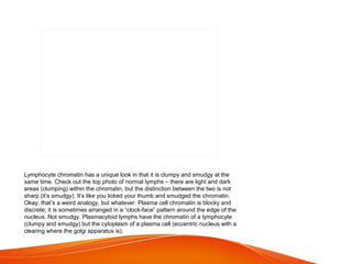
- 1. Lymphocyte chromatin has a unique look in that it is clumpy and smudgy at the same time. Check out the top photo of normal lymphs – there are light and dark areas (clumping) within the chromatin, but the distinction between the two is not sharp (it’s smudgy). It’s like you licked your thumb and smudged the chromatin. Okay, that’s a weird analogy, but whatever. Plasma cell chromatin is blocky and discrete; it is sometimes arranged in a “clock-face” pattern around the edge of the nucleus. Not smudgy. Plasmacytoid lymphs have the chromatin of a lymphocyte (clumpy and smudgy) but the cytoplasm of a plasma cell (eccentric nucleus with a clearing where the golgi apparatus is).
- 2. Reactive lymphocytes – particularly big ones – can look a lot like monocytes. Again, the key is to look at the chromatin. Large reactive lymphocytes are usually immunoblasts, and as such, they have a big nucleolus (or two). In the bottom photo, there is a big reactive lymphocyte (called a Downey 3 cell) on the right. These cells also have fine chromatin (it has to be fine, or you wouldn’t be seeing the nucleolus). Monocyte chromatin is more dense (no nucleoli) and has a “raked” appearance. It is like you dragged a tiny garden rake across the nucleus. Also, the nucleus is often kidney-bean or horse-shoe shaped, or at least has a nice indentation or two. In addition to the chromatin differences, there are cytoplasmic differences (though these are less consistent): monocyte cytoplasm is typically dishwater grey with tiny dust-like granules, whereas reactive lymphocyte cytoplasm is usually light blue (either pale light blue or a relatively bright light blue) and if granules are present, they tend to be larger.
- 3. * demarcates paracortex and medullary areas
- 4. Hemophilus influenzae type b (Hib) Streptococcus pneumoniae (pneumococcus) Neisseria meningitidis (meningococcus) Group B streptococcus (GBS) Klebsiella pneumoniae. Salmonella typhi
- 5. central arteriole with PALS
- 6. The open circulation model is currently the most favored. It provides a more efficient exposure of blood to red pulp macrophages. In the closed circulation model, blood cells would have to leave the sinuses and then reenter them in order to be exposed to macrophages.
- 7. The open circulation model is currently the most favored. It provides a more efficient exposure of blood to red pulp macrophages. In the closed circulation model, blood cells would have to leave the sinuses and then reenter them in order to be exposed to macrophages.