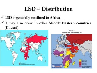
Viral Diseases
Faculty of Veterinary Medicine & Animal
Husbandry
Somali National University
Mogadishu, Gaheyr Campus
OCt. 01. 2022
LUMPY SKIN DISEASE
• Lumpy skin disease was first diagnosed in
northern Rhodesia/Zambia in 1929.
• and then spread to Botswana in 1943, South
Africa in 1944, southern Rhodesia/Zimbabwe in
1945, Kenya in 1957, Sudan in 1971, Chad and
Niger in 1973, Nigeria in 1974, and Ethiopia in
1983.
• Lumpy skin disease spread steadily to almost all
sub-Saharan countries by the end of the 1970s and
remained only in this region till 1987.
From 1988, the diseases spread to Egypt.
Initially it was difficult to know the etiological
agent of LSD.
LSD was first described by MacDonald in
1931 as “pseudo-urticaria”
Lumpy skin disease is an acute disease of
cattle characterized by fever, cutaneous
nodules and generalized lymphadenitis.
Etiology:
LSD is caused by a virus in the genus
Capripoxvirus of the family Poxviridae
related to the arthropod-borne pox virus of
sheep and goats.
Occurrence:
 The endemic area lies in southern Africa.
 In recent years epidemics have occurred
in East and West Africa.
Geographic Distribution and
Economic Impact
• Lumpy skin disease is currently endemic and
widespread in almost all African countries except
for a few northern African countries: Libya,
Tunisia, Algeria, and Morocco.
• After being confined for almost 60 years in the
sub-Saharan African region and Egypt, the disease
has spread to the Middle East countries such as
Israel, the Palestinian Autonomous Territories,
Jordan, Lebanon, Kuwait, Iraq, Iran, Saudi
Arabia, Bahrain, Oman, Yemen, and the United
Arab Emirates
LSD is thought to have originated in Zambia
from where it spread to Zimbabwe,
Mozambique, and South Africa.
In 1957, LSD was first seen in East Africa
in Kenya in the Rift Valley.
epidemics of LSD have occurred irregularly
and have extended to include the whole of
Kenya, and have also been reported from
Uganda, Somalia and Tanzania. The disease
was reported from the Nile basin of southern
Sudan in the early 1970s
• A report estimated a total economic loss of around
USD667,785.60 due to LSD outbreaks between
June and December 2011 in feedlot farms in
central Ethiopia.
• also estimated a milk price loss of USD44.70 per
cow during an average of 20 days when an
outbreak occurs on a dairy farm. Although
mortality rate caused by LSD is usually low, the
disease is of major economic importance owing to
costs incurred for veterinary service, cow death,
abortion, and still birth of calves
LSD – Distribution
 LSD is generally confined to Africa
 It may also occur in other Middle Eastern countries
(Kuwait)
LSD - Species Affected
LSD is primarily a disease of cattle
Bos taurus breeds (Jersey, Guernsey and
Ayrshire) are more susceptible to clinical
disease than zebu cattle (Bos indicus)
Indigenous cattle rarely become sick and
seldom die but exotic cattle react more
severely. A few cases have been reported in
Asian water buffalo (Bubalus bubalis)
In general young calves and lactating cows
appear more susceptible.
Giraffe and impala have been shown
experimentally to be susceptible.
Wild animals are not thought to play an
important role in the spread or
maintenance of LSDV
LSD - Transmission
LSDV is thought to be transmitted primarily
by biting insects
This virus has been found in mosquitoes in the
genera Aedes and Culex during some
outbreaks
Flies and other insects might also be involved
in transmission, but this remains unproven
Direct contact could be a minor source of
infection
LSD - Transmission
LSDV occurs in cutaneous lesions, saliva,
respiratory secretions, milk and semen
Animals can be infected experimentally by
inoculation with material from cutaneous
nodules or blood, or by ingestion of feed
and water contaminated with saliva
LSD - Clinical Signs
CS of LSD range from inapparent to severe (age
and breed)
Fever at the beginning then formation of
nodules on the skin and mucosae (in 2 days)
The size of the nodules vary from 1 cm to 7 cm
and penetrate the full thickness of the skin
Nodules are common on the head, neck, udder,
genitalia, perineum and legs
LSD - Clinical Signs
Nodules develop a characteristic inverted
conical zone of necrosis, which penetrates
the skin layers, subcutaneous tissue, and
sometimes the underlying muscle
The cores of necrotic material become
separated from the adjacent skin and are
called sit-fasts
Secondary bacterial infections occur
LSD - Clinical Signs
Superficial lymph nodes become enlarged
and edematous
Rhinitis, conjunctivitis and keratitis can
also be seen
Abortions and temporary or permanent
sterility may occur in both bulls and cows
A few animals die but the majority slowly
recover
Lumpy skin disease, ulcers from which the "sitfasts" have separated
Bovine, skin. There are disseminated cutaneous papules.
Bovine, skin. There are disseminated cutaneous papules with
necrotic centers (sitfasts).
Bovine, skin. Necrotic centers (sitfasts) of two of these papules
have sloughed.
Bovine, skin. Multiple subcutaneous nodules elevate the skin.
Bovine, muzzle. There are multiple sharply-demarcated
slightly raised papules, often with eroded surfaces, that extend
into the nares.
Pathology:
 The cut surface of the nodules is dull and
pinkish-grey and the subcutis is often
infiltrated by red serous fluid.
 Sit-fasts are usually raised above the level
of the surrounding skin on granulation
tissue.
 Microscopically the nodules are variable in
morphology.
 In animals that die nodules are found to
occur in visceral organs, particularly the
lungs and the tissues of the upper
respiratory and digestive tracts, as well as
cutaneously.
 All the lymph nodes are enlarged.
Bovine, nasal turbinate. Early pox lesions are slightly pale round foci
rimmed by petechiae.
Bovine, nasal turbinate. The centers of well-developed
pox are necrotic.
Bovine, trachea. The mucosa contains a poorly demarcated
round focus rimmed by mild hemorrhage (early pox lesion).
Bovine, trachea. Two coalescing mucosal macules have hyperemic
margins.
Bovine, lung. There is marked generalized interlobular edema,
and there is a small cluster of red nodules on the left side of the
specimen.
Diagnosis:
 Typical cases are easily recognized
clinically during epidemics but subacute
cases are often difficult to diagnose.
 Confirmation is best sought by examining
a biopsy sample from an early skin
nodule by electron microscopy.
Supportive diagnostic aids include:
 virus isolation in cattle or in cell cultures
and
 histopathology.
An early nodule should be excised and
forwarded to the laboratory; half the
nodule should be immersed in ten per cent
formol-saline and the other half should be
sent frozen.
The nodules of the Neethling type of lumpy skin
disease must be differentiated from :
 those of the Allerton type of pseudolumpy skin
disease,
 from the lesions of onchocerciasis,
 streptothricosis,
 globidiosis,
 ringworm,
 skin tuberculosis and urticaria, and
 from tick and insect bites.
Other conditions of differential importance
include:
 malignant catarrhal fever,
 photosensitization and
 sweating sickness.
Immunology:
The duration of immunity in recovered
animals is unknown but it is probably
lifelong.
A hypersensitivity test is used to detect
`immune' animals.
The antigen is injected intradermally and
the test is read 48 hours later, an increase
in the skin thickness of 3 mm or greater
being considered positive.
Control:
Quarantine measures do not prevent
spread of lumpy skin disease.
Prophylactic vaccination is therefore
recommended for the protection of cattle in
endemic areas.
Vaccines are available; an attenuated
Neethling strain of the virus is used in
south Africa and an attenuated sheep-goat
pox vaccine in East Africa.
If the disease occurs affected animals
should be segregated in insect-proof
buildings, and in-contact animals should be
vaccinated. proper disposal of carcasses
cleaning and disinfection of the premises
and insect control
Import restrictions can help prevent the
introduction of LSD
Lumpy skin disease.pptx

Lumpy skin disease.pptx

  • 1.
    Viral Diseases Faculty ofVeterinary Medicine & Animal Husbandry Somali National University Mogadishu, Gaheyr Campus OCt. 01. 2022 LUMPY SKIN DISEASE
  • 2.
    • Lumpy skindisease was first diagnosed in northern Rhodesia/Zambia in 1929. • and then spread to Botswana in 1943, South Africa in 1944, southern Rhodesia/Zimbabwe in 1945, Kenya in 1957, Sudan in 1971, Chad and Niger in 1973, Nigeria in 1974, and Ethiopia in 1983. • Lumpy skin disease spread steadily to almost all sub-Saharan countries by the end of the 1970s and remained only in this region till 1987.
  • 3.
    From 1988, thediseases spread to Egypt. Initially it was difficult to know the etiological agent of LSD. LSD was first described by MacDonald in 1931 as “pseudo-urticaria”
  • 4.
    Lumpy skin diseaseis an acute disease of cattle characterized by fever, cutaneous nodules and generalized lymphadenitis.
  • 5.
    Etiology: LSD is causedby a virus in the genus Capripoxvirus of the family Poxviridae related to the arthropod-borne pox virus of sheep and goats.
  • 6.
    Occurrence:  The endemicarea lies in southern Africa.  In recent years epidemics have occurred in East and West Africa.
  • 7.
    Geographic Distribution and EconomicImpact • Lumpy skin disease is currently endemic and widespread in almost all African countries except for a few northern African countries: Libya, Tunisia, Algeria, and Morocco. • After being confined for almost 60 years in the sub-Saharan African region and Egypt, the disease has spread to the Middle East countries such as Israel, the Palestinian Autonomous Territories, Jordan, Lebanon, Kuwait, Iraq, Iran, Saudi Arabia, Bahrain, Oman, Yemen, and the United Arab Emirates
  • 8.
    LSD is thoughtto have originated in Zambia from where it spread to Zimbabwe, Mozambique, and South Africa. In 1957, LSD was first seen in East Africa in Kenya in the Rift Valley. epidemics of LSD have occurred irregularly and have extended to include the whole of Kenya, and have also been reported from Uganda, Somalia and Tanzania. The disease was reported from the Nile basin of southern Sudan in the early 1970s
  • 9.
    • A reportestimated a total economic loss of around USD667,785.60 due to LSD outbreaks between June and December 2011 in feedlot farms in central Ethiopia. • also estimated a milk price loss of USD44.70 per cow during an average of 20 days when an outbreak occurs on a dairy farm. Although mortality rate caused by LSD is usually low, the disease is of major economic importance owing to costs incurred for veterinary service, cow death, abortion, and still birth of calves
  • 10.
    LSD – Distribution LSD is generally confined to Africa  It may also occur in other Middle Eastern countries (Kuwait)
  • 12.
    LSD - SpeciesAffected LSD is primarily a disease of cattle Bos taurus breeds (Jersey, Guernsey and Ayrshire) are more susceptible to clinical disease than zebu cattle (Bos indicus) Indigenous cattle rarely become sick and seldom die but exotic cattle react more severely. A few cases have been reported in Asian water buffalo (Bubalus bubalis)
  • 14.
    In general youngcalves and lactating cows appear more susceptible. Giraffe and impala have been shown experimentally to be susceptible. Wild animals are not thought to play an important role in the spread or maintenance of LSDV
  • 17.
    LSD - Transmission LSDVis thought to be transmitted primarily by biting insects This virus has been found in mosquitoes in the genera Aedes and Culex during some outbreaks Flies and other insects might also be involved in transmission, but this remains unproven Direct contact could be a minor source of infection
  • 18.
    LSD - Transmission LSDVoccurs in cutaneous lesions, saliva, respiratory secretions, milk and semen Animals can be infected experimentally by inoculation with material from cutaneous nodules or blood, or by ingestion of feed and water contaminated with saliva
  • 20.
    LSD - ClinicalSigns CS of LSD range from inapparent to severe (age and breed) Fever at the beginning then formation of nodules on the skin and mucosae (in 2 days) The size of the nodules vary from 1 cm to 7 cm and penetrate the full thickness of the skin Nodules are common on the head, neck, udder, genitalia, perineum and legs
  • 21.
    LSD - ClinicalSigns Nodules develop a characteristic inverted conical zone of necrosis, which penetrates the skin layers, subcutaneous tissue, and sometimes the underlying muscle The cores of necrotic material become separated from the adjacent skin and are called sit-fasts Secondary bacterial infections occur
  • 22.
    LSD - ClinicalSigns Superficial lymph nodes become enlarged and edematous Rhinitis, conjunctivitis and keratitis can also be seen Abortions and temporary or permanent sterility may occur in both bulls and cows A few animals die but the majority slowly recover
  • 23.
    Lumpy skin disease,ulcers from which the "sitfasts" have separated
  • 24.
    Bovine, skin. Thereare disseminated cutaneous papules.
  • 25.
    Bovine, skin. Thereare disseminated cutaneous papules with necrotic centers (sitfasts).
  • 26.
    Bovine, skin. Necroticcenters (sitfasts) of two of these papules have sloughed.
  • 27.
    Bovine, skin. Multiplesubcutaneous nodules elevate the skin.
  • 28.
    Bovine, muzzle. Thereare multiple sharply-demarcated slightly raised papules, often with eroded surfaces, that extend into the nares.
  • 29.
    Pathology:  The cutsurface of the nodules is dull and pinkish-grey and the subcutis is often infiltrated by red serous fluid.  Sit-fasts are usually raised above the level of the surrounding skin on granulation tissue.  Microscopically the nodules are variable in morphology.
  • 30.
     In animalsthat die nodules are found to occur in visceral organs, particularly the lungs and the tissues of the upper respiratory and digestive tracts, as well as cutaneously.  All the lymph nodes are enlarged.
  • 31.
    Bovine, nasal turbinate.Early pox lesions are slightly pale round foci rimmed by petechiae.
  • 32.
    Bovine, nasal turbinate.The centers of well-developed pox are necrotic.
  • 33.
    Bovine, trachea. Themucosa contains a poorly demarcated round focus rimmed by mild hemorrhage (early pox lesion).
  • 34.
    Bovine, trachea. Twocoalescing mucosal macules have hyperemic margins.
  • 35.
    Bovine, lung. Thereis marked generalized interlobular edema, and there is a small cluster of red nodules on the left side of the specimen.
  • 36.
    Diagnosis:  Typical casesare easily recognized clinically during epidemics but subacute cases are often difficult to diagnose.  Confirmation is best sought by examining a biopsy sample from an early skin nodule by electron microscopy.
  • 37.
    Supportive diagnostic aidsinclude:  virus isolation in cattle or in cell cultures and  histopathology. An early nodule should be excised and forwarded to the laboratory; half the nodule should be immersed in ten per cent formol-saline and the other half should be sent frozen.
  • 38.
    The nodules ofthe Neethling type of lumpy skin disease must be differentiated from :  those of the Allerton type of pseudolumpy skin disease,  from the lesions of onchocerciasis,  streptothricosis,  globidiosis,  ringworm,  skin tuberculosis and urticaria, and  from tick and insect bites.
  • 39.
    Other conditions ofdifferential importance include:  malignant catarrhal fever,  photosensitization and  sweating sickness.
  • 40.
    Immunology: The duration ofimmunity in recovered animals is unknown but it is probably lifelong. A hypersensitivity test is used to detect `immune' animals. The antigen is injected intradermally and the test is read 48 hours later, an increase in the skin thickness of 3 mm or greater being considered positive.
  • 41.
    Control: Quarantine measures donot prevent spread of lumpy skin disease. Prophylactic vaccination is therefore recommended for the protection of cattle in endemic areas. Vaccines are available; an attenuated Neethling strain of the virus is used in south Africa and an attenuated sheep-goat pox vaccine in East Africa.
  • 42.
    If the diseaseoccurs affected animals should be segregated in insect-proof buildings, and in-contact animals should be vaccinated. proper disposal of carcasses cleaning and disinfection of the premises and insect control Import restrictions can help prevent the introduction of LSD