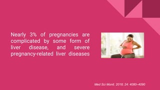
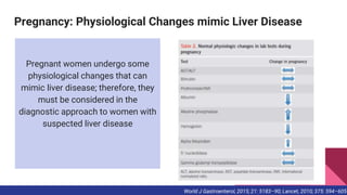
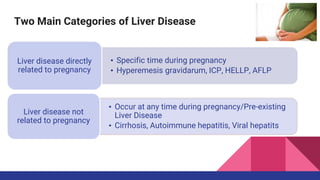
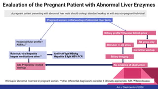
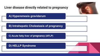
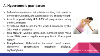
LIVER DISORDERS IN
PREGNANCY
Dr Yashodhara Gaur
Nearly 3% of pregnancies are
complicated by some form of
liver disease, and severe
pregnancy-related liver diseases
Med Sci Monit. 2018; 24: 4080–4090
Pregnancy: Physiological Changes mimic Liver Disease
Pregnant women undergo some
physiological changes that can
mimic liver disease; therefore, they
must be considered in the
diagnostic approach to women with
suspected liver disease
World J Gastroenterol, 2015; 21: 5183–90; Lancet, 2010; 375: 594–605
Liver diseases unique to pregnancy accordingly gestational
period
Best Practice & Research Clinical Gastroenterology 44-45 (2020) 101667
Two Main Categories of Liver Disease
• Specific time during pregnancy
• Hyperemesis gravidarum, ICP, HELLP, AFLP
Liver disease directly
related to pregnancy
• Occur at any time during pregnancy/Pre-existing
Liver Disease
• Cirrhosis, Autoimmune hepatitis, Viral hepatits
Liver disease not
related to pregnancy
Evaluation of the Pregnant Patient with Abnormal Liver Enzymes
A pregnant patient presenting with abnormal liver tests should undergo standard workup as with any non-pregnant individual
Pregnant women: initial workup of abnormal liver tests
Hepatocellular profile?
AST/ALT
Rule out: viral hepatitis
herpes medications other**
Anti-HAV IgM HBsAg
Hepatitis E IgM HSV PCR
See Pregnancy-related
workup
Billiary profile? Elevated bili/alk phos
Bilirubin +/- alk phos
Biliary imaging
No further workup
No evidence of obstruction
Alk phos only
Workup of abnormal liver test in pregnant women. **other differential diagnosis to consider if clinically appropriate. AIH, Wilson disease.
Am J Gastroenterol 2016
Diagnostic Modalities for Liver disease in pregnancy
Hepatology Communications 2020;4:145-156
 Ultrasound is safe and the preferred imaging
modality used in assessment of abnormal liver
tests suggestive of biliary tract disease
 Magnetic resonance imaging without
gadolinium can be used in the second and third
trimester
 Computed tomography scans carry a risk of
teratogenesis and childhood hematologic
malignancies but may be used judiciously with
minimized radiation protocols (2–5 rads)
Evaluation of the Pregnant Patient with Abnormal Liver Enzymes
Am J Gastroenterol 2016
 Endoscopy is safe in pregnancy but should be
deferred until the second trimester if possible
 Meperidine and propofol can be used for endoscopic
sedation
Evaluation of the Pregnant Patient with Abnormal Liver
Enzymes
Am J Gastroenterol 2016
Liver disease directly related to pregnancy
A) Hyperemesis gravidarum
B) Intrahepatic Cholestasis of pregnancy
C) Acute fatty liver of pregnancy (AFLP)
D) HELLP Syndrome
A. Hyperemesis gravidarum
 Defined as nausea and intractable vomiting that results in
dehydration, ketosis, and weight loss >5% of body weight
 Affects approximately 0.3–2.0% of pregnancies during
the first trimester
 Symptoms start before the 9th week & disappear by the
20th week of gestation
 Risk factors: Multiple gestations, increased body mass
index (BMI), pre-existing diabetes, psychiatric illness, past
history
 Complications: Dehydration, increased renal values,
electrolyte abnormalities, metabolic alkalosis,
erythrocytosis J Hepatol. 2016; 64(4):933-45
A. Hyperemesis gravidarum
 Abnormal liver test results are observed in approximately half of all pregnant
women
 Elevations in serum aminotransferases usually go up to 200 U/L and this is
the most common abnormal liver test result
 An abdominal ultrasound must demonstrate normal liver parenchyma
without biliary obstruction, and an obstetrical ultrasound can exclude
hydatidiform mole and multiple gestation
 Management of HG is supportive and includes intravenous fluid
replacement, correction of electrolyte abnormalities, use of antiemetics,
possible parenteral nutrition, and vitamin supplementation
J Hepatol. 2016; 64(4):933-45
B. Intrahepatic Cholestasis of pregnancy
 Most common cause of cholestasis during
pregnancy.
 Most common pregnancy-related liver disease.
 Characterized by a reversible cholestatic condition
that usually occurs during the late second and third
trimester.
 Rapid postnatal resolution, with signs and
symptoms usually disappearing spontaneously
within 6 weeks of delivery.
 Recurs in more than half of subsequent pregnancies.
Dig Liver Dis. 2016; 48(2):120-37
B. Intrahepatic cholestasis of pregnancy
 Risk factors: Maternal age >35 years, multiparity, history of oral contraceptive
use, history of fertility treatment in women, Past h/o ICP
 Symptoms: Pruritus predominates on the palms and soles of the feet and
worsens at night
• Develops after 25 weeks of gestation, with 80% of cases occurring after
the 30th week.
 Other symptoms: Steatorrhea, malabsorption of fat-soluble vitamins, and
weight loss due to cholestasis.
 Jaundice has an incidence of 14–25% and may develop 1 to 4 weeks after the
onset of pruritus, with dark urine and pale feces in some women.
Dig Liver Dis. 2016; 48(2):120-37
B. Intrahepatic cholestasis of pregnancy
 Goals of ICP treatment are to reduce maternal symptoms, improve laboratory
test results, and improve fetal outcome
 First-line therapy for ICP is ursodeoxycholic acid (UDCA) at a dose of 500 mg
twice a day or 15 mg/kg per day
 UDCA is safe in the third trimester because no maternal or fetal adverse
effects have been reported regarding the use of this medication in ICP
 Other medications, such as cholestyramine and S-adenosyl-methionine, have
not had satisfactory result
 In women who do not respond to UDCA, combining rifampicin with UDCA
improves their symptoms and laboratory abnormalities
Dig Liver Dis. 2016; 48(2):120-37
C. Acute fatty liver of pregnancy (AFLP)
 Microvesicular fatty infiltration of hepatocytes
● that is also known as acute yellow atrophy or
● acute fatty metamorphosis.
 Medical and obstetric emergency because it can be fatal for both the mother and
child in the absence of early recognition and appropriate management.
 Rare condition, usually occurring in the third trimester, with an approximate
incidence of 1: 7000 to 1: 16 000 pregnancies.
 Pathogenesis of AFLP is not fully understood, but investigations over the past few
years suggest that it may result from mitochondrial dysfunction in most cases.
J Hepatol. 2016; 64(4):933-45.
C. Acute fatty liver of pregnancy (AFLP)
 Risk factors include multigravidas, preeclampsia, multiple gestation, male
fetus, underweight.
 AFLP usually occurs in the third trimester, rarely before the 30th gestational
week, but up to 20% of cases present postnatally
 Clinical presentation is variable and includes non-specific symptoms such as
nausea, vomiting, abdominal pain, headache, and malaise. The clinical course
can rapidly progress to acute liver failure and its complications, such as
encephalopathy, jaundice, and coagulopathy.
J Hepatol. 2016; 64(4):933-45
C. Acute fatty liver of pregnancy (AFLP)
Swansea criteria for diagnosis of acute fatty liver of pregnancy
• Sensitivity of 100%
• Specificity of 57%
• Positive predictive value 85%
• Negative predictive value 100%
C. Acute fatty liver of pregnancy (AFLP)
 Early recognition of the disease and delivery of the foetus regardless of
gestational age are the keys to successful management, followed by maternal
care, which is mainly supportive
 There are no reports in the literature of recovery before the delivery of the child
 The life-threatening condition associated with AFLP is acute liver failure
 Aggressive maternal stabilization in an intensive care setting is necessary, with
correction of coagulation, hypoglycemia, and other abnormalities
 In some non-randomized reports, the use of plasma exchange (PE), as well as
PE in combination with continuous renal replacement therapy (CRRT), following
delivery was associated with improved clinical outcomes, including reduced
maternal mortality
 Liver transplantation (LT) has been reported sporadically as a last-chance
measure
Gynecol Obstet Invest. 2015; 79(2):97-100.
D. HELLP syndrome
● HELLP syndrome is a variant of severe preeclampsia
that occurs in up to 12% of patients with preeclampsia,
but can also occur in normotensive patients.
● Risk factors for HELLP are advanced maternal age and
multiparity.
● Characterized by the presence of hemolysis, elevated
liver aminotransferases, and low platelet counts.
● Disorder can be diagnosed antepartum (in 70% of
women between 27 and 30 weeks) or postpartum
● In the postpartum period, the HELLP syndrome often
develops within the first 48 h, usually in patients who
had proteinuria and hypertension prior to delivery
D. HELLP syndrome
Diagnostic criteria
considered to be a clinically significant transition phase
of HELLP syndrome, which has the ability to progress
Tennessee HELLP diagnostic criteria Mississippi HELLP diagnostic criteria
Blood Adv. 2017 Sep 12; 1(20):1628-1631
D. HELLP syndrome
● Abdominal imaging methods, such as
ultrasonography, CT, and MRI, should be
performed in all women with HELLP syndrome in
order to investigate liver complications, such as
liver infarction, intraparenchymal hemorrhage,
subcapsular hematoma, and hepatic rupture
● In cases in which liver biopsy was performed,
histological changes in the liver in HELLP
syndrome include periportal changes with
hemorrhage, sinusoidal fibrin deposition, and
hepatocyte necrosis
D. HELLP syndrome
● Differential diagnosis among AFLP and HELLP syndrome
D. HELLP syndrome
 Definitive treatment is delivery of the foetus and stabilizing the maternal
clinical condition
 Steroids should be given only for foetal lung maturity, but their use is unclear
and remains controversial
 A recent meta-analysis, Mao et al. investigated the efficacy of corticosteroid
therapy in patients with HELLP syndrome, reporting that treatment with
corticosteroids significantly improved the PLT count, LDH levels, and ALT
levels, but the decrease in AST levels was not significant
 Corticosteroid treatment was also associated with a significantly lower blood
transfusion rate and shorter hospital/intensive care unit stay
Med Sci Monit. 2015; 21():3777-83
Differential diagnosis of liver diseases unique to pregnancy
Best Practice & Research Clinical Gastroenterology 44-45 (2020) 101667
Liver disease not related to pregnancy
a) Cirrhosis and Portal Hypertension
b) Acute hepatitis in Pregnancy
c) Chronic hepatitis B & C in pregnancy
d) Autoimmune liver disease during pregnancy
e) Liver Transplantation (LT) and Pregnancy
A. Cirrhosis and Portal Hypertension
 Pregnancy is relatively rare in women with liver cirrhosis
 When pregnancy occurs, the risk of spontaneous abortion,
prematurity, and perinatal death are very high
 With advancements in the medical field, pregnancy is not
contraindicated in these women, as previously believed
 Outcome of pregnancy is related to the severity of the
maternal liver disease
○ Model for end-stage liver disease (MELD) has been
shown to be a good model for predicting the risk of
decompensation of maternal liver disease during
pregnancy J Hepatol. 2016; 64(4):933-45
A. Cirrhosis and Portal Hypertension
 In pregnant women with cirrhosis, the most common and most serious
complication is variceal bleeding
 Each episode of variceal bleeding leads to maternal mortality rates as high
as 20–50%, with an even higher risk of fetal loss
 Variceal bleeding during pregnancy is managed very similarly to variceal
bleeding in general given the acute and life-threatening nature of the event,
with a focus on endoscopic hemostasis and supportive care for the mother
and fetus
 Octreotide is a pregnancy category B drug and appears to be safe as an
adjunct treatment in acute variceal bleeding along with antibiotics
 Salvage therapy with transjugular intrahepatic systemic shunts and liver
transplantation has also been described, but are not routinely advocated
J Hepatol. 2016; 64(4):933-45
A. Cirrhosis and Portal Hypertension
 Vaginal delivery with a short second stage of labor has been advocated along
with consideration of forceps or vacuum extraction, if needed
 However, the recognition that prolonged vaginal delivery may be associated
with an increased risk of variceal bleeding owing to repeated Valsalva
maneuvers has led to an interest in considering cesarean sections as an
alternative.
 Cesarean sections may be required for fetal distress or prematurity, but
carries an increased risk of bleeding complications from the surgical site in
the setting of portal hypertension.
 There is no data comparing the benefits of vaginal delivery vs. cesarean
section
J Hepatol. 2016; 64(4):933-45
B. Acute hepatitis in Pregnancy
 Pregnant women presenting with acute hepatitis should be tested for
common etiologies of acute liver injury including viral hepatitis, hepatitis A
virus (HAV), hepatitis B virus (HBV), hepatitis E virus (HEV), and herpes
simplex virus
 No published systematic reviews or meta-analysis on the evaluation and
management of hepatitis A, hepatitis E, or HSV hepatitis during pregnancy
 CDC recommends HAV immunoglobulin treatment for the neonate if the
maternal HAV infection occurs within 2 weeks of delivery
 Pregnant women with acute hepatitis suspected from HSV should be initiated
on acyclovir
Am J Gastroenterol 2016
C. Chronic hepatitis B in pregnancy
 Active–passive immunoprophylaxis with hepatitis B immunoglobulin and the
HBV vaccination series should be administered to all infants born to HBV-
infected mothers to prevent perinatal transmission
 Women chronically infected with HBV and high viral load (>200,000 U/ml or
>10 6 log copies/ml and higher) should be offered antiviral medication with
tenofovir or telbivudine in the third trimester to reduce perinatal transmission
of HBV
 C-section should not be performed electively in HBV-positive mothers to
prevent foetal infection
 Women chronically infected with HBV should be allowed to breastfeed as
recommended for infant health
Am J Gastroenterol 2016
C. Chronic hepatitis C in pregnancy
 All pregnant women with risk factors for HCV should be screened with anti-
HCV antibody. Screening should not be performed in women without risk
factors for HCV acquisition
 Invasive procedures (e.g., amniocentesis, invasive fetal monitoring) should be
minimized in infected mothers and their fetus to prevent vertical transmission
of hevpatitis C
 C-section should not be performed electively in HCV-positive mothers to
prevent fetal infection
 Women chronically infected with HCV should be allowed to breastfeed as
indicated for infant health
 Hepatitis C therapy should not be offered to pregnant women to either treat
HCV or decrease the risk for vertical transmission
Am J Gastroenterol 2016
D. Autoimmune liver disease during pregnancy
 Pregnant women who are found to have large
esophageal varices should be treated with beta-
blockers and/or band ligation
 Pregnant women with a history of liver
transplantation should continue their
immunosuppression except for mycophenolic
acid
Am J Gastroenterol 2016
E. Liver Transplantation (LT) and Pregnancy
● Long-term survival following LT is expected in the majority of patients
● Following LT, fertility is restored in most women with a liver transplant,
especially after the first year
● The first case of successful pregnancy in a patient with transplanted liver was
described by Walcott et al in 1978
● There were several publications on pregnancy in transplanted liver patients
● Most important issues in this context are maternal and graft risk, optimal
immunosuppression, and fetal outcomes
E. Liver Transplantation and Pregnancy
● According to a recently published study, 117 conceptions were achieved in 79
women with liver transplant (median patient age 29 years)
● Maternal adverse effects were preeclampsia/eclampsia (15%), acute cellular
rejection (ACR; 15%), gestational diabetes (7%), graft loss (2%), and bacterial
sepsis (5%)
● Remarkably, more cases of ACR were observed in women who became
pregnant within 12 months after LT
● The LBR was 73%. In addition, the authors reported that 24 (29%) of the
neonates had low birth weight, and prematurity occurred in 26 (31%) cases
● Authors reported that the choice of immunosuppressive therapy
(cyclosporine vs. tacrolimus) had no significant influence on adverse effects
and pregnancy outcomes
Liver Transpl. 2015; 21(9):1153-9
E. Liver Transplantation and Pregnancy
 LT patients require life-long immunosuppression, and it should be continued
throughout pregnancy
 Drugs that can generally be used safely during pregnancy are steroids,
azathioprine, and the calcineurin inhibitor (CNI) tacrolimus
 Use of mycophenolate during pregnancy is not recommended. Moreover,
mycophenolate should be discontinued 6 months before conception
 Pregnant women with LT should be closely and regularly monitored by a
multidisciplinary approach.
Conclusions
 Liver disease during pregnancy is a poorly studied topic and poses a
challenge for both the gynecologist and hepatologist
 Liver disease in pregnancy is a complex issue that deserves a
multidisciplinary approach
 In pregnant women with suspected liver disease, it is essential to distinguish
between the 2 main categories of liver disease: non-pregnancy-related liver
disease and the few diseases that are directly related to pregnancy
 Diagnostic and therapeutic decisions must consider the implications for both,
and rapid diagnosis is indispensable for severe cases because the decision of
immediate delivery is important for maternal and fetal outcomes
Conclusions
 Keep in mind that pregnancy is associated with many normal physiological
changes that should be considered in the diagnosis of liver disease.
 Pregnancy-related liver disorders exhibit trimester-specific characteristics in
their occurrence, whereas non-pregnancy-related liver diseases can occur at
any time.
 The timing of clinical manifestations and liver test result abnormalities can be
critical for determining the diagnosis and treatment strategies.
 More research is needed to understand the epidemiology of pregnancy-
related liver disease and to evaluate the long-term maternal outcome.
Liver disorder at pregnancy

Liver disorder at pregnancy

  • 1.
  • 2.
    Nearly 3% ofpregnancies are complicated by some form of liver disease, and severe pregnancy-related liver diseases Med Sci Monit. 2018; 24: 4080–4090
  • 3.
    Pregnancy: Physiological Changesmimic Liver Disease Pregnant women undergo some physiological changes that can mimic liver disease; therefore, they must be considered in the diagnostic approach to women with suspected liver disease World J Gastroenterol, 2015; 21: 5183–90; Lancet, 2010; 375: 594–605
  • 4.
    Liver diseases uniqueto pregnancy accordingly gestational period Best Practice & Research Clinical Gastroenterology 44-45 (2020) 101667
  • 5.
    Two Main Categoriesof Liver Disease • Specific time during pregnancy • Hyperemesis gravidarum, ICP, HELLP, AFLP Liver disease directly related to pregnancy • Occur at any time during pregnancy/Pre-existing Liver Disease • Cirrhosis, Autoimmune hepatitis, Viral hepatits Liver disease not related to pregnancy
  • 6.
    Evaluation of thePregnant Patient with Abnormal Liver Enzymes A pregnant patient presenting with abnormal liver tests should undergo standard workup as with any non-pregnant individual Pregnant women: initial workup of abnormal liver tests Hepatocellular profile? AST/ALT Rule out: viral hepatitis herpes medications other** Anti-HAV IgM HBsAg Hepatitis E IgM HSV PCR See Pregnancy-related workup Billiary profile? Elevated bili/alk phos Bilirubin +/- alk phos Biliary imaging No further workup No evidence of obstruction Alk phos only Workup of abnormal liver test in pregnant women. **other differential diagnosis to consider if clinically appropriate. AIH, Wilson disease. Am J Gastroenterol 2016
  • 7.
    Diagnostic Modalities forLiver disease in pregnancy Hepatology Communications 2020;4:145-156
  • 8.
     Ultrasound issafe and the preferred imaging modality used in assessment of abnormal liver tests suggestive of biliary tract disease  Magnetic resonance imaging without gadolinium can be used in the second and third trimester  Computed tomography scans carry a risk of teratogenesis and childhood hematologic malignancies but may be used judiciously with minimized radiation protocols (2–5 rads) Evaluation of the Pregnant Patient with Abnormal Liver Enzymes Am J Gastroenterol 2016
  • 9.
     Endoscopy issafe in pregnancy but should be deferred until the second trimester if possible  Meperidine and propofol can be used for endoscopic sedation Evaluation of the Pregnant Patient with Abnormal Liver Enzymes Am J Gastroenterol 2016
  • 10.
    Liver disease directlyrelated to pregnancy A) Hyperemesis gravidarum B) Intrahepatic Cholestasis of pregnancy C) Acute fatty liver of pregnancy (AFLP) D) HELLP Syndrome
  • 11.
    A. Hyperemesis gravidarum Defined as nausea and intractable vomiting that results in dehydration, ketosis, and weight loss >5% of body weight  Affects approximately 0.3–2.0% of pregnancies during the first trimester  Symptoms start before the 9th week & disappear by the 20th week of gestation  Risk factors: Multiple gestations, increased body mass index (BMI), pre-existing diabetes, psychiatric illness, past history  Complications: Dehydration, increased renal values, electrolyte abnormalities, metabolic alkalosis, erythrocytosis J Hepatol. 2016; 64(4):933-45
  • 12.
    A. Hyperemesis gravidarum Abnormal liver test results are observed in approximately half of all pregnant women  Elevations in serum aminotransferases usually go up to 200 U/L and this is the most common abnormal liver test result  An abdominal ultrasound must demonstrate normal liver parenchyma without biliary obstruction, and an obstetrical ultrasound can exclude hydatidiform mole and multiple gestation  Management of HG is supportive and includes intravenous fluid replacement, correction of electrolyte abnormalities, use of antiemetics, possible parenteral nutrition, and vitamin supplementation J Hepatol. 2016; 64(4):933-45
  • 13.
    B. Intrahepatic Cholestasisof pregnancy  Most common cause of cholestasis during pregnancy.  Most common pregnancy-related liver disease.  Characterized by a reversible cholestatic condition that usually occurs during the late second and third trimester.  Rapid postnatal resolution, with signs and symptoms usually disappearing spontaneously within 6 weeks of delivery.  Recurs in more than half of subsequent pregnancies. Dig Liver Dis. 2016; 48(2):120-37
  • 14.
    B. Intrahepatic cholestasisof pregnancy  Risk factors: Maternal age >35 years, multiparity, history of oral contraceptive use, history of fertility treatment in women, Past h/o ICP  Symptoms: Pruritus predominates on the palms and soles of the feet and worsens at night • Develops after 25 weeks of gestation, with 80% of cases occurring after the 30th week.  Other symptoms: Steatorrhea, malabsorption of fat-soluble vitamins, and weight loss due to cholestasis.  Jaundice has an incidence of 14–25% and may develop 1 to 4 weeks after the onset of pruritus, with dark urine and pale feces in some women. Dig Liver Dis. 2016; 48(2):120-37
  • 15.
    B. Intrahepatic cholestasisof pregnancy  Goals of ICP treatment are to reduce maternal symptoms, improve laboratory test results, and improve fetal outcome  First-line therapy for ICP is ursodeoxycholic acid (UDCA) at a dose of 500 mg twice a day or 15 mg/kg per day  UDCA is safe in the third trimester because no maternal or fetal adverse effects have been reported regarding the use of this medication in ICP  Other medications, such as cholestyramine and S-adenosyl-methionine, have not had satisfactory result  In women who do not respond to UDCA, combining rifampicin with UDCA improves their symptoms and laboratory abnormalities Dig Liver Dis. 2016; 48(2):120-37
  • 16.
    C. Acute fattyliver of pregnancy (AFLP)  Microvesicular fatty infiltration of hepatocytes ● that is also known as acute yellow atrophy or ● acute fatty metamorphosis.  Medical and obstetric emergency because it can be fatal for both the mother and child in the absence of early recognition and appropriate management.  Rare condition, usually occurring in the third trimester, with an approximate incidence of 1: 7000 to 1: 16 000 pregnancies.  Pathogenesis of AFLP is not fully understood, but investigations over the past few years suggest that it may result from mitochondrial dysfunction in most cases. J Hepatol. 2016; 64(4):933-45.
  • 17.
    C. Acute fattyliver of pregnancy (AFLP)  Risk factors include multigravidas, preeclampsia, multiple gestation, male fetus, underweight.  AFLP usually occurs in the third trimester, rarely before the 30th gestational week, but up to 20% of cases present postnatally  Clinical presentation is variable and includes non-specific symptoms such as nausea, vomiting, abdominal pain, headache, and malaise. The clinical course can rapidly progress to acute liver failure and its complications, such as encephalopathy, jaundice, and coagulopathy. J Hepatol. 2016; 64(4):933-45
  • 18.
    C. Acute fattyliver of pregnancy (AFLP) Swansea criteria for diagnosis of acute fatty liver of pregnancy • Sensitivity of 100% • Specificity of 57% • Positive predictive value 85% • Negative predictive value 100%
  • 19.
    C. Acute fattyliver of pregnancy (AFLP)  Early recognition of the disease and delivery of the foetus regardless of gestational age are the keys to successful management, followed by maternal care, which is mainly supportive  There are no reports in the literature of recovery before the delivery of the child  The life-threatening condition associated with AFLP is acute liver failure  Aggressive maternal stabilization in an intensive care setting is necessary, with correction of coagulation, hypoglycemia, and other abnormalities  In some non-randomized reports, the use of plasma exchange (PE), as well as PE in combination with continuous renal replacement therapy (CRRT), following delivery was associated with improved clinical outcomes, including reduced maternal mortality  Liver transplantation (LT) has been reported sporadically as a last-chance measure Gynecol Obstet Invest. 2015; 79(2):97-100.
  • 20.
    D. HELLP syndrome ●HELLP syndrome is a variant of severe preeclampsia that occurs in up to 12% of patients with preeclampsia, but can also occur in normotensive patients. ● Risk factors for HELLP are advanced maternal age and multiparity. ● Characterized by the presence of hemolysis, elevated liver aminotransferases, and low platelet counts. ● Disorder can be diagnosed antepartum (in 70% of women between 27 and 30 weeks) or postpartum ● In the postpartum period, the HELLP syndrome often develops within the first 48 h, usually in patients who had proteinuria and hypertension prior to delivery
  • 21.
    D. HELLP syndrome Diagnosticcriteria considered to be a clinically significant transition phase of HELLP syndrome, which has the ability to progress Tennessee HELLP diagnostic criteria Mississippi HELLP diagnostic criteria Blood Adv. 2017 Sep 12; 1(20):1628-1631
  • 22.
    D. HELLP syndrome ●Abdominal imaging methods, such as ultrasonography, CT, and MRI, should be performed in all women with HELLP syndrome in order to investigate liver complications, such as liver infarction, intraparenchymal hemorrhage, subcapsular hematoma, and hepatic rupture ● In cases in which liver biopsy was performed, histological changes in the liver in HELLP syndrome include periportal changes with hemorrhage, sinusoidal fibrin deposition, and hepatocyte necrosis
  • 23.
    D. HELLP syndrome ●Differential diagnosis among AFLP and HELLP syndrome
  • 24.
    D. HELLP syndrome Definitive treatment is delivery of the foetus and stabilizing the maternal clinical condition  Steroids should be given only for foetal lung maturity, but their use is unclear and remains controversial  A recent meta-analysis, Mao et al. investigated the efficacy of corticosteroid therapy in patients with HELLP syndrome, reporting that treatment with corticosteroids significantly improved the PLT count, LDH levels, and ALT levels, but the decrease in AST levels was not significant  Corticosteroid treatment was also associated with a significantly lower blood transfusion rate and shorter hospital/intensive care unit stay Med Sci Monit. 2015; 21():3777-83
  • 25.
    Differential diagnosis ofliver diseases unique to pregnancy Best Practice & Research Clinical Gastroenterology 44-45 (2020) 101667
  • 26.
    Liver disease notrelated to pregnancy a) Cirrhosis and Portal Hypertension b) Acute hepatitis in Pregnancy c) Chronic hepatitis B & C in pregnancy d) Autoimmune liver disease during pregnancy e) Liver Transplantation (LT) and Pregnancy
  • 27.
    A. Cirrhosis andPortal Hypertension  Pregnancy is relatively rare in women with liver cirrhosis  When pregnancy occurs, the risk of spontaneous abortion, prematurity, and perinatal death are very high  With advancements in the medical field, pregnancy is not contraindicated in these women, as previously believed  Outcome of pregnancy is related to the severity of the maternal liver disease ○ Model for end-stage liver disease (MELD) has been shown to be a good model for predicting the risk of decompensation of maternal liver disease during pregnancy J Hepatol. 2016; 64(4):933-45
  • 28.
    A. Cirrhosis andPortal Hypertension  In pregnant women with cirrhosis, the most common and most serious complication is variceal bleeding  Each episode of variceal bleeding leads to maternal mortality rates as high as 20–50%, with an even higher risk of fetal loss  Variceal bleeding during pregnancy is managed very similarly to variceal bleeding in general given the acute and life-threatening nature of the event, with a focus on endoscopic hemostasis and supportive care for the mother and fetus  Octreotide is a pregnancy category B drug and appears to be safe as an adjunct treatment in acute variceal bleeding along with antibiotics  Salvage therapy with transjugular intrahepatic systemic shunts and liver transplantation has also been described, but are not routinely advocated J Hepatol. 2016; 64(4):933-45
  • 29.
    A. Cirrhosis andPortal Hypertension  Vaginal delivery with a short second stage of labor has been advocated along with consideration of forceps or vacuum extraction, if needed  However, the recognition that prolonged vaginal delivery may be associated with an increased risk of variceal bleeding owing to repeated Valsalva maneuvers has led to an interest in considering cesarean sections as an alternative.  Cesarean sections may be required for fetal distress or prematurity, but carries an increased risk of bleeding complications from the surgical site in the setting of portal hypertension.  There is no data comparing the benefits of vaginal delivery vs. cesarean section J Hepatol. 2016; 64(4):933-45
  • 30.
    B. Acute hepatitisin Pregnancy  Pregnant women presenting with acute hepatitis should be tested for common etiologies of acute liver injury including viral hepatitis, hepatitis A virus (HAV), hepatitis B virus (HBV), hepatitis E virus (HEV), and herpes simplex virus  No published systematic reviews or meta-analysis on the evaluation and management of hepatitis A, hepatitis E, or HSV hepatitis during pregnancy  CDC recommends HAV immunoglobulin treatment for the neonate if the maternal HAV infection occurs within 2 weeks of delivery  Pregnant women with acute hepatitis suspected from HSV should be initiated on acyclovir Am J Gastroenterol 2016
  • 31.
    C. Chronic hepatitisB in pregnancy  Active–passive immunoprophylaxis with hepatitis B immunoglobulin and the HBV vaccination series should be administered to all infants born to HBV- infected mothers to prevent perinatal transmission  Women chronically infected with HBV and high viral load (>200,000 U/ml or >10 6 log copies/ml and higher) should be offered antiviral medication with tenofovir or telbivudine in the third trimester to reduce perinatal transmission of HBV  C-section should not be performed electively in HBV-positive mothers to prevent foetal infection  Women chronically infected with HBV should be allowed to breastfeed as recommended for infant health Am J Gastroenterol 2016
  • 32.
    C. Chronic hepatitisC in pregnancy  All pregnant women with risk factors for HCV should be screened with anti- HCV antibody. Screening should not be performed in women without risk factors for HCV acquisition  Invasive procedures (e.g., amniocentesis, invasive fetal monitoring) should be minimized in infected mothers and their fetus to prevent vertical transmission of hevpatitis C  C-section should not be performed electively in HCV-positive mothers to prevent fetal infection  Women chronically infected with HCV should be allowed to breastfeed as indicated for infant health  Hepatitis C therapy should not be offered to pregnant women to either treat HCV or decrease the risk for vertical transmission Am J Gastroenterol 2016
  • 33.
    D. Autoimmune liverdisease during pregnancy  Pregnant women who are found to have large esophageal varices should be treated with beta- blockers and/or band ligation  Pregnant women with a history of liver transplantation should continue their immunosuppression except for mycophenolic acid Am J Gastroenterol 2016
  • 34.
    E. Liver Transplantation(LT) and Pregnancy ● Long-term survival following LT is expected in the majority of patients ● Following LT, fertility is restored in most women with a liver transplant, especially after the first year ● The first case of successful pregnancy in a patient with transplanted liver was described by Walcott et al in 1978 ● There were several publications on pregnancy in transplanted liver patients ● Most important issues in this context are maternal and graft risk, optimal immunosuppression, and fetal outcomes
  • 35.
    E. Liver Transplantationand Pregnancy ● According to a recently published study, 117 conceptions were achieved in 79 women with liver transplant (median patient age 29 years) ● Maternal adverse effects were preeclampsia/eclampsia (15%), acute cellular rejection (ACR; 15%), gestational diabetes (7%), graft loss (2%), and bacterial sepsis (5%) ● Remarkably, more cases of ACR were observed in women who became pregnant within 12 months after LT ● The LBR was 73%. In addition, the authors reported that 24 (29%) of the neonates had low birth weight, and prematurity occurred in 26 (31%) cases ● Authors reported that the choice of immunosuppressive therapy (cyclosporine vs. tacrolimus) had no significant influence on adverse effects and pregnancy outcomes Liver Transpl. 2015; 21(9):1153-9
  • 36.
    E. Liver Transplantationand Pregnancy ● According to a recently published study, 117 conceptions were achieved in 79 women with liver transplant (median patient age 29 years) ● Maternal adverse effects were preeclampsia/eclampsia (15%), acute cellular rejection (ACR; 15%), gestational diabetes (7%), graft loss (2%), and bacterial sepsis (5%) ● Remarkably, more cases of ACR were observed in women who became pregnant within 12 months after LT ● The LBR was 73%. In addition, the authors reported that 24 (29%) of the neonates had low birth weight, and prematurity occurred in 26 (31%) cases ● Authors reported that the choice of immunosuppressive therapy (cyclosporine vs. tacrolimus) had no significant influence on adverse effects and pregnancy outcomes Liver Transpl. 2015; 21(9):1153-9
  • 37.
    E. Liver Transplantationand Pregnancy  LT patients require life-long immunosuppression, and it should be continued throughout pregnancy  Drugs that can generally be used safely during pregnancy are steroids, azathioprine, and the calcineurin inhibitor (CNI) tacrolimus  Use of mycophenolate during pregnancy is not recommended. Moreover, mycophenolate should be discontinued 6 months before conception  Pregnant women with LT should be closely and regularly monitored by a multidisciplinary approach.
  • 38.
    Conclusions  Liver diseaseduring pregnancy is a poorly studied topic and poses a challenge for both the gynecologist and hepatologist  Liver disease in pregnancy is a complex issue that deserves a multidisciplinary approach  In pregnant women with suspected liver disease, it is essential to distinguish between the 2 main categories of liver disease: non-pregnancy-related liver disease and the few diseases that are directly related to pregnancy  Diagnostic and therapeutic decisions must consider the implications for both, and rapid diagnosis is indispensable for severe cases because the decision of immediate delivery is important for maternal and fetal outcomes
  • 39.
    Conclusions  Keep inmind that pregnancy is associated with many normal physiological changes that should be considered in the diagnosis of liver disease.  Pregnancy-related liver disorders exhibit trimester-specific characteristics in their occurrence, whereas non-pregnancy-related liver diseases can occur at any time.  The timing of clinical manifestations and liver test result abnormalities can be critical for determining the diagnosis and treatment strategies.  More research is needed to understand the epidemiology of pregnancy- related liver disease and to evaluate the long-term maternal outcome.