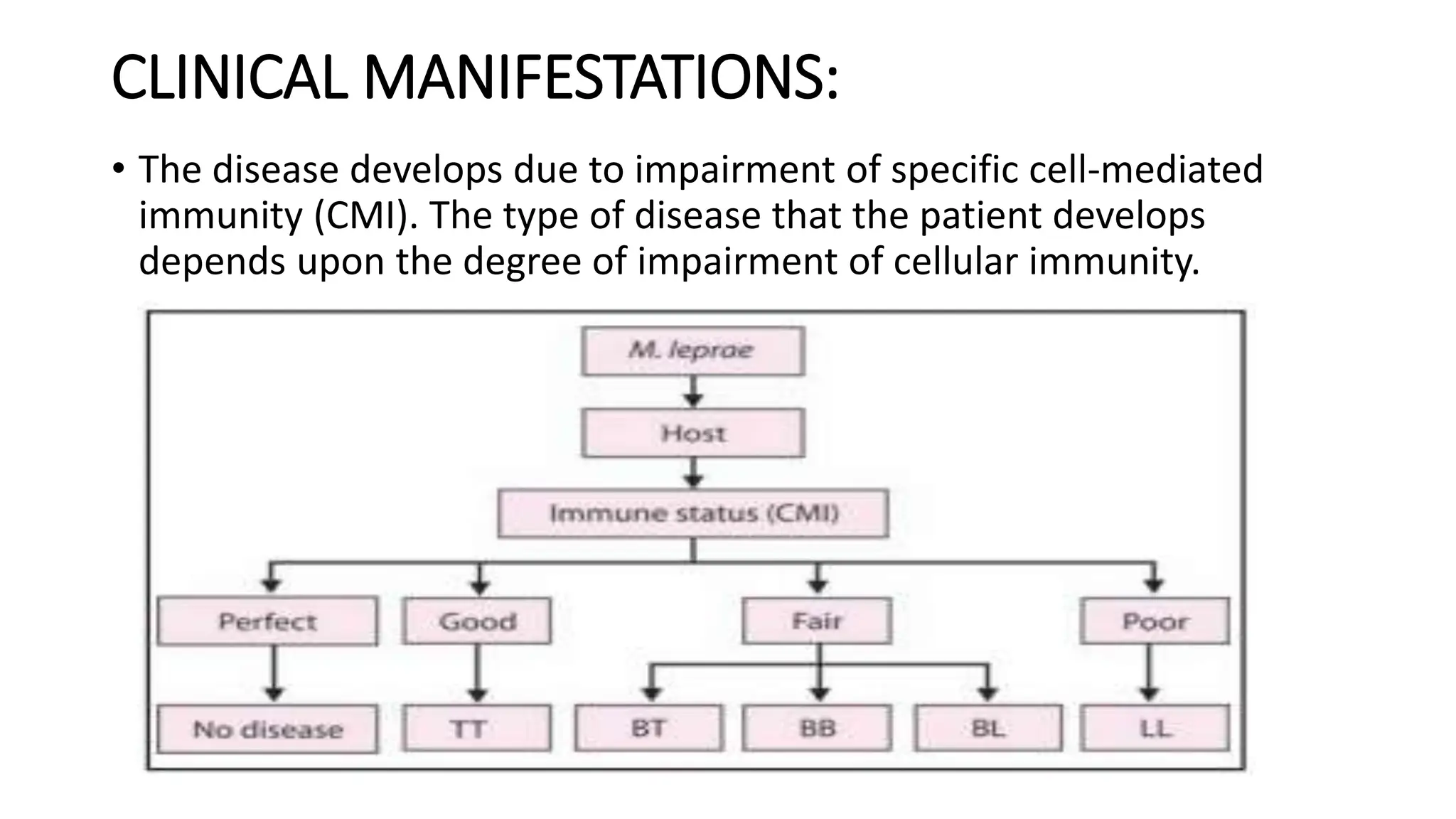
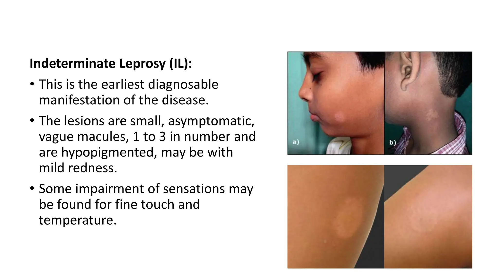
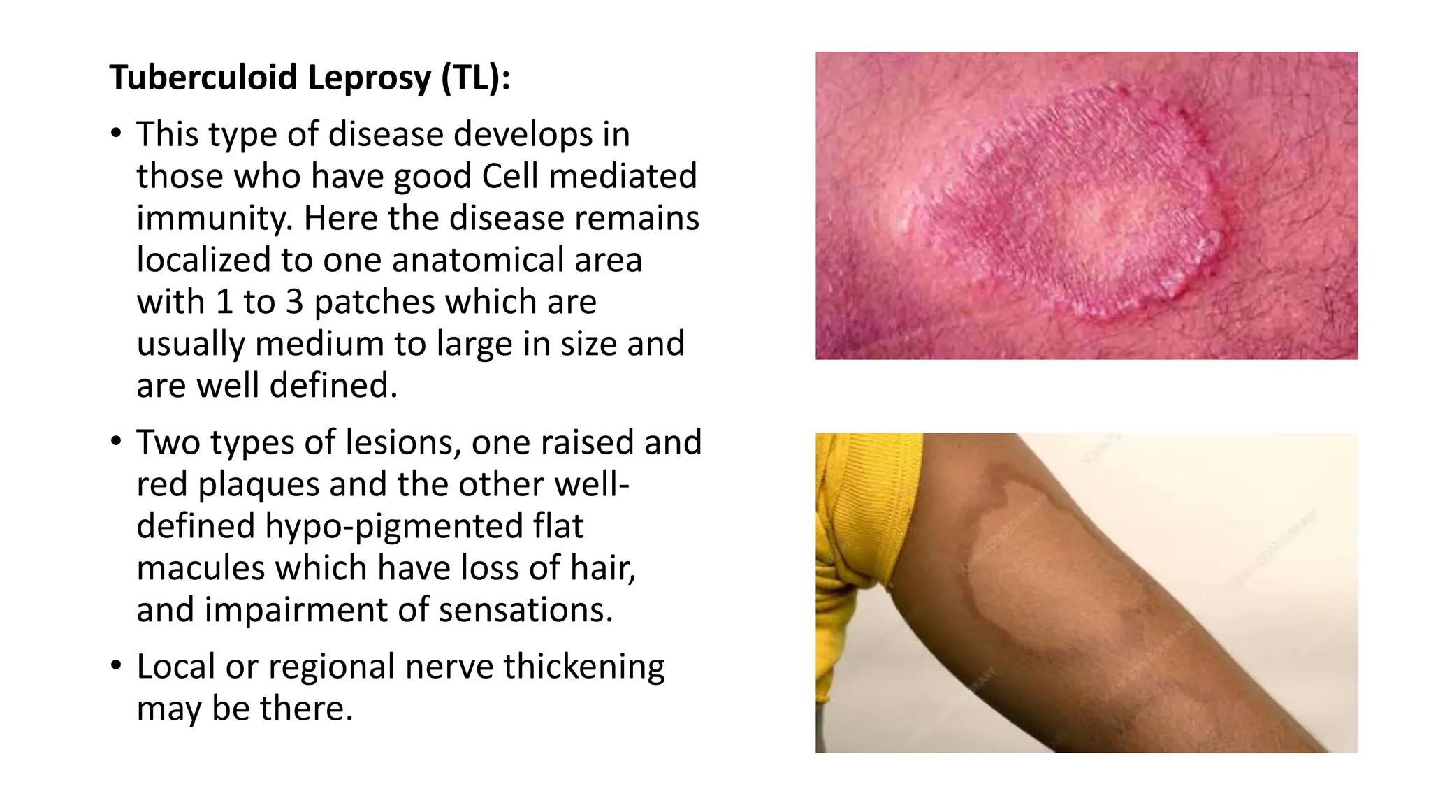
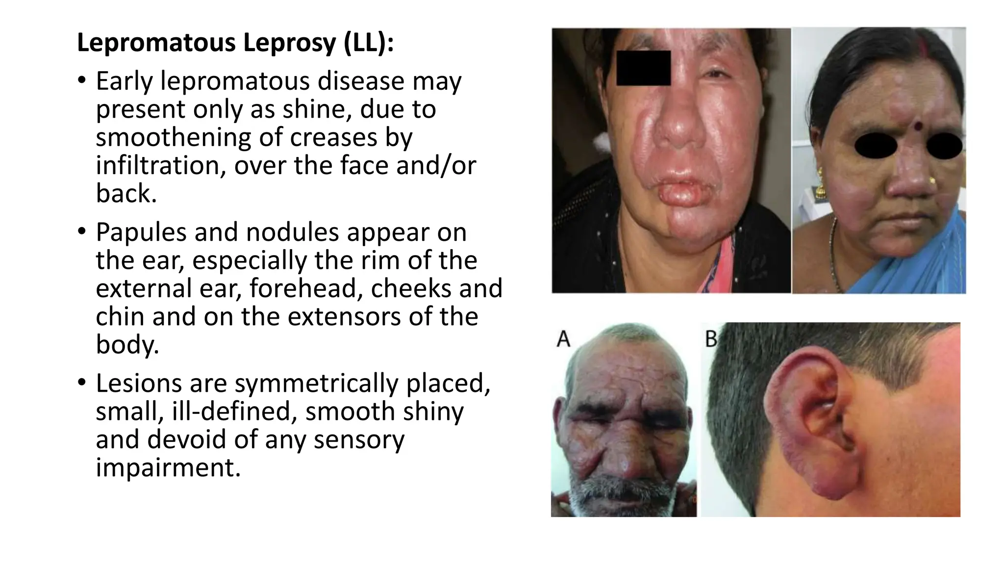
SEMINAR PRESENTATION
LEPROSY
PRESENT BY
Dr Ragul S
MD Part 1
LEPROSY
• Leprosy (Hansen’s disease) is a chronic infectious disease caused by
Mycobacterium leprae.
• The organism mainly affects the skin and the peripheral nerves.
• EPIDEMIOLOGY: In India, seven states (Bihar, Chattisgarh, Jharkhand,
Maharashtra, Orissa, Uttar Pradesh and West Bengal) continue to
have higher prevalence.
• CAUSATIVE ORGANISM: Mycobacterium leprae, a Gram-positive
bacillus which is weekly acid-fast.
• TRANSMISSION: Man is the only natural host and reservoir of this
infection.
• It is mainly the untreated lepromatous patients who are infectious
and spread the disease in the community.
• Aerosol from mouth or nose and close contact are important source
of transmission. The organisms enter the healthy contacts either
through microscopic breaches in the skin or more likely through
upper respiratory tract, in particular the nasal mucosa.
• INCUBATION PERIOD: from six months to more than 20 years (in
most instances 2 to 5 years.
CLINICAL MANIFESTATIONS:
• The disease develops due to impairment of specific cell-mediated
immunity (CMI). The type of disease that the patient develops
depends upon the degree of impairment of cellular immunity.
Indeterminate Leprosy (IL):
• This is the earliest diagnosable
manifestation of the disease.
• The lesions are small, asymptomatic,
vague macules, 1 to 3 in number and
are hypopigmented, may be with
mild redness.
• Some impairment of sensations may
be found for fine touch and
temperature.
Tuberculoid Leprosy (TL):
• This type of disease develops in
those who have good Cell mediated
immunity. Here the disease remains
localized to one anatomical area
with 1 to 3 patches which are
usually medium to large in size and
are well defined.
• Two types of lesions, one raised and
red plaques and the other well-
defined hypo-pigmented flat
macules which have loss of hair,
and impairment of sensations.
• Local or regional nerve thickening
may be there.
Lepromatous Leprosy (LL):
• Early lepromatous disease may
present only as shine, due to
smoothening of creases by
infiltration, over the face and/or
back.
• Papules and nodules appear on
the ear, especially the rim of the
external ear, forehead, cheeks and
chin and on the extensors of the
body.
• Lesions are symmetrically placed,
small, ill-defined, smooth shiny
and devoid of any sensory
impairment.
Borderline Leprosy:
• This is a large group with variable and unstable immune system.
i. Borderline tuberculoid (BT) – Features closeness to the tuberculoid type
ii. Borderline lepromatous (BL) - Features more akin to the lepromatous
form
iii. Mid borderline (BB) – Features of both types
• Lesions are relatively more localised, fewer, often larger with definite sensory
impairment in patients toward the BT group.
• Progressive wide spread, more in number, smaller and vague with partial or
no loss of sensations as one goes towards the BL type.
• Neuritic Leprosy (NL): There are no skin but only nerve thickening.
There may be only one nerve thickening (mono-neuritis) or multiple
nerves may be clinically involved (mono-neuritis multiplex).
• Nerve damage is the main cause for the deformities.
• Primary deformities - these are due to direct damage by the disease
process, e.g. muscle paralysis, nasal deformity, eye damage, etc.
• Secondary deformities are a consequence of sensory loss and thereby
loss of tissue due to burns, trauma and secondary infection, e.g. post-
burn blisters, ulcers, scars, contractures and loss of digits on hands,
trophic ulcers on feet and corneal ulcers due to affection of trigeminal
nerve.
LABORATORY INVESTIGATION:
• Skin biopsy
• Nerve biopsy
• Lepromin test
• Serological tests for leprosy (fluorescent antibody absorption test,
PGL-1 antibody and monoclonal antibody based tests)
HOMOEOPATHIC THERAPEUTICS:
HYDROCOTYLE:
• Spots almost circular, with raised scaly edges.
• Erysipelatous redness.
• Red spots, covered with whitish scales, on I. side of neck.-
• Lilac- coloured spot, shaped like an ear, on sole; the skin over it is
depressed, and walking is painful.
• Erythema on face, neck, back, chest, arms, and thighs; with much
itching; with copious sweat.
• Miliary eruptions on neck, back, and chest.
ARSENIC IOD:
• Dry, scaly, itching.
• Marked exfoliation of skin in large scales, leaving a raw exuding
surface beneath.
• Ichthyosis.
• Enlarged scrofulous glands.
• Debilitating night- sweats.
GRAPHITES:
Rough, hard, persistent dryness of portions of skin unaffected by
eczema.
Rawness in bends of limbs, groins, neck, behind ears.
Unhealthy skin; every little injury suppurates.
Swelling and induration of glands.
Phlegmonous erysipelas of face; burning and stinging pain.
THYROIDINUM:
• Leprosy with syphilitic eruptions.
• Scleroderma, intense redness and scaling of legs.
HOANG NAN:
• Leprosy with numbness and tingling in hands and feet.
HURA BRAZ:
• Leprosy when the skin is felt to be hide bound

LEPROSY (Hansen disease) types, clinical features.pptx

  • 1.
  • 2.
    LEPROSY • Leprosy (Hansen’sdisease) is a chronic infectious disease caused by Mycobacterium leprae. • The organism mainly affects the skin and the peripheral nerves. • EPIDEMIOLOGY: In India, seven states (Bihar, Chattisgarh, Jharkhand, Maharashtra, Orissa, Uttar Pradesh and West Bengal) continue to have higher prevalence. • CAUSATIVE ORGANISM: Mycobacterium leprae, a Gram-positive bacillus which is weekly acid-fast.
  • 3.
    • TRANSMISSION: Manis the only natural host and reservoir of this infection. • It is mainly the untreated lepromatous patients who are infectious and spread the disease in the community. • Aerosol from mouth or nose and close contact are important source of transmission. The organisms enter the healthy contacts either through microscopic breaches in the skin or more likely through upper respiratory tract, in particular the nasal mucosa. • INCUBATION PERIOD: from six months to more than 20 years (in most instances 2 to 5 years.
  • 4.
    CLINICAL MANIFESTATIONS: • Thedisease develops due to impairment of specific cell-mediated immunity (CMI). The type of disease that the patient develops depends upon the degree of impairment of cellular immunity.
  • 5.
    Indeterminate Leprosy (IL): •This is the earliest diagnosable manifestation of the disease. • The lesions are small, asymptomatic, vague macules, 1 to 3 in number and are hypopigmented, may be with mild redness. • Some impairment of sensations may be found for fine touch and temperature.
  • 6.
    Tuberculoid Leprosy (TL): •This type of disease develops in those who have good Cell mediated immunity. Here the disease remains localized to one anatomical area with 1 to 3 patches which are usually medium to large in size and are well defined. • Two types of lesions, one raised and red plaques and the other well- defined hypo-pigmented flat macules which have loss of hair, and impairment of sensations. • Local or regional nerve thickening may be there.
  • 7.
    Lepromatous Leprosy (LL): •Early lepromatous disease may present only as shine, due to smoothening of creases by infiltration, over the face and/or back. • Papules and nodules appear on the ear, especially the rim of the external ear, forehead, cheeks and chin and on the extensors of the body. • Lesions are symmetrically placed, small, ill-defined, smooth shiny and devoid of any sensory impairment.
  • 8.
    Borderline Leprosy: • Thisis a large group with variable and unstable immune system. i. Borderline tuberculoid (BT) – Features closeness to the tuberculoid type ii. Borderline lepromatous (BL) - Features more akin to the lepromatous form iii. Mid borderline (BB) – Features of both types • Lesions are relatively more localised, fewer, often larger with definite sensory impairment in patients toward the BT group. • Progressive wide spread, more in number, smaller and vague with partial or no loss of sensations as one goes towards the BL type.
  • 10.
    • Neuritic Leprosy(NL): There are no skin but only nerve thickening. There may be only one nerve thickening (mono-neuritis) or multiple nerves may be clinically involved (mono-neuritis multiplex). • Nerve damage is the main cause for the deformities. • Primary deformities - these are due to direct damage by the disease process, e.g. muscle paralysis, nasal deformity, eye damage, etc. • Secondary deformities are a consequence of sensory loss and thereby loss of tissue due to burns, trauma and secondary infection, e.g. post- burn blisters, ulcers, scars, contractures and loss of digits on hands, trophic ulcers on feet and corneal ulcers due to affection of trigeminal nerve.
  • 11.
    LABORATORY INVESTIGATION: • Skinbiopsy • Nerve biopsy • Lepromin test • Serological tests for leprosy (fluorescent antibody absorption test, PGL-1 antibody and monoclonal antibody based tests)
  • 12.
    HOMOEOPATHIC THERAPEUTICS: HYDROCOTYLE: • Spotsalmost circular, with raised scaly edges. • Erysipelatous redness. • Red spots, covered with whitish scales, on I. side of neck.- • Lilac- coloured spot, shaped like an ear, on sole; the skin over it is depressed, and walking is painful. • Erythema on face, neck, back, chest, arms, and thighs; with much itching; with copious sweat. • Miliary eruptions on neck, back, and chest.
  • 13.
    ARSENIC IOD: • Dry,scaly, itching. • Marked exfoliation of skin in large scales, leaving a raw exuding surface beneath. • Ichthyosis. • Enlarged scrofulous glands. • Debilitating night- sweats.
  • 14.
    GRAPHITES: Rough, hard, persistentdryness of portions of skin unaffected by eczema. Rawness in bends of limbs, groins, neck, behind ears. Unhealthy skin; every little injury suppurates. Swelling and induration of glands. Phlegmonous erysipelas of face; burning and stinging pain.
  • 15.
    THYROIDINUM: • Leprosy withsyphilitic eruptions. • Scleroderma, intense redness and scaling of legs. HOANG NAN: • Leprosy with numbness and tingling in hands and feet. HURA BRAZ: • Leprosy when the skin is felt to be hide bound