
Recommended
More Related Content
Similar to Lecture101210.ppt
Similar to Lecture101210.ppt (20)
More from UmaShanksr
More from UmaShanksr (17)
Recently uploaded
Recently uploaded (20)
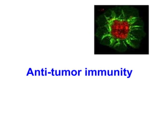
Lecture101210.ppt
- 2. Malignant transformation Failure of regulation of cell division and regulation of "social" behavior of the cells The uncontrollable proliferation, dissemination to other tissues Mutations in protoonkogenes and antionkogenes
- 3. Mutagens (carcinogens) - physical (eg various forms of radiation) - chemical (eg aromatic hydrocarbons) - biological (mainly various oncogenic viruses)
- 4. Protoonkogens promitotic (promoting cell division) for the malignant transformation is enough mutation in one copy of the gene protoonkogen (dominant oncogenes)
- 5. Antionkogens tumor-suppressor genes regulation of cell cycle for the malignant transformation should be excluded from function both copies of the gene (recessive oncogenes) TP53, RB1
- 6. Anti-tumor immune mechanisms Hypothesis of immune control tumor cells normally arise in tissues and are eliminated by T lymphocytes (probably wrong hypothesis) Defensive immune response tumor cells are weakly immunogenic occurs when tumor antigens are presented to T lymphocytes by activated dendritic cells in defense may be involved: non-specific mechanisms (neutrophilic granulocytes, macrophages, NK cells, interferons) and antigen-specific mechanisms (complement activating antibodies or ADCC, TH1 and TC)
- 7. cancer-associated antigens are processed by APC and recognized by T lymphocytes in complex with HLA I. and II. class with providing costimulus signals predominance of TH1 (IFN TNF) specific cell-mediated cytotoxic reactivity – TC activation TH2 → support B lymphocytes→ tumor specific antibodies (involved in the ADCC) tumor cells are destroyed by cytotoxic NK cells (low MHC gpI expression on tumor cells) interferons - antiproliferative, cytotoxic effect on tumor cells - INFγ - DC maturation
- 8. Cytotoxic mechanisms of NK cells
- 9. Tumor antigens Antigens specific for tumors (TSA) a) complexes of MHCgp I with abnormal fragments of cellular proteins - chemically induced tumors - leukemia with chromosomal translocation b) complexes of MHC gp with fragments of proteins of oncogenic viruses - tumors caused by viruses (EBV, SV40, polyomavirus) c) abnormal forms of glycoproteins - Sialylation of surface proteins of tumor cells d) idiotypes of myeloma and lymphoma - clonotyping TCR and BCR
- 10. Antigens associated with tumors (TAA) - also on normal cells - differences in quantity, time and local expression - auxiliary diagnostic markers a) onkofetal antigens - on normal embryonic cells and some tumor cells - -fetoprotein (AFP) - hepatom - canceroembryonal antigen (CEA) - colon cancer b) melanoma antigens - MAGE-1, Melan-A
- 11. c) antigen HER2/neu - receptor for epithelial growth factor - mammary carcinoma d) EPCAM - epithelial adhesion molecule - metastases e) differentiation antigens of leukemic cells - present on normal cells of leukocytes linage - CALLA -acute lymphoblastic leukemia (CD10 pre-B cells)
- 12. Mechanisms of tumors resistance to immune system high variability of tumor cells low expression of tumor antigens sialylation tumor cells do not provide costimulus signals → T lymphocyte anergy some anticancer substances have a stimulating effect production of factors inactivating T lymphocytes expression of FasL → T lymphocyte apoptosis inhibition of the function or durability dendritic cells (NO, IL-10, TGF-
- 13. Tumor immunotherapy Therapy - surgical removal of tumor - chemotherapy or radiotherapy - immunotherapy Immunotherapy - induction of anti-tumor immunity, or the use of immune mechanisms to targeting drugs to the tumor site
- 14. Immunotherapy using antibodies Antibodies functions - opsonization - activation of complement - induction of ADCC - carriers of drugs or toxins
- 15. 1) Monoclonal antibodies - against TAA - mouse and humanised antibodies - imunotoxins, radioimunotoxins - the possibility of damage surrounding tissues - HERCEPIN - Ab against HER2/neu, breast cancer - RITUXIMAB - Ab against CD20, lymphoma 2) Bispecific antibodies - bind a tumor antigen and the T lymphocyte or NK cell - Fc fragment of antibody binds to Fc receptors on phagocytes and NK cells 3) Elimination of tumor cells from the suspension of bone marrow cells using monoclonal antibodies for autologous transplantation
- 16. Immunotherapy using cell-mediated mechanisms 1) stimulation of inflammation at the tumor site 2) stimulation of LAK and TIL - isolation of T and NK cells, stimulation by cytokines, and return to the patient - LAK (lymphokine activated killers) - TIL (tumor infiltrating lymphocytes) 3) improving of tumor cells antigenpresenting function - genetic modification of tumor cells - expression of CD80, CD86 - production of IL-2, GM-CSF - modified cells are irradiated and returned to the patient
- 17. 4) tumor vaccines - in vitro stimulation of TH1 cells and TC with tumor antigens 5) the dendritic cell immunotherapy - in vitro cultivation of monocytes in an appropriate cytokine environment (GM-CSF, IL-4) → transformation into dendritic cells - cultivation of dendritic cells with tumor antigens 6) immunotherapy by donor T lymphocytes - after allogeneic transplantation - causing graft-versus-host disease 7) immunotherapy by immune system products - IL-2 - renal cell carcinoma - IFN - hematoonkology
- 18. Transplantation
- 19. Transplantation = transfer of tissue or organ ● autologous - donor = recipient ● syngeneic - genetically identical donor recipient (identical twins) ● allogeneic - genetically nonidentical donor of the same species ● xenogenic - the donor of another species ● implant - artificial tissue compensation
- 20. Allogeneic - differences in donor-recipient MHC gp and secondary histocompatibility Ag - alloreactivity of T lymphocytes - the risk of rejection and graft-versus-host - direct detection of alloantigens – recipient T lymphocytes recognize the different MHC gp and non-MHC molecules on donor cells - indirect recognition of alloantigens - APC absorb different MHC gp from donor cells and present the fragments to T lymphocytes - CD8+ T cells recognize MHC gp I. - CD4+ T cells recognize MHC gp II.
- 21. Testing before transplantation Compatibility in the system ABO -risk of hyperacute or accelerated rejection = formation of Ab against A or B Ag on graft vascular endothelium) HLA typing (determining of MHC gp alelic forms) phenotyping and genotyping by PCR Cross-match - lymfocytotoxic test - testing preformed Ab (after blood transfusions, transplantation, repeated childbirth) Mixed lymphocyte test - testing of alloreactivity T lymphocytes monitor for reactivity of lymphocytes to allogeneic HLA
- 22. HLA typing a) phenotyping: Evaluation of HLA molecules using typing serums Typing antiserums = alloantiserums of multipar (created cytotoxic Ab against paternal HLA Ag of their children), serum of patients after repeated blood transfusions, monoclonal Ab - molecules HLA class I: separated T lymphocytes - molecules HLA class II: separated B lymphocytes b) genotyping: evaluation of specific alleles DNA typing of HLA class II: DR, DP, DQ by PCR.
- 23. Cross-match test ● determination of preformed antibodies ● recipient serum + donor lymphocytes + rabbit complement → if cytotoxic Ab against donor HLA Ag are present in recipient serum (called alloantibodies = Ab activating complement) → lysis of donor lymphocytes. Visualization of dye penetration into lysis cells. ● positive test = the presence of preformed Ab → risk of hyperacute rejection! → contraindication to transplantation
- 24. Mixed lymphocyte reaction (MRL) ● determination of alloreactivity T lymphocytes ● mixed donor and recipient lymphocytes → T lymphocytes after recognition allogeneic MHC gp activate and proliferate One-way MRL ● determination of recipient T lymphocytes reactivity against donor cells ● donor cells treated with chemotherapy or irradiated lose the ability of proliferation
- 25. Rejection Factors: The genetic difference between donor and recipient, especially in the genes coding for MHC gp (HLA) Type of tissue / organ - the strongest reactions against vascularized tissues containing much APC (skin) The activity of the immune system of the recipient - the immunodeficiency recipient has a smaller rejection reaction; immunosuppressive therapy after transplantation – suppression of rejection Status transplanted organ - the length of ischemia, the method of preservation, traumatization of organ at collection
- 26. Hyperacute rejection ● minutes to hours after transplantation ● antibodies type of immune response mechanism: ● in recipients blood are present before transplantation preformed or natural Ab (IgM anti-carbohydrate Ag) → Ab + Ag of graft (MHC gp or endothelial Ag) → graft damage by activated complement (lysis of cells) ● the graft endothelium: activation of coagulation factors and platelets, formation thrombi, accumulation of neutrophil granulocytes prevention: ● negat. cross match before transplantation, ABO compatibility
- 27. Accelerated rejection ● 3 to 5 days after transplantation ● caused by antibodies that don´t activate complement ● cytotoxic and inflammatory responses activated by antibodies binding to Fc-receptors on phagocytes and NK cells prevention: ● negative cross match before transplantation, ABO compatibility
- 28. Acute rejection ● days to weeks after the transplantation or after a lack of immunosuppressive treatment ● cell-mediated immune response mechanism: ● recipient TH1 and TC cells response against Ag of graft tissue ● infiltration of lymphocytes, mononuclears, granulocytes around small vessels → destruction of transplant tissue
- 29. Chronic rejection ● from 2 months after transplantation ● the most common cause of graft failure mechanism is not fully understood: ● non-immunological factors (tissue ischemia) and TH2 responses with production alloantibodies, pathogenetic role of cytokines and growth factors (TGF β) ● replacement of functional tissue by connective tissue, endothelial damage →impaired perfusion of graft → gradual loss of its function dominating findings: vascular damage
- 30. Graft-versus-host disease (GVHD) ● after bone marrow transplantation ● GVHD also after blood transfusion to immunodeficiency recipients ● T-lymphocytes in the graft bone marrow recognize recipient tissue Ag as foreign (alloreactivity) Acute GVHD ● days to weeks after the transplantation of stem cells ● damage of liver, skin and intestinal mucosa ● Prevention: appropriate donor selection, T lymphocytes removal from the graft and effective immunosuppression
- 31. Chronic GVHD ● months to years after transplantation ● TH2 lymphocytes infiltration of tissues and organs, production of alloantibodies and production of cytokines → fibrotization ● process like autoimmune disease: vasculitis, scleroderma, sicca-syndrome ● chronic inflammation of blood vessels, skin, internal organs and glands, which leads to fibrotization, blood circulation disorders and loss of function
- 32. Graft versus leukemia effect (GVL) ● donor T lymphocytes react against residual leukemick cells of recipient ● mechanism is consistent with acute GVHD ● associated with a certain degree of GVHD (adverse reactions)
- 33. Immunologic relationship of mother and allogenic fetus ● fetal cells have on the surface alloantigens inherited from his father Tolerance of fetus by mother allow the following mechanisms: ● the relative isolation of the fetus from maternal immune system (no mixing of blood circulation) ● trophoblast - immune barrier witch protect against mother alloreaktive T lymphocytes - don‘t express classical MHC gp, expresses non-classical HLA-E and HLA-G ● depression of TH1 immune mechanisms in pregnancy Complications in pregnancy: production of anti-RhD antibodies by RhD- mother carrying RhD+ fetus (hemolytic disease of newborns)
- 35. Classification by Coombs and Gell Immunopathological reactions: immune response, which caused damage to the body (secondary consequence of defense responses against pathogens, inappropriate responses to harmless antigens, autoimmunity) IV types of immunopathological reactions: Type I reaction - response based on IgE antibodies Type II reaction - response based on IgG and IgM antibodies Type III reaction - response based on the formation of immune complexes Type IV reaction - cell-mediated response
- 36. Immunopathological reaction based on IgG and IgM antibodies (reaction type II) Cytotoxic antibodies IgG and IgM: ● complement activation ● ADCC ● binding to phagocytes and NK cells Fc receptors Haemolytic reactions after transfusion of ABO incompatible blood: Binding of antibodies to antigens of erythrocytes → activation of the classical way of complement → cell lysis Hemolytic disease of newborns: Caused by antibodies against RhD antigen
- 37. Autoimmune diseases: ● organ-specific cytotoxic antibodies (antibodies against erythrocytes, neutrophils, thrombocytes, glomerular basement membrane ...) ● blocking or stimulating antibodies Graves - Basedow disease - stimulating antibodies against the TSH receptor Myasthenia gravis - blocking of acetylcholin receptor→ blocking of neuromuscular transmission Pernicious anemia - blocking of vitamin B12 absorption Antiphospholipid syndrome - antibodies against fosfolipids Fertility disorders - antibodies against sperms or oocytes
- 38. Immunopathological reactions based on immune complex formation (reaction type III) ● caused by IgG antibodies → bind to antigen → creation of immunecomplexes ● immunocomplexes - bind to Fc receptors on phagocytes - activate complement ● immune complexes (depending on the quantity and structure) are eliminated by phagocytes or stored in tissues ● pathological immunocomplexes response arises when is a large dose of antigen, or antigen in the body remains ● immune complexes are deposited in the kidneys (glomerulonephritis), on the endothelial cells surface (vasculitis) and in synovial joints (arthritis)
- 39. Serum sickness ● after therapeutic application of xenogeneic serum (antiserum to snake venom) ● creation of immune complexes and their storage in the vessel walls of different organs ● clinical manifestations: urticaria, arthralgia, myalgia Systemic lupus erythematosus ● antibodies against nuclear antigens, ANA, anti-dsDNA Farmer's lung ● IgG antibodies against inhaled antigens (molds, pollens) Poststreptococcal glomerulonephritis
- 40. Immunopathological delayed-type reaction (reaction type IV) ● delayed-type hypersensitivity (DTH) ● local reaction caused by TH1 cells and monocytes / macrophages Experimental model (testing of cellular immunity): ● intradermal immunization by antigen → creation of antigen- specificTH1 cells ● after a few weeks intradermal administration of antigen → creates local reaction (granuloma) - TH1 cells and macrophages Tuberculin reaction Tissue damage in tuberculosis and leprosy Sarcoidosis
- 41. THANK YOU FOR YOUR ATTENTION