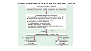
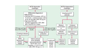
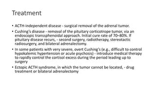
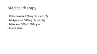
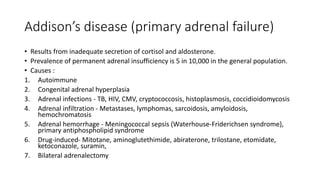
Cushing's syndrome and Addison's disease are both endocrine disorders caused by issues with cortisol production and regulation. Cushing's syndrome results from excessive cortisol levels due to conditions like pituitary or adrenal tumors. Its symptoms are treated through surgical removal of the tumor or medical therapy to control cortisol levels. Addison's disease is caused by inadequate cortisol production, often due to autoimmune destruction of the adrenal glands. Its treatment involves lifelong glucocorticoid and mineralocorticoid hormone replacement therapy. Both conditions require monitoring and medication adjustments in response to stress or illness to prevent adrenal insufficiency or crisis.

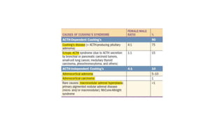
![• clinical features that result from chronic exposure to excess
glucocorticoids of any etiology.
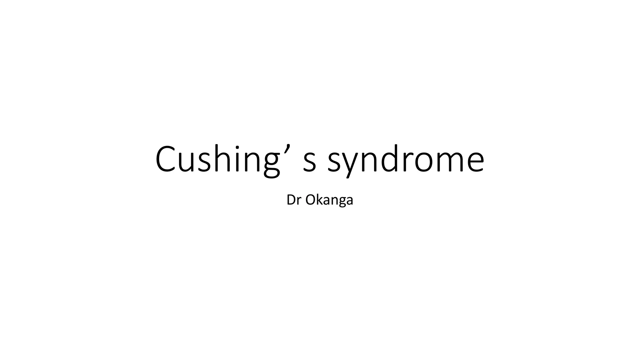
• Causes - ACTH-dependent (e.g., pituitary corticotrope adenoma,
ectopic secretion of ACTH by nonpituitary tumor) or ACTH-
independent (e.g., adrenocortical adenoma, adrenocortical carcinoma
[ACC], nodular adrenal hyperplasia), as well as iatrogenic (e.g.,
administration of exogenous glucocorticoids to treat various
inflammatory conditions).
• Cushing’s disease - Cushing’s syndrome caused by a pituitary
corticotrope adenoma.](https://image.slidesharecdn.com/lec7cushingssyndrome-230616055120-2812fd80/85/lec-7-Cushing-s-syndrome-pptx-2-320.jpg)


![CTEV [ clubfoot] DR ARUN LAL ,DR MOHAMED ASHRAF travancore medical college k...](https://cdn.slidesharecdn.com/ss_thumbnails/ctevclubfootdrarunlaldrmohamedashraftravancoremedicalcollegekollamkeralaindia-260208063247-18fc466c-thumbnail.jpg?width=640&height=640&fit=bounds)


