
Recommended
More Related Content
Viewers also liked
Viewers also liked (15)
Similar to Layered Learning
Similar to Layered Learning (20)
More from meducationdotnet
More from meducationdotnet (20)
Layered Learning
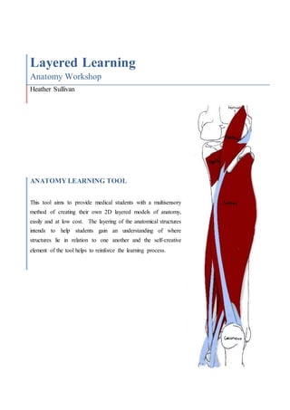
- 1. Layered Learning Anatomy Workshop Heather Sullivan ANATOMY LEARNING TOOL This tool aims to provide medical students with a multisensory method of creating their own 2D layered models of anatomy, easily and at low cost. The layering of the anatomical structures intends to help students gain an understanding of where structures lie in relation to one another and the self-creative element of the tool helps to reinforce the learning process.
- 2. 2 Contents Page Content Page Title Page 1 Contents sheet 2 Explanatory guide 3 Work Sheets 4-11 Base Layer 4 Popliteus 5 Tibialis Posterior 6 Flexor Digitorum Longus 7 Flexor Hallucis Longs 8 Soleus 9 Plantaris 10 Gastrocnemius 11 Reference sheets 12-13
- 3. 3 Explanatory Guide to the Anatomy Learning Tool In order to create the 2D layered model of the posterior lower leg follow the instructions below. First of all ensure you have all the necessary materials and equipment: The complete workbook, containing the base-layer and seven pages of muscles. Scissors A set of 25 sticky attachments. (ask for more if needed) Making your Model: Step 1: Take out and study the base-layer (this will help to keep orientated to the area of anatomy during the making of the model). Step 2: Read through the first muscle sheet (popliteus), taking note of the origin and insertion as this will help you place the muscle correctly on the base-layer. Step 3: Cut out the muscle, ensuring to keep track of which end is the insertion and which the origin. Step 4: Attach the square(s) of the sticky foam to the reverse side of where the hashed colour tab is. Step 5: Matching the colour of the attaching end to the colour on the base-layer, peel off the sticky cover and attach the muscle to the base-layer. Step 6: Repeat steps 2 through 5 with each muscle until you have layered up to the final sheet (Gastrocnemius). Step 7: Once the layered model has been completed the accuracy can be checked with stage seven of the Reference Sheet on page 16. If you have any problems, consult the Reference Sheets on pages 12 and 13.
- 4. 4
- 5. 5
- 6. 6
- 7. 7
- 8. 8
- 9. 9
- 10. 10
- 11. 11
- 12. 12 Reference Sheets The following sheets show the order and positions of the muscles. This can be used for reference if there are difficulties with following the guidelines or at the end to check the accuracy of the muscle positioning. The images are displayed in the order that the muscles are attached, beginning with the popliteus in image 1. 1 2 3
- 13. 13 6 4 5 7