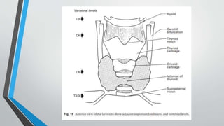
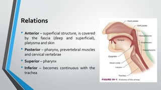
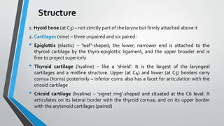
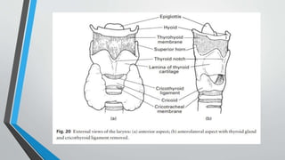
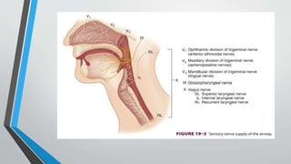
2. The Larynx
• The larynx is an organ of voice extending from root of tongue to trachea.
• It consists of a complex arrangement of muscles, cartilages, membranes
and ligaments.
• It extends from C3 to C6 in the midline (adult).
3.
4. Relations
• Anterior – superficial structure, is covered
by the fascia (deep and superficial),
platysma and skin
• Posterior – pharynx, prevertebral muscles
and cervical vertebrae
• Superior – pharynx
• Inferior – becomes continuous with the
trachea
5. Structure
1. Hyoid bone (at C3) – not strictly part of the larynx but firmly attached above it
2. Cartilages (nine) – three unpaired and six paired:
• Epiglottis (elastic) – ‘leaf’-shaped; the lower, narrower end is attached to the
thyroid cartilage by the thyro-epiglottic ligament, and the upper broader end is
free to project superiorly
• Thyroid cartilage (hyaline) – like a ‘shield’. It is the largest of the laryngeal
cartilages and a midline structure. Upper (at C4) and lower (at C5) borders carry
cornua (horns) posteriorly – inferior cornu also has a facet for articulation with the
cricoid cartilage
• Cricoid cartilage (hyaline) – ‘signet ring’-shaped and situated at the C6 level. It
articulates on its lateral border with the thyroid cornua, and on its upper border
with the arytenoid cartilages (paired)
6.
7. Structure (cont.)
• Arytenoid cartilages (paired) – pyramidal in shape, each with a lateral
muscular process (for insertion of both crico-arytenoid muscles) and an
anterior vocal process (being the posterior attachment of the vocal
ligament)
• Corniculate cartilages (paired) – two small conical-shaped cartilages,
articulate with the apices of arytenoid cartilages, give attachment to
aryepiglottic folds (the fibro-elastic membrane between the epiglottis and
arytenoids – lower border of which is free and forms the vestibular ligament
or false cord)
• Cuneiform cartilages (paired) – two small rod-shaped cartilages are in
aryepiglottic folds and serve to strengthen them
11. Structure (cont.)
3. Ligaments and Membranes:
• Thyrohyoid membrane – between the upper border of the thyroid and the hyoid
bone. Strengthened anteriorly and laterally
• Hyo-epiglottic ligament – connects the hyoid bone to the lower part of the
epiglottis
• Cricothyroid ligament –interconnects cricoid, thyroid and arytenoid cartilages;
upper free margin composed almost entirely of elastic tissue forms vocal ligament
on each side which form interior of the vocal folds (vocal cords); between the
thyroid above and the cricoid below, the preferred site for cricothyrotomy
• Cricotracheal ligament – connects the cricoid to the first ring of the trachea
12.
13.
14. Structure (cont.)
• Quadrangular membrane – between epiglottis and arytenoid cartilages,
thickened inferior margin form vestibular ligament which form interior of
vestibular folds
15. Structure (cont.)
4. Muscles – three extrinsic (connect larynx to its neighbours) and six intrinsic:
● Extrinsic:
• Sternothyroid – depresses the larynx, connects the posterior manubrium sterni to
the lateral thyroid lamina
• Thyrohyoid – elevates the larynx, connects the lateral thyroid lamina to the
inferior greater horn of the hyoid bone
• Inferior constrictor – constricts the pharynx, origins from the thyroid lamina, the
tendinous arch over the cricothyroid and the side of the pharynx
● Intrinsic:
• Posterior crico-arytenoid – opens the glottis by the abducting cords
• Lateral crico-arytenoid – closes the glottis by the adducting cords (principle
adductors)
16. Structure (cont.)
• Interarytenoid (unpaired) – closes the glottis (especially posteriorly) by
connecting the arytenoids. Some fibers become the aryepiglottic muscle
laterally, which constricts the laryngeal inlet somewhat
• Thyro-arytenoid – relaxes the cords by shortening, thus pulling the
arytenoids anteriorly
• Vocalis – fine adjustment of vocal cord tension (fibers come from the thyro-
arytenoid)
• Cricothyroid – only true tensor and the only muscle that lies outside the
cartilages. It works by tilting the cricoid and putting stretch on the vocal
cords
17. Vascular supply
The blood supply of the larynx is derived from branches of the thyroid arteries. The
cricothyroid artery arises from the superior thyroid artery itself, the first branch
given off from the external carotid artery, and crosses the upper cricothyroid
membrane (CTM), which extends from the cricoid cartilage to the thyroid cartilage.
The superior thyroid artery is found along the lateral edge of the CTM.
Arterial:
• Superior laryngeal (from superior thyroid artery) – accompanies the internal
branch of the superior laryngeal nerve
• Inferior laryngeal (from inferior thyroid artery) – accompanies the recurrent
laryngeal nerve
Venous – into the corresponding superior and inferior thyroid veins
Lymph vessels drain into deep cervical group of nodes
18.
19. Nerve supply
Branches of vagus (X) nerve:
● Superior laryngeal nerve – passes deep to the internal and external carotid
arteries and then divides into:
• External branch (small) – motor to cricothyroid
• Internal branch (larger) – sensory above the vocal cords
● Recurrent (inferior) laryngeal nerve – It supplies: Motor to all intrinsic
muscles of the larynx (except cricothyroid)
• Sensory supply below the level of vocal cords by inferior laryngeal nerve
20.
21. Laryngeal nerve injuries
• Unilateral denervation of a cricothyroid muscle causes very subtle clinical findings.
Bilateral palsy of the superior laryngeal nerve may result in hoarseness or easy
tiring of the voice, but airway control is not jeopardized.
• Unilateral paralysis of a recurrent laryngeal nerve results in paralysis of the
ipsilateral vocal cord, causing deterioration in voice quality. Assuming intact
superior laryngeal nerves, acute bilateral recurrent laryngeal nerve palsy can
result in stridor and respiratory distress because of the remaining unopposed
tension of the cricothyroid muscles. Airway problems are less frequent in chronic
bilateral recurrent laryngeal nerve loss because of the development of various
compensatory mechanisms (e.g, atrophy of the laryngeal musculature).
• Bilateral injury to the vagus nerve affects both the superior and the recurrent
laryngeal nerves. Thus, bilateral vagal denervation produces flaccid, mispositioned
vocal cords similar to those seen aft er administration of succinylcholine. Although
phonation is severely impaired in these patients, airway control is rarely a problem.