Prophages, phage genomes integrated into a bacterium, offer a potential gene repository for synthetic-biology applications
Streptococcus suis is a zoonotic pig pathogen currently only effectively controlled by antibiotics(1)
Phage-bacteria infection networks (PBINs) allow us to gauge the pathogenicity and host range of phages, however they are limited by phages which can be isolated into pure culture(2)
In silico PBINs could offer a more ‘complete’ analysis by using predicted prophages that have successfully gained entry to both the host and its genome
Shared prophage-clusters across isolates could reveal efficacious genetic components for use in targeted phage-therapies
Large-scale prophage-bacterial infection networks reveal the host-phage signatures of coevolution in Streptococcus suis
1. Large-scale prophage-bacterial infection networks reveal the host-phage
signatures of coevolution in Streptococcus suis
Emmet Campbell(1)
| Nicholas J. Dimonaco(2) Timofey Skvortsov(3)|Christopher J. Creevey(1)
Background
§ Prophages, phage genomes integrated into a
bacterium, offer a potential gene repository
for synthetic-biology applications
§ Streptococcus suis is a zoonotic pig
pathogen currently only effectively controlled
by antibiotics(1)
§ Phage-bacteria infection networks (PBINs)
allow us to gauge the pathogenicity and host
range of phages, however they are limited by
phages which can be isolated into pure
culture(2)
§ In silico PBINs could offer a more ‘complete’
analysis by using predicted prophages that
have successfully gained entry to both the
host and its genome
§ Shared prophage-clusters across isolates
could reveal efficacious genetic components
for use in targeted phage-therapies
Aim
To create an in-silico PBIN for S. suis that
can be used to identify phages of
therapeutic interest by simulating
infections based on community formation
Method
GitHub
(1) Queen’s University Belfast – School of Biological Sciences | (2) McMaster University – Department of Medicine | (3) Queen’s University Belfast – School of Pharmacy
§ In silico PBINs were successful in finding groups
of genetically/functionally homologous isolates,
suitable for use in downstream network
analyses
§ If PCs are functionally homologous based on k-
mer pairwise similarity, we can simulate
infection potential on large-scale phage-
bacteria infection networks
§ Promiscuous PCs could inform broad-range
therapy applications, and specific PCs could
expedite therapies for targets with difficult-to-
isolate or no known phages
Future Works
§ Compare receptor-binding proteins within PCs
to reveal how similar prophages attack the host
§ Investigate associations/dissociations between
presence/absence of PCs and antiviral defense
systems
@the_phagemage
@CreeveyLab
ecampbell50@qub.ac.uk
Conclusions
References: (1) Uruén et al. How Streptococcus suis escapes antibiotic
treatments, 2022. (2) Weitz et al. Phage-bacteria infection networks. 2013. (3)
Vötsch et al. Streptococcus suis – The “Two Faces” of a Pathobiont in the
Porcine Respiratory Tract. 2018. Tools: all tools used (Prokka, PADLOC,
Sourmash, Roary, geNomad) are available on github.
§ S. suis is known to be a heterogenous
species(3)
, which is reflected in its low
percentage of core genes in the pangenome.
However, communities formed through k-mer
based similarity networks show a dramatic
increase in their core genome size, and lower
total pangenome.
§ Members of PC-80 are grouped by k-mer
similarity yet predicted from dissimilar BCs.
This suggests small, evolutionary differences
in an otherwise identical phage, or common
lack of resistance in the host.
§ The presence of a prophage within an isolate
does not necessarily indicate successful
infection, as it may be cryptic or inherited
before gaining resistance (e.g. superinfection
exclusion)
§ Core-genome differences in antiviral defense
systems show BCs functionally differ in their
bacteriophage-resistance. Further suggesting
PCs may be isolated from hosts lacking
common resistance mechanisms
Discussion
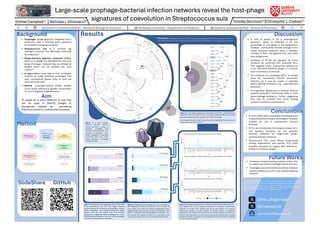
Results
Figure 1: In silico phage-
bacteria infection network.
Nodes represent a group of
isolates/ prophages that
formed a community from their
pairwise comparisons. Blue
nodes represent a bacterial
community (BC), and purple
nodes represent a prophage
community (PC).
Figure 2: Tree shows grouping of Bacterial Community representatives
‘infected’ by PC-80, based on their core gene alignment. BCs show broad
grouping despite all being ‘infected’ by a similar prophage.
BC-2 (largest) and BC-14 (4th largest) have been highlighted for downstream
analysis (see figures 3,4 and 5)
PC-80
Infections: An edge between a PC and BC indicates a prophage from that community was identified in
one of the isolates from the connected BC. For the purposes of this study, this is considered an ‘infection’
PC-80
0.03
0
10
20
30
AbiD
AbiO−Nhi_family
AbiQ
AbiU
AbiZ
AVAST_type_II
Borvo
DRT_class_I
DRT_class_II
HEC−05
HEC−06
Mokosh_TypeII
PD−Lambda−1
PD−T4−6
PD−T7−4
PDC−S02
PDC−S04
PDC−S05
PDC−S06
PDC−S07
PDC−S11
PDC−S13
PDC−S14
PDC−S15
PDC−S16
PDC−S18
PDC−S19
PDC−S22
PDC−S25
PDC−S28
PDC−S30
PDC−S32
PDC−S35
PDC−S37
PDC−S38
PDC−S42
PDC−S47
PDC−S49
PDC−S50
PDC−S51
PDC−S53
PDC−S56
PDC−S58
PDC−S60
PDC−S73
ppl
PrrC
retron_I−C
RM_type_IIG
RM_type_IV
SEFIR
shedu
SoFic
Stk2
Tiamat
TIR−NLR
tmn
Uzume
viperin_solo
DM
Gene
Count
Pangenome Distribution of Defense Systems across All 2,119 isolates
0
10
20
30
AbiD
AbiO−Nhi_family
AbiQ
AbiU
AbiZ
AVAST_type_II
Borvo
DRT_class_I
DRT_class_II
HEC−05
HEC−06
Mokosh_TypeII
PD−Lambda−1
PD−T4−6
PD−T7−4
PDC−S02
PDC−S04
PDC−S05
PDC−S06
PDC−S07
PDC−S11
PDC−S13
PDC−S14
PDC−S15
PDC−S16
PDC−S18
PDC−S19
PDC−S22
PDC−S25
PDC−S28
PDC−S30
PDC−S32
PDC−S35
PDC−S37
PDC−S38
PDC−S42
PDC−S47
PDC−S49
PDC−S50
PDC−S51
PDC−S53
PDC−S56
PDC−S58
PDC−S60
PDC−S73
ppl
PrrC
retron_I−C
RM_type_IIG
RM_type_IV
SEFIR
shedu
SoFic
Stk2
Tiamat
TIR−NLR
tmn
Uzume
viperin_solo
DM
Gene
Count
Pangenome Distribution of Defense Systems across PC−80 Infected BCs (1,097 isolates)
0
10
20
30
AbiD
AbiO−Nhi_family
AbiQ
AbiU
AbiZ
AVAST_type_II
Borvo
DRT_class_I
DRT_class_II
HEC−05
HEC−06
Mokosh_TypeII
PD−Lambda−1
PD−T4−6
PD−T7−4
PDC−S02
PDC−S04
PDC−S05
PDC−S06
PDC−S07
PDC−S11
PDC−S13
PDC−S14
PDC−S15
PDC−S16
PDC−S18
PDC−S19
PDC−S22
PDC−S25
PDC−S28
PDC−S30
PDC−S32
PDC−S35
PDC−S37
PDC−S38
PDC−S42
PDC−S47
PDC−S49
PDC−S50
PDC−S51
PDC−S53
PDC−S56
PDC−S58
PDC−S60
PDC−S73
ppl
PrrC
retron_I−C
RM_type_IIG
RM_type_IV
SEFIR
shedu
SoFic
Stk2
Tiamat
TIR−NLR
tmn
Uzume
viperin_solo
DM
Gene
Count
Community Cloud Core Shell SoftCore
Pangenome Distribution of Defense Systems across BC−14 (46 isolates)
1281
1155
736
136
83
308
340
421
1287
611
1055
2146
2109
7345
20064
55778
0% 10% 20% 30% 40% 50% 60% 70% 80% 90% 100%
BC -
14
BC -
2
PC -
80 BCs
Al l
Pangenome Distribution of Different Sets of Bacterial Isolates
Cor e Sof t
- C or
e Shel l Cl oud
2,119 isolates
1,097 isolates
963 isolates
46 isolates
Figure 3: Pangenome distribution of defense systems within three sets of S.
suis isolates: All (2,119 isolates), members of bacterial communities
‘infected’ by PC-80 (1,097 isolates) and BC-14 (46 isolates). The species
pangenome contains a larger number of defense genes as expected,
however PD-T4-6 moves from soft-core to core in both subsets, and in BC-14,
PDC-S73 becomes soft-core and several cloud genes become shell, showing
that these subsets of isolates are increasingly functionally similar in regards
to their antiviral potential
Figure 4: Pangenome of 4 isolate sets: All, members of
BCs ‘infected’ by PC-80, BC-2 and BC-14. BC-2 and BC-14
have similar core sizes, but different pangenome sizes,
suggesting BCs group based on their core genome as it
does not decrease when the pangenome increases. PC-
80 ‘infected’ BCs appear to share more core genes than
the species, though less than k-mer based communities
Figure 5: Pangenome of 4 prophage sets: PC-80 is the
most promiscuous, and PC-2 the largest. Despite
genetic diversity and mosaicism of prophages, subsets
share core-genes. PC-2, despite being the largest
subset, also has the largest core and smallest
pangenome, suggesting these prophages are clonal.
PC-80 share terminases in the core, while prophages
from BC-2 in PC-80 share an endolysin.
SlideShare