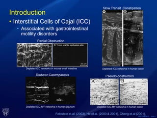
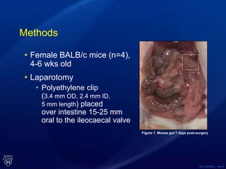
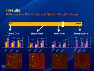
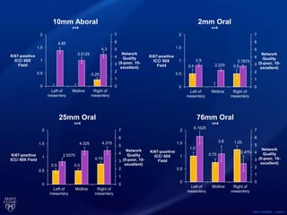
This document summarizes a study on the effects of partial obstruction on networks of Interstitial Cells of Cajal (ICC) in the mouse small intestine. The study found depleted and altered ICC networks near the site of obstruction compared to regions further away. Specifically, ICC density, proliferation and network quality decreased while the size of ICC-negative spaces increased near the obstruction. This supports previous findings and suggests ICC loss may contribute to motility disorders caused by obstruction.
Proposed Structure of Siamese CNN for Fusion face and palm veins at Feature L...Husam I. Abdulrazzaq
A Proposed Modification of Siamese Convolutional Neural Network Structure for Fusion face and palm veins multi biometrics at Feature Level, which is a presentation of the thesis titled "Robust authorization based on multi-biometric techniques"
A new software has been developed to measure the hip-knee condylar angle using digital radiological images and computer analysis. In a study comparing the new computerized method to conventional manual goniometry, 60 knee x-rays following total knee arthroplasty were measured twice by the same observer using both methods. Statistical analysis showed the computerized method produced highly reproducible results with a mean difference of 0.324 degrees between readings and a Pearson correlation of 0.999. The new software allows for precise, reliable and inexpensive measurement of joint angles that can be performed by non-medical personnel.
An experimental model has been developed to characterize closed soft tissue trauma (CSTT) and its impact on the microvasculature in rats. An impact device was used to create standardized CSTT in the thighs of anesthetized rats. Micro-CT imaging of the injured and uninjured limbs after perfusing with a contrast agent allows evaluation of changes to vessel density, diameter and other morphological parameters during healing. This provides a basis for quantitatively assessing CSTT and optimizing clinical treatment.
3d ultrasound-guided-robotic-needle-steering-in-biological-tissueieeepondy
3d ultrasound-guided-robotic-needle-steering-in-biological-tissue
Contact:+91-9994232214,8144199666
Email:ieeeprojectchennai@gmail.com
www.projectsieee.com
Dr. Coşkunseven "The New Keraring 340 Degree Segment"Mediphacos
This document summarizes a presentation on KeraRing 340 implantation for treating keratoconus. It includes patient inclusion/exclusion criteria, pre-op and post-op data from one patient, and conclusions. KeraRing 340 implantation provided over 11 diopters of flattening, and improved the patient's UCVA and BCVA. The procedure is effective and safe for improving vision in central or paracentral keratoconic eyes, with about 7D of flattening from a 200 micron segment and 13D from a 300 micron segment. However, long-term follow up of more patients is still needed to fully validate the approach.
This document summarizes 7 articles related to orthodontic miniscrews. The articles studied factors that influence miniscrew stability and success rates, such as insertion method/tool, miniscrew design, bone characteristics, and insertion torque values. Key findings included that motorized insertion tools and thicker cortical bone promoted higher success rates and stability. Recommended insertion torque was below 20 Ncm to avoid fractures. Miniscrew diameter, length, and design also impacted stability measurements like insertion, removal and fracture torques. The studies provided information to optimize miniscrew performance and outcomes.
Medcross Imaging LLC is an ACR accredited facility and has been in business in Georgia since 1980 with offices located in Macon, Eastman, and Johns Creek, Riverdale, Stockbridge, Conyers, Monroe, GA.
- Partial obstruction of the small intestine in mice resulted in distance-dependent disruption of interstitial cells of Cajal (ICC) networks, with number of proliferating ICC appearing to be lower closest to the occlusion.
- Proliferating ICC were unexpectedly low 10mm from the occlusion.
- This study aims to improve quantification methods of ICC networks and examine proliferation as a potential factor in ICC network depletion using a mouse model of partial small intestine obstruction.
Scandinavian Total Ankle Replacement System (STAR™ Ankle) | Enovis DJO®
The Scandinavian Total Ankle Replacement System (STAR Ankle) is comprised of a Tibial Plate, Mobile Bearing, and Talar Component. The STAR Ankle is designed to replace a portion of the distal tibial and proximal talar bones of the natural ankle joint. The device is designed to allow the patient to regain and/or retain some of his/her normal ankle mobility and function.
Reach us through https://www.djoglobal.com/contact-us.
Scandinavian Total Ankle Replacement System (STAR™ Ankle) | DJO GlobalDJO®
Comprised of a Tibial Plate, Mobile Bearing, and Talar Component, the STAR Ankle is designed to replace a portion of the distal tibial and proximal talar bones of the natural ankle joint. The device is designed to allow the patient to regain and/or retain some of his/her normal ankle mobility and function. Visit us at https://www.djoglobal.com/sites/default/files/IFU/V15154_eIFU_EN%20AA%20STAR%20Ankle%20IFU%20OT-IFU-33.pdf to know more.
Athens-Comparison of Laser In Situ Keratomileusis Flaps Created by 3 Femtosec...Sunčana Kovačić
The document compares corneal flap morphology created by three femtosecond lasers: Intralase, Ziemer LDV, and VisuMax. Key findings include:
1) Central flap thickness was highest for Intralase (111 μm), followed by VisuMax (105.58 μm) and lowest for Ziemer LDV (94.04 μm).
2) Flap thickness was most uniform across meridians and between center and periphery for VisuMax, followed by Intralase, with the most asymmetry for Ziemer LDV.
3) Ziemer LDV had the lowest mean deviation between achieved and intended flap thickness, making it the most predictable for flap thickness.
This document provides an overview of a company focused on developing transformative therapies for chronic eye diseases. It discusses the company's leadership which has expertise in ophthalmics, retinal disease, and regulatory matters. It also outlines the company's lead product NT-503, an encapsulated cell therapy for wet age-related macular degeneration, and its versatile drug delivery platform which aims to provide long-term continuous production of therapeutic proteins in the eye to treat a broad range of eye diseases.
Micro CT settings for caries detection: how to optimizeIJERA Editor
Some important items that can influence micro CT image were reviewed in this study. Different settings were
optimized for the assessment of early caries lesions. There are several researches on bone using micro CT but not
too much on dental hard tissues when assessing mineral loss. Different kinds of micro CT devices and
technologies are taking place today, each requiring unique settings, and this consists one of the greatest obstacles
for the use of micro CT on dental hard tissues. Achieving the settings for an ideal dental image is therefore a
challenge. The purpose of this study was to evaluate different micro CT settings to optimize the assessment of
early caries lesions aiming the integrity of the dental specimen thus, making possible to reuse it for further
studies. Three teeth with early caries lesions were submitted to different micro CT settings and different
reconstruction settings, aiming a better image. The final image was compared visually through different densities
and attenuation coefficients. The best setting for teeth tissues was achieved regarding contrast, definition, noise
reduction and the larger difference between sound enamel and early lesions attenuation coefficient.
JOURNAL CLUB ON THE OUTCOME OF ORAL IMPLANTS PLACED IN BONE WITH LIMITED BU...Shilpa Shiv
This document summarizes a study that evaluated the outcomes of oral implants placed in bone with limited bucco-oral dimensions over a 3-year period. 100 implants were placed in 28 patients. The study found that the implants had a 100% survival rate over 3 years and that the marginal bone levels around the implants remained stable. The results indicate that implants can successfully be placed in sites with up to 4.5mm of bucco-oral bone width without the need for bone grafting, providing patients maintain good oral hygiene.
There is no question that given the current state of the art in dentistry, that dental implants are pretty much the best way to replace teeth; they are stand alone tooth replacement systems that look and function just like natural teeth. They do not attach to adjacent teeth like a fixed bridge and don't have to be taken in and out like removable partial dentures.
A dental implant is a tooth root replacement made of titanium, which has the unique property of being osteophilic (osteo-bone, philic-loving) and actually fuses to bone. A crown, the part of the tooth that you see in your mouth, is attached to the implant. And the great thing about implants is they are not susceptible to decay or periodontal (gum) disease in the same way that teeth are.
Now here are a few important pointers, which hold for implants generally and are especially important in your case when replacing a front tooth for an imminent event . Dr Harshavardhan Patwal
Tecnología para niños con parálisis cerebral y otras discapacidades - Dra. De...Teletón Paraguay
This document discusses technology for children with cerebral palsy and other disabilities. It begins by providing historical context of the Rehabilitation Institute of Chicago and outlines the objectives of quantifying impairments, increasing practice through therapy, and discussing substitution technologies. Ultrasound is discussed as a way to quantify muscle properties and stiffness. Robotic gait training using Lokomat is presented as a method to increase practice intensity and improve activities like walking. Challenges and opportunities for data collection across centers to improve guidelines for robotic device use are also covered.
Rotator cuff tears are a very common cause of shoulder pain. Surgery is very successful in improving pain but biological augmentation is aimed at improving the healing rate. Amniotic membrane allografts, PRP (platelet rich plasma) and stem cells are all currently popular options.
This study is an In vitro analysis of amniotic membrane allograft as a potential agent for biological augmentation of rotator cuff repair performed by Dr Adnan Saithna, Orthopedic Surgeon, AZBSC Orthopedics
Three-Years Results After Femtosecond Laser-Assisted Circular Keratotomy As a Treatment for Keratoconus Stages I-II:
Comparison with (Iontophoresis-Assisted) CXL, presented at ASCRS 2018 – by Detlev Breyer, H. Kaymak, K. Klabe, P. Hagen, T. Ax, F. Kretz, G. Auffarth
HIGH-FIELD MRS methods to Study Human Body ZOOM MRSI 2-hg 2-hydroxyglutarate...Uzay Emir
This document discusses the use of high field MRI methods to study the human body. It provides background on how MRS has evolved from obtaining phosphate spectra in the 1970s to playing a pivotal role in human brain mapping. It describes comparative histories of automobile and smartphone societies from 2001-2012. Methods discussed include 1H MRS at high fields to study cancer metabolism, detect biomarkers for precision medicine in gliomas, and measure neurochemical profiles from childhood to adulthood. Ongoing work includes using ZOOM-MRSI, MRS-fMRI, and ultra-high field sodium and UTE imaging to study the brain, kidneys, cartilage and lungs.
Traditional shortening experiments utilizing isolated myocytes have provided scientists a valuable functional assay. These studies, however, have been limited to techniques that could not replicate the mechanical environment of the heart. New technology now provides investigators the ability to reliably attach myocytes, mechanically stretch them, make direct force measurements and control cell length intelligently. This webinar reviews best-practices and techniques for attaching, stretching, and studying isolated cells.
During this exclusive webinar sponsored by IonOptix, presenters Ben Prosser and Michiel Helmes discuss methodology, best-practices, and show attendees how to attach isolated myocytes to ensure accurate force measurements. In addition, Ben Prosser reviews an application of myocyte stretching and loading. Michiel Helmes discusses the importance of both mechanical loading and measuring force, and how controlling myocyte length to regulate force development enables generation of work loops and a host of mechanical studies.
Key Topics:
Why it is important to mechanically load single myocytes and how to do master the technique
The value of measuring force in single cell applications of cardiac function research
Why it is important to control myocyte length
The value of combining force measurements with other indices of contraction and cellular function
Presenters:
Benjamin L. Prosser, PhD
Department of Physiology
Perelman School of Medicine
University of Pennsylvania
Michiel Helmes, PhD
Department of Physiology
VU University Medical Center Amsterdam & IonOptix
This document presents a case study of a patient with branch retinal vein occlusion (BRVO) who was treated with multiple injections of Avastin and Ozurdex over 27 months. The study compares measurements from the patient's optical coherence tomography (OCT) scans, including central subfield thickness (CST), cube volume (CV), cube average thickness (CAT), and thickness in the superior 3mm and 6mm quadrants. It finds that changes in the superior 6mm quadrant measurement provided the greatest sensitivity in detecting progression or regression of intraretinal fluid, ranging 325 microns over the course of treatment, compared to smaller ranges of change for the other measurements. The study concludes that physicians should consider using OCT quadrant measurements in addition to C
Comparison of the two treatments for Keratoconus shows: iCXL is only a treatment for grade I (and II)
Increase irradiation period: from 30 min to 40 min
- more time for oxygen diffusion
- compensates for epithelial UV-absorbance
ABSTRACT- The purpose of this study was to access the outcome of modified manual small incision cataract surgery (M-MSICS) in terms of postoperative visual recovery (Best Corrected Visual Acuity). In this prospective study, the patients having cataracts with nuclear sclerosis not more than early grade 3 were randomly assigned in 2-groups with 50- patients in each group [Group A (C-MSICS), Group B (M-MSICS)]. Both techniques were compared for each stage in terms of postoperative visual recovery (Best Corrected Visual Acuity). Follow ups in postoperative period were carried out on 1st and 3rd postoperative days, 2 weeks, 4 weeks and 6 weeks. Significant early postoperative visual recovery was observed in Modified manual small incision cataract surgery (M-MSICS) as compare to conventional technique. Postoperative surgical induced astigmatism at 6 weeks was significantly less in M-MSICS group (p<0.05%). So it can be concluded that M-MSICS is better technique than C-MSICS in terms of early postoperative visual recovery & less postoperative surgical induced astigmatism.
Key-words- Conventional manual small incision cataract surgery (C-MSICS), Modified manual small incision cataract surgery (M-MSICS), Postoperative visual outcome
The document discusses implant failure, its classification, diagnosis and management. It defines different types of implant failures such as ailing, failing and failed implants. The most common implant failures discussed are malpositioning, improper occlusal scheme, cantilevers that are too long, and implant abutment misfit. Management strategies focus on identifying the cause, nonsurgical debridement, and antiseptic treatment to resolve inflammation and prevent further bone loss.
This document describes the development of a method for identifying cyclic dinucleotides (CDNs) in bacterial cultures. The author synthesized 10 variants of CDNs and optimized an LC/MS method using C18 columns to separate and identify the CDNs. Testing on synthetic standards showed good separation and detection of CDNs. The method was then applied to bacterial samples, identifying several known CDNs and detecting potential new ones in various bacteria. The method provides a way to preliminarily identify CDNs and study their roles in bacterial physiology and pathogenesis.
Articulo de revision de otorrinolaringologiaAxel Prez G
This study evaluated the efficacy of using bioresorbable plating systems for treating mandibular fractures in pediatric patients. Sixty patients aged 8-15 years with mandibular fractures were treated using 2.5mm resorbable plates and screws. All fractures healed successfully with adequate stability as measured by increasing bite force over time. Minor complications occurred in 6 patients (10%) including soft tissue infections, nerve injuries, and plate exposure, all of which resolved without long term effects. The study concluded that bioresorbable plating provides effective treatment of mandibular fractures in pediatric patients.
Biomechanics of extra alveolar mini-implantsAshok Kumar
1) Extra-alveolar mini-implants placed in the infrazygomatic crest and mandibular buccal shelf areas provide effective anchorage for orthodontic tooth movement and treatment of complex malocclusions.
2) These mini-implants allow en masse retraction of the entire maxillary or mandibular arch in a single step using statically determinate biomechanics.
3) Retraction forces generated rotate the dental arch, causing intrusion of posterior teeth and extrusion of anterior teeth, which can assist in treating open bites and sagittal discrepancies.
This document describes a retrospective study that aimed to create corneal graft tissues with parallel faces using excimer laser and microkeratome. 15 donor corneas were used. The corneas were first regularized to a uniform 550um thickness using excimer laser ablation guided by optical tomography. Then a 400um thick stromal flap was cut using a microkeratome, leaving a 150um thick endothelial lenticule. Histological analysis found the lenticules had the intended thicknesses. The study concluded the procedure successfully created graft tissues with parallel faces and uniform thicknesses.
Hey everyone, I’m Kayleigh and I’m a rising junior at Grinnell College in Iowa. My project this summer focused on alterations in the Interstitial cells of Cajal networks in the small intestine in mice after partial obstruction.
Interstitial Cells of Cajal (ICC) are cells located within the smooth muscle tissue of the gastrointestinal tract, which interact with enteric nerve cells and smooth muscle cells to control gastrointestinal motility. They function as a network of electrical pacemaker cells that transmit a series of slow wave signals to smooth muscle, regulating smooth muscle function and initiating contractile movement.
The depletion of ICC have become an area of great interest as it is associated with several gastrointestinal motility disorders in humans such as diabetic gastroparesis, slow transit constipation, and intestinal pseudo-obstruction. Past studies of ICC have focused on how electrical slow waves are altered in motility disorders, how those slow waves correlate with contractile patterns, and how they relate to changes in ICC networks. My project specifically relates to a paper by Chang and his group published in 2001, in which partial obstruction showed loss of ICC in mouse small intestine.
One of the ultimate goals of my project is to improve the methods of quantifying ICC networks. Particularly because it is not well understood how ICC are lost in motility disorders and what the consequences are of the alterations in ICC networks on contractile activity. Jerry Gao and his group at the Auckland Bioengineering Institute in New Zealand have formulated metrics used to quantitatively determine ICC network changes, which are listed here: the density, thickness, size of holes in the network, amount of contact between ICC and non-ICC, connectivity, and anisotropy, all of which are variables involved in determining the phenotypes of depleted ICC networks. However, quantification of ICC networks still remains difficult and mostly subjective.
We also wanted to look at the role of proliferation in the development of ICC networks- does a decrease in proliferating ICC’s correlate with more depleted ICC networks? Is proliferation a potential factor in ICC depletion?
For the experiments, we used four female BALB/c mice aged between 4-6 wks. They were fully weaned and on liquid diets. I assisted Gary in performing a laparotomy on them in which a polyethylene clip was placed over piece of intestine 15-25 mm oral to the ileocaecal valve so that we could get enough data oral and aboral of the clip.
The mice were killed by CO2 inhalation 7 days following the surgery, at which time I dissected out the ileum and distal jejunum. I obtained a 1mm cross section of the ileum just oral of the clip for H&E staining and then removed the mucosa and mesentery from the rest of the intestine, leaving the smooth muscle tissue for staining.
I then did the immunohistochemistry in order to view the cells. We used a goat anti-c-Kit antibody as a marker for ICC and a rabbit anti-Ki67 antibody as a marker for proliferation. DAPI was also used as a nuclear counter stain.
And then we collected the images by confocal microscopy and a 60X water immersion objective.
Tissue was collected from 10mm aboral to the clip and 2, 25, and 76mm oral to the clip.
And at each of these distances, we collected images of both sides of the mesentery and the midline, which is just opposite of the mesentery.
The ICC networks were then assessed by 2 independent blinded reviewers.
So these are the four distances we took images from. And you can qualitatively see here that the most faint tissue is the one just oral of the occlusion, which was expected and hoped for. And the others look relatively normal and healthier.
And if you take a closer look at the graph, the overall scores of the networks at 2mm oral of the occlusion were scored the lowest in quality, while other scores for 10mm aboral, 25mm oral, and 76mm oral report better-looking networks. In terms of network quality being dependent on the specific area (left, right, midline), we hypothesized that the networks closer to the mesentery would be healthier since it has easier access to nutrients. More data would be needed to prove this hypothesis since we only see the midline having the lowest network quality in 10mm aboral and 2mm oral. The average number of proliferating ICC’s seem to increase as we move oral from the occlusion, which would be consistent with our hypothesis that ICC proliferation is positively correlated with network quality. However, there is an unexpected low amount of proliferating ICC 10mm aboral of the clip, which would need more investigation if we really wanted to prove our hypothesis.
This study was further evidence of what was reported in the Chang 2001 paper: that partial obstruction of the small intestine resulted in a distance dependent disruption of the ICC networks. However, we still need to confirm this because when we opened the mouse for dissection after a week, there was no visible distension of the intestine. So H&E staining is still in progress to confirm that the muscle layer was morphologically altered.
With some data supporting the hypothesis that the regions closer to the mesentery might have healthier networks and the midline more depleted networks, I think that if we gathered more data from more mice, and increase n, we might see this overall trend in all areas of the intestine we obtained tissue.
We also have some evidence that loss of proliferation may be contributing to the ICC loss, but I think gathering data on this would really confirm this and make this finding more pronounced.
One other future direction that we just didn’t have time for is to insert the clip, wait a week, remove the clip, wait another week, and then harvest tissues to study how proliferating ICC’s are altered after the mouse has had a chance to recover from partial obstruction.
I would like to thank the Mayo Clinic SURF program for having me this summer. I would like to thank my mentor, Simon, and my PI, Gianrico, for giving me such an interesting project and for their guidance. I would like to thank Seth for helping me with nearly every aspect of the project, Gary for performing the surgeries, Sha Lei for taking pictures for me, Kmu for showing me how to make the scoring sheets, and Cheryl for answering my questions and helping me with H&E staining.