Download to read offline
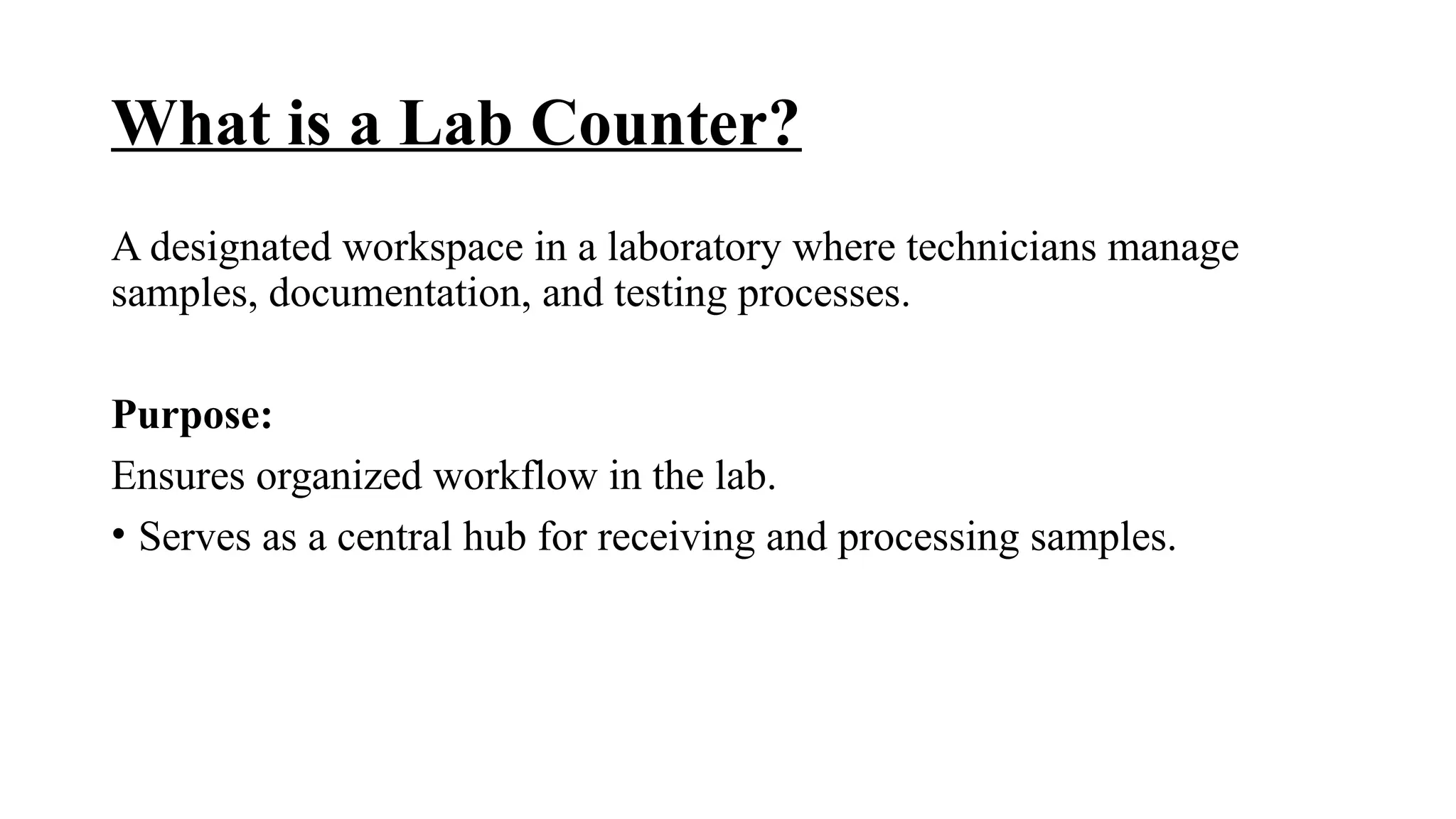


The document outlines the roles, responsibilities, and skills required for lab technicians working in laboratory settings, emphasizing the importance of sample management, documentation, quality control, and safety practices. It details the processes involved in blood drawing, sample labeling, and the correct use of various anticoagulant tubes, alongside the workflow for sample distribution to laboratory departments for timely and accurate diagnosis. Additionally, it highlights common challenges faced in lab operations and best practices for addressing them.