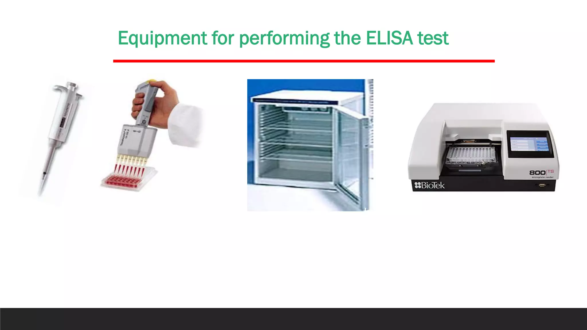
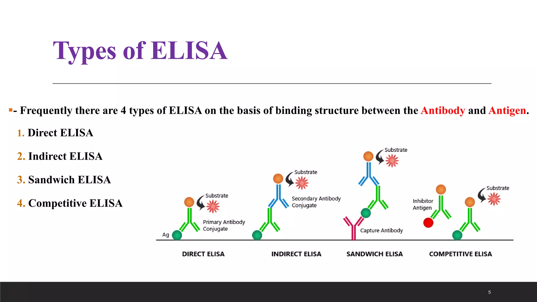
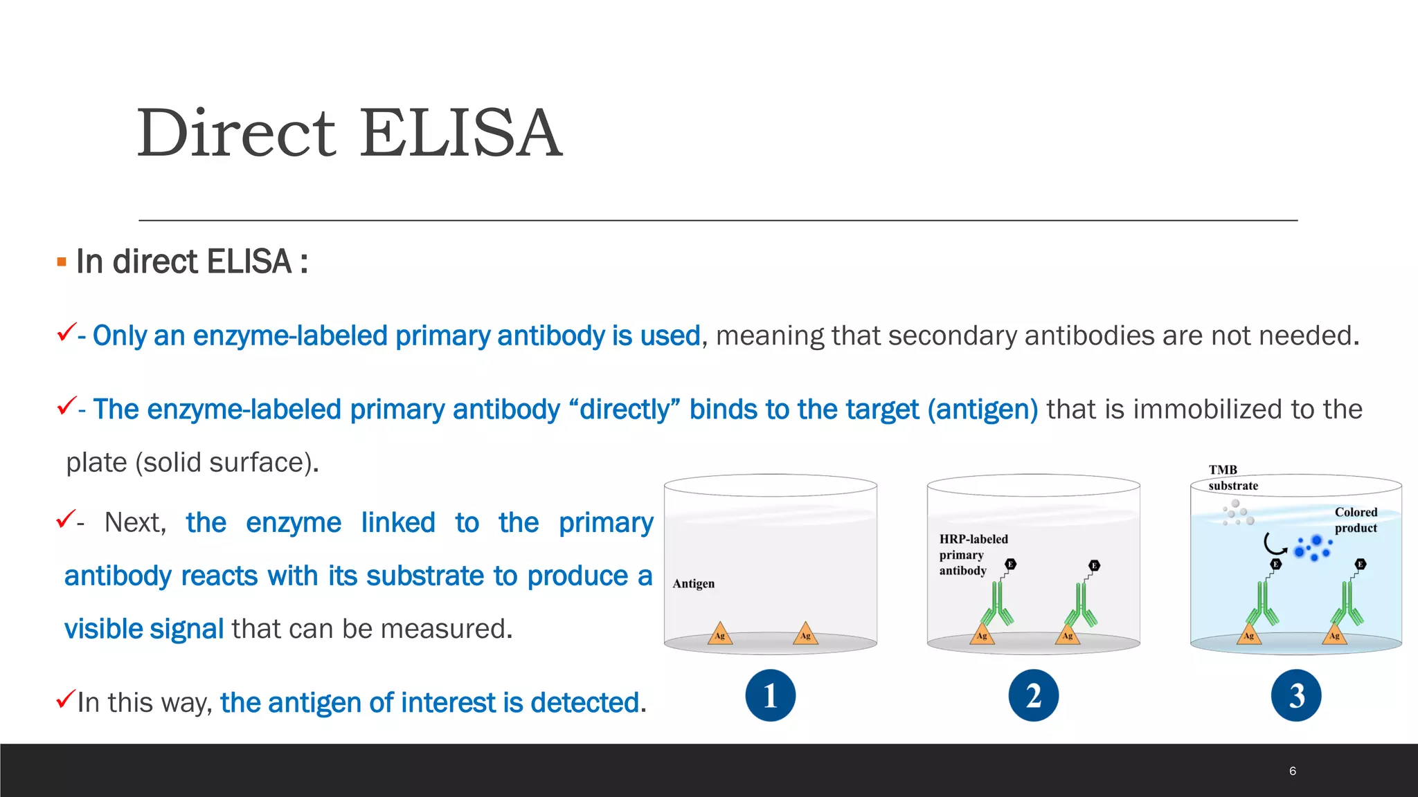
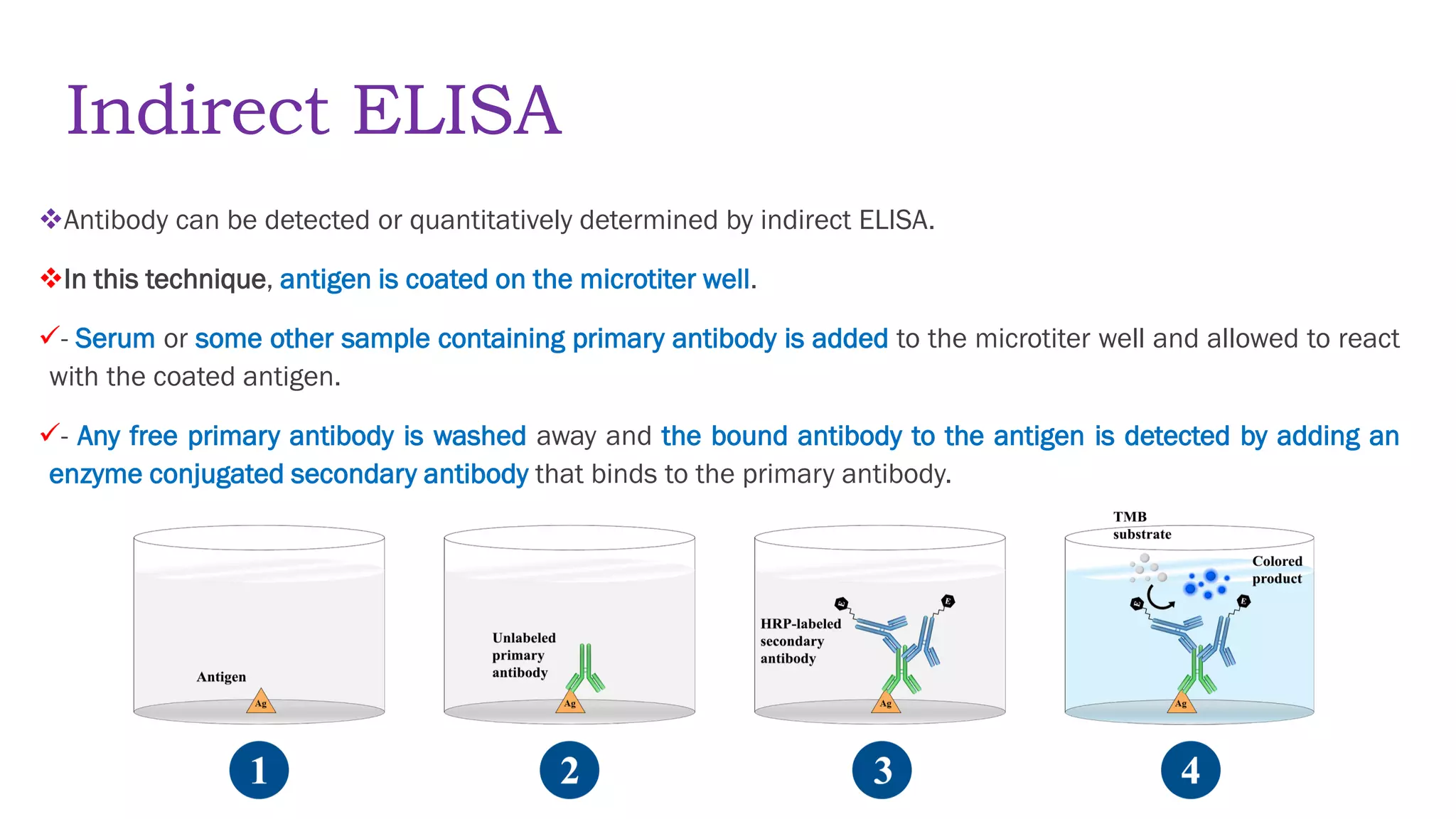
this region is termed the posterior perforated substance.
The oculomotor nerve emerges from a groove on the medial side of the crus cerebri and passes forward in the lateral wall of the cavernous sinus
1. 1
Dr. Omeed Akbar Ali
PhD. Clinical Biochemistry
Tikrit University - College of Medicine
2. Enzyme-linked immunosorbent assay
▪ ELISA is an antigen-antibody reaction.
✓ It is a common laboratory technique which is usually used to
measure the concentration of antibodies or antigens in the blood.
▪ELISA is a plate-based assay technique which is used for
detecting and quantifying substances such as peptides, proteins,
antibodies, and hormones.
✓An enzyme conjugated with an antibody reacts with colorless
substrate to generate a colored product.
✓ Such substrate is called chromogenic substrate.
2
1971
Peter Perlmann & Eva Engvall
Stockholm University
Sweden
4. Principle
✓ELISAs are typically performed in 96-well polystyrene plates.
✓The serum is incubated in a well, and each well contains a different serum.
✓A positive control serum and a negative control serum would be included among the 96 samples being tested.
✓Antibodies or antigens present in serum are captured by corresponding antigen or antibody-coated onto the solid
surface.
✓After some time, the plate is washed to remove serum and unbound antibodies or antigens with a series of wash
buffer.
✓To detect the bound antibodies or antigens, secondary antibody that are attached to an enzyme such as peroxidase or
alkaline phosphatase are added to each well.
✓After an incubation period, the unbound secondary antibodies are washed off.
✓When a suitable substrate is added, the enzyme reacts with it to produce a color.
✓This color produced is measurable as a function or quantity of antigens or antibodies present in the given sample.
✓The intensity of color/ optical density is measured at 450nm.
✓The intensity of the color gives an indication of the amount of antigen or antibody. 4
5. Types of ELISA
▪- Frequently there are 4 types of ELISA on the basis of binding structure between the Antibody and Antigen.
1. Direct ELISA
2. Indirect ELISA
3. Sandwich ELISA
4. Competitive ELISA
5
6. Direct ELISA
▪ In direct ELISA :
✓- Only an enzyme-labeled primary antibody is used, meaning that secondary antibodies are not needed.
✓- The enzyme-labeled primary antibody “directly” binds to the target (antigen) that is immobilized to the
plate (solid surface).
6
✓- Next, the enzyme linked to the primary
antibody reacts with its substrate to produce a
visible signal that can be measured.
✓In this way, the antigen of interest is detected.
8. Indirect ELISA
❖Antibody can be detected or quantitatively determined by indirect ELISA.
❖In this technique, antigen is coated on the microtiter well.
✓- Serum or some other sample containing primary antibody is added to the microtiter well and allowed to react
with the coated antigen.
✓- Any free primary antibody is washed away and the bound antibody to the antigen is detected by adding an
enzyme conjugated secondary antibody that binds to the primary antibody.
8
9. Indirect ELISA Cont….
9
✓- Unbound secondary antibody is then washed away and a specific substrate for the enzyme
is added.
✓- Enzyme hydrolyzes the substrate to form colored products.
✓- The amount of colored end product is measured by spectrophotometric plate readers that
can measure the absorbance of all the wells of 96-well plate.
11. Sandwich ELISA
❖- Antigen can be detected by sandwich ELISA.
❖- In this technique, antibody is coated on the microtiter well.
✓- A sample containing antigen is added to the well and allowed to react with the antibody
attached to the well, forming antigen-antibody complex.
✓- After the well is washed, a second enzyme-linked antibody specific for a different epitope on the
antigen is added and allowed to react with the bound antigen.
✓- Then after unbound secondary antibody is removed by washing.
✓- Finally substrate is added to the plate which is hydrolyzed by enzyme to form colored products.
11
14. Competitive ELISA
❖- This test is used to measure the concentration of an antigen in a sample.
❖- In this test, antibody is first incubated in solution with a sample containing antigen.
✓- The antigen-antibody mixture is then added to the microtitre well which is coated with antigen.
✓- The more the antigen present in the sample, the less free antibody will be available to bind to
the antigen-coated well.
✓- After the well is washed, enzyme conjugated secondary antibody specific for isotype of the
primary antibody is added to determine the amount of primary antibody bound to the well.
✓- The higher the concentration of antigen in the sample, the lower the absorbance.
14
17. Applications
1-Screening donated blood for evidence of viral contamination by
◦ HIV-1 and HIV-2 (presence of anti-HIV antibodies)
◦ hepatitis C (presence of antibodies)
◦ hepatitis B (testing for both antibodies and a viral antigen)
2-Measuring hormone levels
◦ HCG (as a test for pregnancy)
◦ LH (determining the time of ovulation)
◦ TSH, T3 and T4 (for thyroid function)
18. ◦ sexually-transmitted agents like HIV, syphilis and chlamydia
◦ hepatitis B and C
◦ Toxoplasma gondii
Detecting allergens in food and house dust
Measuring "rheumatoid factors" and other autoantibody in autoimmune diseases like
lupus erythematosus
Measuring toxins in contaminated food
3-Detecting infections
Applications