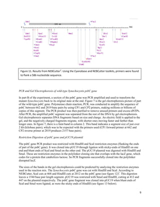
This experiment transformed the cyanobacterium Synechocystis sp. PCC 6803 in two parts. Part A introduced a mutation to the psbC gene, which encodes a chlorophyll-binding protein, via a plasmid. This disrupted photosystem II and allowed selection of transformed cells. Part B amplified the wild-type psbC gene, cloned it into a plasmid, and transformed mutant cells to restore photosystem function. Various DNA manipulations, transformations, and selections were performed to characterize and select transformed cells at each step.
Genetically engineered E. coli were designed to express enhanced green fluorescent protein (EGFP). The EGFP gene was PCR amplified from a plasmid and inserted into the expression vector pET-41a. This recombinant DNA was transformed into E. coli. While some colonies were observed, none exhibited green fluorescence under UV light. Errors in PCR amplification and potential issues with the recombinant DNA inserts suggest the hypothesis that E. coli transformants would express EGFP was not supported.
Through genetic barcoding of the tufA gene, an unknown green algae specimen was identified as belonging to the Ulva compressa clade. DNA was isolated from the specimen and the tufA gene was amplified via PCR. The gene was cloned and the nucleotide sequence analyzed and compared to sequences in GenBank. Phylogenetic analysis indicated the specimen was most similar to Ulva sp. BER-2007, but could not be identified to the species level. While in the U. compressa clade, further testing is needed to confirm its identity as a potential new species of Ulva in Narragansett Bay.
This laboratory report summarizes an experiment exploring RNA splicing in Drosophila melanogaster. Genomic DNA and total RNA were extracted from fruit flies and used to study the rngo gene. PCR and RT-PCR were performed on the genomic DNA and cDNA samples. The genomic PCR product was cloned and sequenced. Bioinformatics analysis showed the genomic sequence was longer, containing introns absent from the cDNA, indicating splicing of the rngo pre-mRNA. Future work could investigate other splicing sites and homology to human genes.
This document summarizes an experiment that aimed to change both the expression level and color of the fluorescent protein mCherry. The experiment involved:
1) Using restriction digestion and ligation to swap the promoter of mCherry from low to high expression, resulting in more mCherry colonies.
2) Attempting site-directed mutagenesis to change mCherry to mOrange but this was unsuccessful, as no orange colonies were observed.
3) Characterizing the fluorescence of mCherry, mOrange from a partner, and a negative control colony, finding mOrange emitted better at 500nm.
The document summarizes a student laboratory experiment attempting to genetically transform E. coli bacterial cells with a GFP plasmid (pGLO) using heat shock. The expected results were that bacterial cells transformed with the plasmid would grow on ampicillin-containing media and glow under black light. However, the actual results found that all bacterial cells died on ampicillin-containing plates, contradicting expectations. Possible sources of error that could have caused transformation failure are discussed.
The document describes a laboratory experiment on recombinant DNA techniques. The goal was to create a recombinant plasmid containing genes for resistance to both ampicillin and kanamycin. This was accomplished by purifying plasmids containing each resistance gene (pAMP and pKAN), digesting them with restriction enzymes, ligating the fragments together, and transforming E. coli bacteria with the recombinant plasmid. Success was indicated by bacterial growth on plates containing both antibiotics. The procedures and results are described in detail, demonstrating the process of creating and cloning recombinant DNA.
Gateway cloning is a molecular biology technique that allows efficient transfer of DNA fragments between plasmids using proprietary enzyme mixes and recombination sequences called "Gateway att sites". It involves two recombination reactions: BP reaction to create an entry clone, and LR reaction to create an expression clone in 1 hour at room temperature with over 95% efficiency, avoiding the need for restriction enzymes, ligation, or subcloning. The system enables rapid shuttling of DNA between vectors for applications like protein expression and RNAi.
The document describes protocols for preparing competent E. coli cells, transforming those cells with plasmid DNA, growing cultures of E. coli containing plasmids, and purifying plasmid DNA from the bacterial cells. Specifically, it provides detailed multi-step protocols for making competent cells, transforming the cells, preparing glycerol stocks and stab cultures for long-term storage of bacterial strains, recovering single colonies, monitoring bacterial growth, and lysing the bacterial cells to release plasmid DNA.
This document describes experiments aimed at isolating bacteriophage mutants with altered tolerance for plating when the gpD capsid protein is fused to foreign proteins. The author isolated "IPDF" mutants of phage i434Dam123 that could not plate when gpD-fusions were expressed from a plasmid. These IPDF mutants were then used to select "supIPDF" mutants that regained the ability to plate equally in the presence or absence of gpD-fusions. Sequencing showed the mutations responsible for the IPDF and supIPDF phenotypes were located outside of the D and E genes. This suggests there are extragenic mutations that can enhance or suppress the toxicity of gpD-fusions towards viable phage
Cell culture is the process by which prokaryotic, eukaryotic or plant cells are grown under controlled conditions. Mammalian cell culture technology has become a major field in modern biotechnology; mammalian cell culture refers to the cells of a mammalian, isolated from specific tissues (i.e. skin, liver, glands, etc.) and further cultivated and reproduced in an artificial medium. Cell culture technology is currently playing a major role in toxicity testing, cancer research, virology, genetic engineering, and gene therapy.
OBJECTIVE:
To observe the transfection of CHO and HEK cells with GFP
To observe the recombinant GFP using Western Blotting
To purify the transfected HEK and CHO cells using AKTA Pure Purification
The purpose of the experiment was to create a mutant cancer gene by fusing the Jaz-f1 and Su(z)-12 genes using E. coli bacteria. The gene would then be injected into fruit flies to study its effects. Initial attempts to fuse the genes through PCR were unsuccessful due to errors. A second trial showed two positive results but the bands disappeared unexpectedly. While the specific goal was not achieved, the experiment provided insights and pointed to ways to improve fusion methods, such as forced splicing. It also validated that the gene transfer method could work with further refinement.
- The study investigated the effect of calcium chloride concentration on the transformation efficiency of E. coli with plasmids pUC19 and pBR322, which differ in size.
- Maximum transformation efficiency was observed at 0.15M CaCl2 for pUC19 and 0.1M CaCl2 for the larger pBR322 plasmid.
- Increasing calcium chloride concentration above these levels decreased transformation efficiency for both plasmids, with no transformants observed above 0.2M, possibly due to decreased cell viability in hypertonic conditions.
ReedWoyda_Introducing Green Fluorescence Into Homo sapiens And Escherichia Co...Reed Woyda
This study aimed to introduce the green fluorescent protein (GFP) gene into E. coli and human cells. GFP was successfully inserted into E. coli and shown to be expressed under control of the L-arabinose promoter. Addition of restriction sites to GFP was also successful, allowing for digestion of the GFP and pcDNA plasmid. However, ligation of the digested GFP fragment into pcDNA was unsuccessful, likely due to nuclease contamination. Expression of GFP in human cells could not be verified due to a technical error during immunoblotting. While some goals were achieved, such as GFP expression in E. coli, further optimization is needed to fully introduce GFP into human cells via this methodology.
This proposal seeks funding to develop an assay to determine the efficiency of the E. coli γ-complex clamp loader loading the E. coli β-clamp onto DNA. The researcher will purify proteins and assemble the γ-complex. An oligonucleotide will be biotinylated and annealed to bind to streptavidin beads. The γ-complex will load β-clamp onto the DNA using varying ratios and times. Analyzing samples by SDS-PAGE will optimize conditions and verify loading. Developing this assay will allow future study of the β-clamp-clamp loader complex role in DNA replication. Challenges include optimizing protein ratios and times to achieve sufficient loading without unloading.
Urja Bhatt undergraduate 8th sem project pptUrja Bhatt
1) The document discusses the importance of central pair apparatus proteins in the flagella of the unicellular alga Chlamydomonas reinhardtii. It focuses on generating mutants through RNAi to study the role of proteins like hydin that contain an adenylate kinase domain.
2) Experiments generated hydin mutants in wild type and cpc1 backgrounds through transformation. Prospective double mutants were screened through transcript analysis and phenotype observation.
3) A reactivation assay showed that the cpc1 mutant had a ten-fold decrease in speed compared to wild type, indicating central pair proteins are required for optimal motility.
4) While RNAi was unstable, the study provides a framework
The document summarizes several biology lab experiments conducted by the author:
1. They performed DNA extraction from samples, PCR, and Western blot techniques. For DNA extraction, their sample did not produce the expected results.
2. They learned aseptic technique and used Gram staining to identify bacteria samples as gram positive or negative.
3. PCR was used to amplify genomic DNA between primers over multiple cycles. Controls were included.
4. Nested PCR with more specific primers was used to further amplify portions of DNA. Exonuclease treated samples before nested PCR.
5. Gel electrophoresis separated DNA fragments by size. PCR products from two plant samples were analyzed, with one showing bands.
1. The document describes constructing deletion mutants of several genes (spr, prc, eco293-sinI and fimS) in the uropathogenic E. coli strain UTI89 using PCR and homologous recombination.
2. PCR was used to generate gene disruption cassettes containing antibiotic resistance genes flanked by FRT sites. These were electroporated into UTI89 cells expressing Red recombinase to disrupt the target genes.
3. Successful disruptions were checked by PCR and colonies were grown on antibiotic plates. The antibiotic resistance genes were then evicted using FLP recombinase, leaving only the gene deletions.
Glass bead transformation method for gram positive bacteriaCAS0609
This study developed a simple glass bead transformation method for introducing DNA into Gram-positive bacteria. The method involves treating bacterial protoplasts with glass beads, DNA, and polyethylene glycol. Using this method, the plasmid pGK12 was successfully introduced into several Gram-positive bacteria, including Enterococcus faecalis, Lactobacillus casei, Lactococcus lactis, Leuconostoc dextranicum, Listeria innocua, Staphylococcus aureus, and Streptococcus pneumoniae. Transformation frequencies ranged from 3.56 x 10³ to 6.62 x 10³ colonies per microgram of pGK12. This glass bead method provides an inexpensive and reproducible way to transform Gram
In Cell-Western Antibody Customer Review for Anti-LC3A Antibody (STJ97755)St John's Laboratory Ltd
Microtubule-associated proteins 1A/1B light chain 3A is a protein that in humans is encoded by the MAP1LC3A gene
Whereas LC3s are involved in elongation of the phagophore membrane, the GABARAP/GATE-16 subfamily is essential for a later stage in autophagosome maturation.
Anti-LC3A - http://www.stjohnslabs.com/lc3a-antibody?filter_name=STJ97755
Join our Antibody Validation Project - http://www.stjohnslabs.com/services/antibody-validation
Ubiquitin-like modifier involved in formation of autophagosomal vacuoles (autophagosomes) . Whereas LC3s are involved in elongation of the phagophore membrane, the GABARAP/GATE-16 subfamily is essential for a later stage in autophagosome maturation.
Anti-LC3A -http://www.stjohnslabs.com/lc3a-antibody
Join our Antibody Validation Project - http://www.stjohnslabs.com/services/antibody-validation
Molecular Cloning of the Structural Gene for ExopolygalacturonateAlan Brooks
This document summarizes research on the cloning and characterization of a gene (pelX) from Erwinia chrysanthemi that encodes an exopolygalacturonate lyase (ExoPL). The pelX gene was cloned from a mutant strain lacking known pectate lyase genes. ExoPL was purified from a recombinant E. coli strain and characterized. A pelX mutant was constructed in E. chrysanthemi but retained pathogenicity, indicating ExoPL does not contribute to tissue maceration ability.
Frederick Griffith discovered that heat-killed virulent strains of Streptococcus pneumoniae could transform nonvirulent strains into virulent ones when mixed together and injected into mice. This suggested that some genetic material was being transferred. Avery, MacLeod and McCarty showed that the transforming principle was DNA. Hershey and Chase used radioactive labeling to show that when bacteriophage infect bacteria, only the viral DNA enters the host cell. This provided strong evidence that DNA is the genetic material. Watson and Crick deduced the double-helix structure of DNA using evidence from Chargaff, Franklin and others. Meselson and Stahl's experiment supported the semiconservative model of DNA replication.
Ubiquitin-like modifier involved in formation of autophagosomal vacuoles (autophagosomes). Plays a role in mitophagy which contributes to regulate mitochondrial quantity and quality by eliminating the mitochondria to a basal level to fulfill cellular energy requirements and preventing excess ROS production. Whereas LC3s are involved in elongation of the phagophore membrane, the GABARAP/GATE-16 subfamily is essential for a later stage in autophagosome maturation. Promotes primary ciliogenesis by removing OFD1 from centriolar satellites via the autophagic pathway.
Anti-MAP LC3β -http://www.stjohnslabs.com/map-lc3b-antibody
Join our Antibody Validation Project - http://www.stjohnslabs.com/services/antibody-validation
In Cell-Western Antibody Customer Review for Anti-MAP LC3β antibody (STJ97398)St John's Laboratory Ltd
Microtubule-associated proteins 1A/1B light chain 3A is a protein that in humans is encoded by the MAP1LC3A gene. MAP1A and MAP1B are microtubule-associated proteins which mediate the physical interactions between microtubules and components of the cytoskeleton. Plays a role in mitophagy which contributes to regulate mitochondrial quantity and quality by eliminating the mitochondria to a basal level to fulfill cellular energy requirements and preventing excess ROS production.
Anti-MAP LC3β - http://www.stjohnslabs.com/map-lc3b-antibody?filter_name=STJ97398
Join our Antibody Validation Project - http://www.stjohnslabs.com/services/antibody-validation
Ubiquitin-like modifier involved in formation of autophagosomal vacuoles (autophagosomes). Plays a role in mitophagy which contributes to regulate mitochondrial quantity and quality by eliminating the mitochondria to a basal level to fulfill cellular energy requirements and preventing excess ROS production. Whereas LC3s are involved in elongation of the phagophore membrane, the GABARAP/GATE-16 subfamily is essential for a later stage in autophagosome maturation. Promotes primary ciliogenesis by removing OFD1 from centriolar satellites via the autophagic pathway.
Anti-MAP LC3β-http://www.stjohnslabs.com/map-lc3b-antibody
Join our Antibody Validation Project - http://www.stjohnslabs.com/services/antibody-validation
Ubiquitin-like modifier involved in formation of autophagosomal vacuoles (autophagosomes) . Whereas LC3s are involved in elongation of the phagophore membrane, the GABARAP/GATE-16 subfamily is essential for a later stage in autophagosome maturation .
Anti-LC3A-http://www.stjohnslabs.com/lc3a-antibody
Join our Antibody Validation Project - http://www.stjohnslabs.com/services/antibody-validation
Genetic transformation is a technique used to introduce new genes into organisms to produce desired proteins. This lab explored transformation in E. coli to express green fluorescent protein (GFP) when exposed to arabinose. Bacteria without the GFP plasmid only grew in media containing LB, while those with the plasmid grew in all media due to antibiotic resistance. The bacteria appeared white in LB and ampicillin but turned green under UV light in LB, ampicillin and arabinose, showing arabinose activated GFP production.
This document summarizes a study on the heterologous expression and characterization of the c1 dioxygenase enzyme from Tetranychus urticae. Key points:
- The c1 dioxygenase gene was inserted into a plasmid and transformed into E. coli cells for expression. However, initial expression attempts did not show overexpression of the protein.
- Additional expression trials were conducted by varying culture conditions and bacterial clones. SDS-PAGE analysis showed some potential overexpression in new clones induced overnight at 37°C, though further optimization is still needed.
- Once expression is optimized, the goal is to purify the recombinant protein using its His-tag and proceed with structural characterization through crystallization,
This document describes methods for stably transfecting cell lines to create new cell lines. It compares the calcium phosphate precipitation method, spontaneous transfection method, and lipofection method. It also details monitoring transfection efficiency by fluorescence microscopy and spectroscopy. Results show the calcium phosphate method worked best for most cell lines tested except HeLa cells. Stable transfection of HeLa cells with a plasmid increased fluorescence over 21 days then plateaued. Plasmid DNA concentration did not correlate with fluorescence yield.
1. The document describes methods for stably transfecting cell lines, including calcium phosphate transfection, lipofection, and spontaneous transfection.
2. It examines the efficiency of transfection for different cell lines using green fluorescent protein and red fluorescent protein plasmids.
3. Results show that calcium phosphate transfection was more efficient than spontaneous transfection for most cell lines tested, except HeLa cells. Stable transfection of HeLa cells over 21 days resulted in gradually increasing fluorescent intensity that then remained constant.
Isolation and characterization of an extracellular antifungal protein from an...Maulik Kamdar
The document describes a thesis project that aims to isolate and characterize an extracellular antifungal protein (exAFP-C28) from an endophytic fungal isolate. The objectives are to isolate the fungal strain from a medicinal plant, optimize culture conditions, purify the protein, and determine its antifungal activity and mechanism of action against Candida albicans through assays and microscopy. Results showed that the protein was effective against C. albicans by disrupting cell membranes, had a molecular weight of 28.2 kDa, and likely forms amphipathic helices contributing to its antifungal activity.
This document describes experiments performed to sequence the human Apolipoprotein B (ApoB) gene. A portion of the ApoB gene was amplified via PCR and subcloned into E. coli plasmid vectors. The plasmid vectors containing the inserted ApoB fragment were then purified and sequenced. Sequence analysis revealed that human ApoB is highly similar to Canis lupus familiaris (dog) ApoB, indicating evolutionary conservation. The experiments aimed to accurately insert, track, and sequence the ApoB gene to better understand its structure, function, and evolutionary relationships.
This document describes experiments to express and purify the enzyme CelB2 from E. coli for studying its functionality in cellulosic biofuel production. CelB2 was expressed as a His6MBPCelB2 fusion to improve expression and purification. Purification using amylose affinity chromatography was successful, but further purification by DEAE ion exchange chromatography and Factor Xa digest was needed. Fractions from ion exchange chromatography were tested for CelB2 activity to determine the most active fractions for further study.
The document summarizes steps taken to clone a drug resistant gene from Bacillus subtilis using TA cloning technology. Key steps included isolating genomic DNA from B. subtilis, amplifying the gene of interest via PCR, isolating and digesting a plasmid vector, and ligating the insert into the vector. The ligation mixture was then used to transform competent E. coli cells. Gene cloning has applications in developing diagnostic tests, vaccines, and antibiotics.
i need a tutor to complete 3 results conclusion sections.pdfbkbk37
The student needs help completing the results and conclusion sections for 3 lab reports. The student has performed a thermofluor assay and enzyme assay and needs help analyzing the results and drawing conclusions. The student will provide the raw results from the lab sessions for the assistant to complete the write up.
This document summarizes research purifying the Streptomyces lividans endoglucanase CelB2 enzyme expressed in E. coli. CelB2 mutants were designed to be more thermally stable. CelB2 was expressed in E. coli BL21(DE3) as a fusion protein with maltose binding protein (MBP) to aid purification. CelB2 was purified using affinity chromatography on amylose resin, Factor Xa cleavage of MBP, and ion exchange chromatography. While CelB2 was purified, only low levels were recovered, suggesting further work is needed to improve CelB2 recovery and stability.
The study aimed to test the specificity of two antibodies (033 and 1-9E10) for binding the c-myc protein. Human colon carcinoma cells were extracted to obtain cytosolic, nuclear, and nuclear pellet extracts. The 033 antibody bound c-myc in the cytosolic extract at approximately 110 kDa, but the 9E10 antibody did not bind c-myc. This suggests c-myc is not restricted to the nucleus and more research is needed to fully understand c-myc and its role in cancer.
The researchers aimed to purify cellular retinol binding protein (CRBP) from bovine liver. Through a process involving homogenization, centrifugation, cation exchange chromatography, gel filtration, and concentration, they obtained a final product. However, characterization through SDS-PAGE and absorption spectroscopy identified the protein as catalase rather than CRBP. Despite initial absorption at 350nm for CRBP, the maximal absorption and thermal/pH profiles matched those of catalase. The purification resulted in the isolation of catalase rather than the intended CRBP.
Expression Purification and Immunodetection of a fusion protein Glutathione S...iosrjce
Glutathione S Transferase(GST) is an enzyme of a multi gene family which is involved in reducing
oxidative damage to cells and detoxification of Xenobiotic compounds and plays critical role in life processes.
The entire work was completely qualitative and the objective of my work was to deal with the induction,
extraction and purification of the GST fusion protein from pGEX 3X vector.In order to achieve high degree of
transformed cells,the E.Coli BL21 host strain was made competent using 0.1M CaCl2 and adding of pGEX 3X
vector into host made it transformed.With the induction of GST protein by 0.1mM IPTG,the desired protein was
purified through glutathione Cl agarose column and was detected by immunoblotting method with the use of
anti GST HRP conjugate Ab which expressed the desired protein.
Expression Purification and Immunodetection of a fusion protein Glutathione S...iosrjce
IOSR Journal of Biotechnology and Biochemistry (IOSR-JBB) covers studies of the chemical processes in living organisms, structure and function of cellular components such as proteins, carbohydrates, lipids, nucleic acids and other biomolecules, chemical properties of important biological molecules, like proteins, in particular the chemistry of enzyme-catalyzed reactions, genetic code (DNA, RNA), protein synthesis, cell membrane transport, and signal transduction. IOSR-JBB is privileged to focus on a wide range of biotechnology as well as high quality articles on genetic engineering, cell and tissue culture technologies, genetics, microbiology, molecular biology, biochemistry, embryology, cell biology, chemical engineering, bioprocess engineering, information technology, biorobotics.
B. Pharm. (Honours) Part-IV Practical,Molecular biology & Biotechnology, MANIKImran Nur Manik
Molecular Biology & Biotechnology: (Marks –35)
a) Isolation of plasmid DNA
b) Estimation of DNA, RNA and oligonucleotides
c) Agarose-gel electrophoresis of nucleic acids
d) Determination of bacterial drug resistance by disk diffusion method.
e) Estimation of protein concentration by Lowry method
Presentation on increased bioavailability of breviscapine via a pluronic p85 ...2503jyoti
Breviscapine is a hydrophobic drug used in cerebovascular diseases.It's bioavailability due to it's efflux by P-gp system.pluronics are non-ionic tri-bolic co-polymers made up of central part of PPO(hydrophobic) which is flanked by two PEO(hydrophilic) molecules
Western Blot Antibody Customer Review for Anti-Peroxin 2 Antibody (STJ95040)St John's Laboratory Ltd
PEX2 is gene on chromosome 8q21.1 that encodes a peroxin, an integral membrane protein involved in peroxisome biosynthesis and integrity, which assembles membrane vesicles before translocation of matrix proteins.
Defects of PEX2 cause peroxisome biogenesis disorder complementation group 5, Zelleger syndrome and a form of Refsum disease.
Anti-Peroxin 2 - http://www.stjohnslabs.com/peroxin-2-antibody?filter_name=STJ95040
Join our Antibody Validation Project - http://www.stjohnslabs.com/services/antibody-validation
This study investigated the effects of neuropeptide Y (NPY) on signaling proteins in C. elegans. NPY is a neurotransmitter that regulates anxiety in humans. The researchers extracted proteins from C. elegans exposed to NPY and analyzed them using ELISA. They found a statistically significant upregulation of Akt1 protein levels in the presence of NPY, but no significant effects on other proteins like p38 MAP kinase and Stat3. This suggests NPY may activate Akt1 signaling in C. elegans. The results help validate the proposed pathway of NPY's anxiolytic effects.
This study investigated the effects of neuropeptide Y (NPY) on signaling proteins in C. elegans. NPY is a neurotransmitter that regulates anxiety in humans. The researchers extracted proteins from C. elegans exposed to NPY and analyzed them using ELISA. They found a statistically significant upregulation of Akt1 protein levels in the presence of NPY, but no significant effects on other proteins like p38 MAP kinase and Stat3. This suggests NPY specifically increases Akt1, a regulator of cell growth and survival, in C. elegans. The results help validate the proposed pathway of NPY's anxiolytic effects.
Similar to Karen Hatten Experiment II Final Report (20)
2. 2
Introduction
Synechocystis sp. PCC 6803 is a cyanobacterium with two significant features relevant to this experiment.
It has natural transformability meaning that it is readily available for the uptake of foreign DNA, in this
case the pKCP43 plasmid. However, only a single genome copy out of the possible 6-12 copies may be
transformed. The pKCP43 plasmid confers a kanamycin resistance gene which enables selection of
transformed cells. After a series of gradual but steep additions of kanamycin to the growth medium,
eventually the transformed Synechocystis will be completely mutant in all genome copies.1
Additionally,
Synechocystis has the gene psbC which encodes for CP43, a chlorophyll-binding protein responsible for
photosystem II. This enables the bacterium to grow without an added carbon source because it uses light
for energy to produce sugars from carbon dioxide and water.1
The experiment is designed around this
concept.
Two concurrent experiments are eventually integrated. The first part (A) introduces a mutation to the
psbC gene via double homologous recombination with the pKCP43 plasmid. The kanamycin resistance
gene disrupts the psbC gene. This change results in two important consequences, first the mutation
inhibits photosystem II so that the transformed Synechocystis cells need added sugar to survive, second
the cells are resistant to the antibiotic kanamycin. These two factors help to select transformed cells.
The second part (B) of the experiment entails PCR amplifying and cloning part of the wild type psbC
gene into the pUC19 plasmid. E. coli is transformed with the recombinant plasmid because it synthesizes
beta-galactosidase which cleaves lactose. This detail enables identification and selection with a blue-
white screening system, because transformed E. coli. cannot catabolize the color-inducing substrate X-gal
in the medium. The isolated plasmid is then sequenced and eventually used to transform the mutant
Synechocystis bacteria from the first part of the experiment. This restores the photosystem II functionality.
At the end of the experiment, the re-transformed mutant Synechocystis is plated on a medium without
glucose and is observed to confirm the occurrence of photosynthesis. Growth indicates that the mutant
cells have been transformed with the psbC gene.
Materials and Methods
Transformation of wild-type Synechocystis
Part A of experiment II consists of transforming Synechocystis with the pKCP43 plasmid and selecting
for transformants by gradually increasing the antibiotic concentration of the growth medium. First, 2 mcL
of pKCP43 plasmid was added to a prepared culture of Synechocystis sp. PCC 6803 in growth medium.
The mixture was incubated at room temperature for 30 minutes. The suspension was then spread onto a
filter on a BG–11+5 mM glucose agar plate in a vertical flow hood. The plate was incubated at 30°C
under light. It was then transferred to a plate containing glucose and 5 mcg/mL kanamycin. After two
weeks, the filter with the cell culture was transferred to a plate with a higher antibiotic concentration (BG-
11+glucose+kanamycin 20 mcg/mL) in sterile flow hood. The plate was again incubated at 30°C in the
light. The colonies were transferred to a BG-11+glucose+kanamycin 50 mcg/mL plate after another two
weeks, then to BG-11+glucose+kanamycin 75 mcg/mL after two more weeks, in the same fashion. After
yet another two weeks, the colonies were transferred to BG-11+glucose+kanamycin 100 mcg/mL by
making zigzag streaks in each of four quadrants of the new plate. Sterile loops were used to continue one
quadrant’s streak into the next quadrant. Consistent with preceding transfers, the plate was incubated at
30°C in the light.
3. 3
Analysis of Synechocystis wild-type and psbC-transformant
Part A of the experiment was continued with genomic DNA extraction. A loopful of cells from the psbC-
mutant plate, as well as a loopful from a plate with wild type, were each mixed with 200 mcL of TE. 150
mcL glass beads and 150 mcL more TE were added. They were blended and settled, and the supernatant
was removed. An additional 100 mcL TE was added, mixed and settled. Supernatant was again removed.
The samples were centrifuged, and the supernatant from these was removed. 250 mcL of a phenol:
chloroform: isoamyl alcohol concoction was added to each sample, and they were vortexed and
centrifuged. The upper layers of the samples were removed, and 125 mcL of 7.5 M ammonium acetate
and 325 mcL ethanol were added to them and mixed. The samples were centrifuged and the supernatant
was discarded. The pellets were washed with 400 mcL 70% ethanol. Supernatant was again discarded and
the pellet was dried in the Speed Vac for 10 minutes. The pellets were resuspended in 20 mcL of TE.
2 mcL of each sample was added to 50 mcL of a PCR reaction mixture of buffers, dNTPs, Taq
Polymerase, and primers that flank the interruption region of the psbC gene. The entire mixtures were run
through the Thermal Cycler as follows:
1. 94°C x 3 min
2. 94°C x 1 min
3. 58°C x 1 min
4. 72°C x 1 min
Repeat steps 2-4 x 30 cycles
5. 4°C soak
10 mcL of each PCR product was mixed with 2 mcL of loading dye and loaded into wells 2 and 3 of an
agarose gel. 120 volts were run through the gel electrophoresis for 30 minutes. The gel was viewed under
u.v. light and photographed.
PCR and Gel Electrophoresis of wild-type Synechocystis psbC gene
Part B of the experiment consists of amplifying the wild-type psbC gene and using it to transform the
mutant Synechocystis from part A, restoring it to its natural state. First, 2 mcL of Synechocystis genomic
DNA was added to a prepared PCR reaction mix, and the samples went through the 4 PCR cycles and a
soak cycle.
5 mcL of the PCR product was added to 1 mcL of loading dye. This mixture was placed into a well and
ran against 4 mcL of the kb DNA ladder at 70 volts for 40 to 50 minutes. The ladder was viewed and
photographed under the Alphamager UV light. The PCR product was purified using the QIAquick PCR
Purification Kit. To bind the DNA, Buffer PBI was added to the PCR sample at a 5:1 ratio and
centrifuged in a QIAquick column for 30 to 60 seconds. The mixture was drained and 0.75 mL Buffer PE
was added to wash the sample. It was again centrifuged and drained. After a third centrifuge, a pure DNA
fragment was eluted by adding 30 mcL Buffer EB and centrifuging.
Restriction Digestion of psbC gene and pUC19 plasmid
4 mcL of pUC19 plasmid was mixed with a total 16 mcL of ddH20, NEB CutSmart 10X buffer mix, and
the SmaI and HindIII-HF restriction enzymes. The vector plasmid digest sample was incubated at room
temperature for an hour, then at 37°C for another hour.
4. 4
15 mcL of the PCR product of psbC gene was mixed with 5 mcL digest mix (also containing ddH20,
NEB buffer mix and ScaI-HF/HindIII-HF restriction enzymes). This sample was incubated at 37°C for an
hour and a half.
Both digestion reaction mixes were then incubated at 70°C for 20 minutes to inactivate the restriction
enzymes. Then, 4 mcL of the pUC19 digestion mix was added to 8.75 mcL of the PCR digestion mix, 1.5
mcL of ligase buffer and 0.75 mcL of ligase enzyme. The sample was mixed and incubated at room
temperature for 2 hours.
Transforming E. coli with recombinant plasmid pUC19
15 mcL of the ligation mixture was carefully mixed with 50 mcL of NEB 5-alpha competent E. coli cells.
The sample was iced for 30 minutes, heat shocked at 42°C for 30 seconds, then iced again for 5 minutes.
1 mL of SOC was added and the sample was rotated at 37°C for an hour. 450 mcL of the transformation
cell mixture was spread with a sterilized glass spreader onto an LB-AMP-Xgal-IPTG plate. The plates
were stored inverted at 37°C for one day. Two white colonies were picked and put in 5 mL of LB-Amp
liquid medium and incubated at 37°C for another day.
Preparing the recombinant plasmid pUC19
1.4 mL of the E. coli cell culture was centrifuged for one minute and the supernatant discarded. The
sample was microfuged for 5 seconds and the rest of the supernatant was removed. The remaining pellet
was resuspended in 50 mcL sterile water, and 300 mcL of NS solution (0.5% SDS and 0.1M NaOH) was
added. The sample was gently mixed and incubated at room temperature for 90 seconds. 200 mcL of
7.5M ammonium acetate was mixed in and the sample was put on ice for 3 minutes. The sample tube was
centrifuged for 5 minutes and the supernatant was added to 330 mcL isopropanol. This mixture was
centrifuged for 7 minutes and the supernatant was discarded. The remaining pellet was washed with 0.5
mL of 70% ethanol. It was then centrifuged for 2 minutes and decanted. The pellet was dried in a heat
block at 50°C for 5 minutes. The pellet was resuspended in 30 mcL water, and 1 mcL RNase was added.
The tube was incubated at 37°C for 15 minutes.
The sample was mixed with 15 mcL of 7.5M ammonium acetate and 45 mcL isopropanol. The mixture
was iced for 3 minutes and centrifuged for 5 minutes. The supernatant was discarded and the pellet was
washed with 0.5 mL of 70% ethanol. The tube was centrifuged for 2 minutes and decanted. Again, the
pellet was heat-blocked for 5 minutes. The DNA pellet was resuspended with 20 mcL of sterile water.
Restriction digest and electrophoresis of recombinant plasmid
5 mcL of the sample was added to 15 mcL of an enzyme premix containing ddH2O, NEB 10X CutSmart
Buffer, HindIII-HF, and EcoRI-HF. The mixture was incubated at 37°C for an hour. 2 mcL of loading dye
was added and the sample was pipeted into lane 5 of an agarose gel. The gel was electrophoresed at 80
volts for 30 minutes, then photographed under u.v. light.
Transform psbC- Synechocystis with recombinant plasmid and wild-type DNA
A liquid culture of psbC- Synechocystis from part A was spun down and resuspended in 1/50 of the
original culture volume of BG – 11+ glucose. 200 mcL was transferred into 4 different tubes. The first
tube served as a negative control and no DNA was added. 1 mcL wild-type Synechocystis DNA was
added to a second tube, 1 mcL psbC recombinant plasmid DNA from part B was added to a third, and 1
mcL psbC- Synechocystis DNA was added to the fourth tube. After 30 minutes, the cells were plated on
BG – 11 agar plates, with no added glucose. The next week plates were observed and photographed.
5. 5
Results
Transformation of wild-type Synechocystis
In part A, observations of transformed Synechocystis colonies were made over time (about every two
weeks). Figures 1 through 5 capture images of the surviving colonies from a gradual increase in the
kanamycin antibiotic. There is considerable growth in each phase.
Figure 1.Week 3 Part A. Synechocystis
transformation plate: green colonies of
cyanobacteria are observed. The plate contains
BG – 11+glucose growth medium with 5
mcg/mL kanamycin.
Figure 2.Week 6 Part A. Synechocystis
transformation plate: individual colonies of
cyanobacteria are observed. The plate contains
BG – 11+glucose growth medium with 20
mcg/mL kanamycin.
Figure 3.Week 8 Part A. Synechocystis
transformation plate: individual colonies of
cyanobacteria are observed. The plate contains
BG – 11+glucose growth medium with 50
mcg/mL kanamycin.
Figure 4.Week 10 Part A. Synechocystis
transformation plate: individual colonies of
cyanobacteria are observed. The plate contains
BG – 11+glucose growth medium with 75
mcg/mL kanamycin.
6. 6
Analysis of Synechocystis wild-type and psbC-transformant
Samples were placed in lanes 2 and 3 of the agarose gel. Lane 2 has the wild-type DNA and lane 3 has the
psbC- DNA. The sample washed out of lane 3 and there is no visible band because of this misstep. Both
samples should have displayed a band at 586, and the sample in lane 3 should have a band at about 1500.
PCR and Gel Electrophoresis of wild-type Synechocystis psbC gene
In part B, PCR and gel electrophoresis of the psbC gene were successful. The band above the arrow
indicated the size just above 2 kilobase pairs (see figure 7 below).
Figure 7. Week 3 Part B. Gel
electrophoresis picture for the PCR of
part of the wild-type psbC.
CP1 forward primer (662) and CP2
reverse primer (2819) = 2157 bp.
Figure 5. Week 12 Part A.
Synechocystis transformation plate:
individual colonies of cyanobacteria
are observed. The plate contains BG –
11+glucose growth medium with 100
mcg/mL kanamycin.
Figure 6. Week 14 Part A. Gel
electrophoresis of PCR products from
DNA of wild type and psbC-
transformant of Synechocystis.
Sample is in lane two under red arrow.
7. 7
Transforming E. coli with Recombinant plasmid pUC19
Many E. coli cells were successfully transformed with the recombinant pUC19 plasmid. Both blue and
white colonies grew on the LB-AMP-Xgal-IPTG medium plate (see figure 8 below). White colonies were
picked after a week and cultured.
Gel Electrophoresis of Recombinant pUC19 Plasmid
The purified recombinant pUC19 plasmid was isolated from the culture. When the plasmid was digested
with HindIII and EcoRI, it left the cloned 1544 base pair fragment from the psbC gene plus another 16
base pairs to the EcoRI restriction site (see figure 13 in the discussion section). The remaining section of
the HindIII/EcoRI digestion should be about 1560 base pairs long. Results confirmed the DNA segment
size. The digest sample was electrophoresed and photographed under UV light (see figure 9 below). The
ladder in gel 1 is not visible, so the picture of gel 2 is shown for comparison. The sample in lane 5 of gel
1 shows a band at about 1.5 kb.
Figure 8. Week 9 Part B. LB-AMP-Xgal-
IPTG plate with many blue and white
colonies, representing untransformed
and transformed E. coli cells.
Figure 9. Comparison 1
Kb ladder and gel
electrophoresis pictures
in Alphalmager of
recombinant plasmids.
Arrow points to sample
in gel 1, lane 5. Ladders
are in first lanes of the
two gels.
Gel 1 Gel 2
8. 8
Transform psbC- Synechocystis with recombinant plasmid and wild-type DNA
There are cells in each of the four quadrants, however quandrant 3 has the greatest concentration of cells.
Quandrant 1 is the negative control, with no added DNA; quandrant 2 has wild-type Synechocystis DNA;
quandrant 3 has psbC recombinant plasmid DNA; and quadrant 4 has psbC- Synechocystis DNA.
Discussion
Transformation of wild-type Synechocystis
In part A of the experiment, Synechocystis was transformed by double-homologous recombination with
the pKCP43 plasmid. The plasmid has a kanamycin resistance gene disrupting part of the psbC gene.
Figure 1 shows growth of Synechocystis in week 3 (1 week after transformation). The BG – 11+glucose
plate provided the carbon source for the transformed Synechocystis which no longer encodes the protein
for photosystem II and cannot grow photoautotrophically. Figures 1 through 5 show growth of
Synechocystis over time, with a gradual increase in the kanamycin antibiotic. The gradual increase in
antibiotic concentration allows for only the transformed Synechocystis with the resistance to kanamycin to
grow. Eventually, the surviving Synechocystis cells are fully mutant in every genome copy.
Analysis of Synechocystis wild-type and psbC-transformant
Using the Cyanobase and NEBCutter toolkits, the PCR primers used were found to flank a 586 nucleotide
sequence (See figure 11 below), so both the wild-type and psbC- PCR samples should have bands at that
size. Also, because the primers flank the interruption region of the psbC gene, the psbC- sample should
also have an additional band at about 1500 base pairs.
Figure 10. Week 14. Parts A and B.
Testing restoration of
photoautotrophic phenotype in
transformed psbC- mutant.
9. 9
PCR and Gel Electrophoresis of wild-type Synechocystis psbC gene
In part B of the experiment, a section of the psbC gene was PCR amplified and used to transform the
mutant Synechocystis back to its original state at the end. Figure 7 is the gel electrophoresis picture of part
of the wild-type psbC gene. Polymerase chain reaction, PCR, was conducted to amplify the sequence of
psbC between 662 and 2819 base pairs by using CP1 and CP2 primers, making millions or billions of
copies of this segment. The PCR product was then purified to remove unused primers and excess dNTPs.
After PCR, the amplified psbC segment was separated from the rest of the DNA by gel electrophoresis.
Gel electrophoresis separates DNA fragments based on size and charge. An electric field is applied to the
gel, and the negatively charged fragments migrate, with shorter ones moving faster and farther than
longer ones. In figure 7, there is a faint band in column 2. This band indicates a segment size of just over
2 kb (kilobase pairs), which was to be expected with the primers used (CP1 forward primer at 662 and
CP2 reverse primer at 2819 produces 2157 base pairs).
Restriction Digestion of psbC gene and pUC19 plasmid
The psbC gene PCR product was restricted with HindIII and ScaI restriction enzymes (flanking the ends
of part of the psbC gene). It was cloned into pUC19 through ligation with sticky-ends of HindIII on one
end and blunt ends of ScaI and SmaI on the other end. The pUC19 plasmid was digested with HindIII and
SmaI. These are restriction enzymes in the polylinker cloning site that overlaps with the lacZ gene, which
codes for a protein that catabolizes lactose. So PCR fragments successfully cloned into the polylinker
disrupted lacZ.
The sizes of the bands in the gel electrophoresis could be predicted by analyzing the restriction enzymes
used in the reaction mix. The Synechocystis psbC gene was cut with HindIII and ScaI. According to
NEBCutter, ScaI cuts at 468 and HindIII cuts at 2012 on the psbC gene (see figure 12)2
. This digestion
leaves a 1544 base pair length segment. pUC19 was restricted with SmaI and HindIII, cutting at 412 and
447 on the plasmid respectively. The psbC gene fragment was cloned into pUC19 when blunt ends of
ScaI and SmaI were ligated, as were the sticky ends of HindIII (see figure 13 below).
Figure 11. Results from NEBCutter2
. Using the Cyanobase and NEBCutter toolkits, primers were found
to flank a 586 nucleotide sequence.
10. 10
Transforming E. coli with Recombinant plasmid pUC19
The recombinant pUC19 plasmids were then used to transform E. coli. Transformed cells were selected
using the “blue-white” screening procedure. The cells with intact pUC19 effectively catabolized the X-gal
in the medium and colonies turned blue, whereas the transformed cells had the recombinant plasmid with
a disrupted lacZ gene and the colonies stayed white. White colonies were then chosen with this “blue-
white” screening procedure because they contained part of the psbC gene (see figure 8). An alkaline lysis
plasmid prep and reprecipitation were performed to isolate the DNA for sequencing. A high-quality
purified sample is important to the success of automated sequencing reactions.
Restriction digestion and electrophoresis of recombinant plasmid
When the isolated recombinant plasmid was digested with EcoRI and HindIII, the resulting fragment was
about 1560 base pairs. This includes the 1544 base pairs inserted from the psbC gene during the previous
digestion (see figure 12), plus a few more base pairs to the EcoRI site (see figure 13 below).
Figure 13. pUC19 plasmid
polylinker site where psbC
gene fragment is cloned
into. Restriction enzyme
sites and resulting fragment
sizes from digestion are
noted. Polylinker site map is
from the lab protocol.
Figure 12. Results from NEBCutter2
. With the forward primer, 5' GTTGGATCATCAGTGTCAACAACATGG 3'
and reverse primer 5' GCTACCTAAACAGAGTATCTAACG 3', PCR amplified a psbC gene segment 2158 bp
long. The CyanoBase toolkit calculated the DNA sequence based on the primers3
. The sequence was then
input into the NEBCutter toolkit which mapped it out with corresponding restriction enzymes sites. The
ScaI and HindII enzymes cut at 468 and 2012 respectively, creating a fragment 1544 bp long.
11. 11
Transform psbC- Synechocystis with recombinant plasmid and wild-type DNA
Parts A and B of the semester-long experiment merge together in this last section. The purpose is to
observe if the psbC- Synechocystis cells become re-transformed, and regain their ability to grow
autotrophically. Since the final plate contained no glucose, any surviving cells would have the psbC gene
that encodes for CP43, a chlorophyll-binding protein that enables photosystem II functionality.
Conversely, there was growth in the negative control sample with no added DNA to complement the
psbC- mutation. This was unexpected because in the psbC- mutant, part of the psbC gene had been
interrupted by a kanamycin resistance gene. A series of steep increases of kanamycin in the growth
medium helped to segregate mutants. Nevertheless, Synechocystis has multiple copies of the genome in its
cells, and if there was not a complete segregation then there would be an existing copy that carries the
wild-type psbC gene. This would allow the cells to grow on a medium without glucose. Because of this
reason, there was cell growth in all four quandrants, including the negative control section.
The most growth occurred in the third quandrant, which has the psbC recombinant plasmid DNA added.
This makes sense, because the recombinant plasmid has the psbC gene re-inserted into pUC19. The psbC
gene is complementary to the mutant, and restores the cell’s ability to live without glucose in the medium.
Synechocystis cells were directly observed to lose their photosynthetic capabilities and regain them by
systematically disrupting the psbC gene and reintroducing it through genetic recombination methods.
References
1. Shen, G., Vermaas W. (1994) Chlorophyll in a Synechocystis sp. PCC 6803 Mutant without
Photosystem I and Photosystem II Core Complexes. The Journal of Biological Chemistry 269(19), 13904-
13910.
2. NEBcutter V2.0. New England Biolabs Inc. Retrieved from http://nc2.neb.com/NEBcutter2.
3. CyanoBase Genome Annotation Database. Retrieved from http://genome.kazusa.or.jp/cyanobase.