1. Molecular Cell Biology 341 October 7, 2014
1
Analysis of MYC Antibody Specificity
Cameron McInnes
Vancouver Island University
Abstract
The over-expression of c-myc has been connected with a variety of human cancers,
therefore being able to isolate c-myc is the first step in understanding it. The predicted molecular
weight of the c-myc gene product is 49 kDa, but antibodies have bound this gene product
indicating fragment ranges of 32-89 kDa. This study aims to test two antibodies, 033 and 1-
9E10, and their specificity for binding c-myc. We washed cytosolic, nuclear, and nuclear pellet
extracts from human colon carcinoma cells with these two antibodies, as well as a control, to
determine binding affinity and location of myc. We found that the 033 antibody bound to myc in
the cytosolic extract at a weight of approximately 110 kDa. No binding of myc by the 9E10
antibody was observed.
Introduction
C-myc is the cellular homolog of the retroviral v-myc oncogene (Dang, 1999). C-myc
belongs to the myc family which includes B-myc, L-myc, N-myc, and S-myc, (Dang, 2009). C-
myc is estimated to be involved in over 70,000 deaths per year in the United States, based on the
frequency of genetic alterations that occurs within c-myc (Dang, 2009). In order to further study
c-myc, antibodies that recognize different antigens are tested for their affinity to bind to myc and
provide a method which allows us to isolate the protein for study. We will examine two specific
monoclonal antibodies, 9E10 and 033, to test whether they are successful at binding to myc,
2. Molecular Cell Biology 341 October 7, 2014
2
more specifically the myc gene product p62, which is a 62 kDa proto-oncogene found in the
nucleus (Hilpert et. at., 2001).
9E10 is a monoclonal antibody isolated from mice and is very successful at tagging c-
myc using immuno-staining of blots or in ELISAs (Hilpert et. al., 2001). 9E10 has an affinity for
myc peptides 408-439. Another antibody for molecular tagging of myc is 033. 033 recognizes
amino acids 42-55 of the myc sequence. We will test these antibodies to see whether or not they
are useful in tagging myc proteins from Colo 320HSR cells. Finding effective antibodies for myc
is crucial to further our studies of myc for cancer research.
Materials and Methods
Cells lines and antibodies. Human colon carcinoma cells were provided by American Type
Culture Collection (Colo 320HSR). Anti-c-MYC antibody 1-9E10 was supplied by Santa Cruz
Biotechnology. Anti-c-MYC antibody 033 was made by Robert Wager at Vancouver Island
University. The alkaline phosphatase-conjugated goat polyclonal antibody used for tagging 9E10
and 033 was supplied by the Jackson Laboratory.
Preparation of nuclei, extracts, and purified c-MYC. A 9 ml homogenous suspension of Colo
320HSR cells (~5 X10⁷ cells) was transferred to a 15 ml conical tube using a cell scraper. The
cells were then counted with a haemocytometer supplied by VIU. The cells were then
centrifuged at 500 x g for 5 minutes, and the medium poured away as waste. The cell pellets
were resuspended in 1 ml of cold phosphate-buffered saline (PBS) which was supplied by VIU.
The cells were transfered to a 1.5 ml plastic tube and centrifuged at 1000 x g for 1 minute and
supernatant was poured away as waste. The cells were resuspended with 420 µl low-salt buffer
3. Molecular Cell Biology 341 October 7, 2014
3
(LSB) (20 mM HEPES, pH 6.8, 5 mM KCl, 5mM MgCl2, 0.5% Triton X-100, 0.1% Sodium
deoxycholate)(supplied by VIU) to obtain a concentration of 2.5x10⁷ cells. This suspension is
aspirated using a narrow gauge glass pipette (VIU), and centrifuged a third time at 500 x g for 2
minutes. The supernatant is saved as cytosolic extract in a 1.5 ml tube and placed on ice. LSB
was added to resuspend the pellet, and centrifuged again at 500 x g for 2 minutes, and the
supernatant was discarded. The pellet is then suspended in high-salt buffer (HSB) (low-salt
buffer with 250 mM NaCl) (VIU) and incubated on ice for 12 minutes while flicking the tube to
resuspend the nuclei every 3 minutes. The tube was then centrifuged at 500 x g for 2 minutes;
with the supernatant stored as nuclear extract. The nuclear pellet was then washed with HSB.
The cytosolic extract, nuclear extract, and nuclear pellet were centrifuged at maximum g for 10
minutes. The supernatant was saved from the two extracts. The supernatant was poured off from
the nuclear pellet and the pellet was saved.
SDS-PAGE and electroblotting. 40 µl of cytosolic extract was mixed with 30 µl of SDS
reducing sample buffer (62.5 mM Tris-HCl, pH 6.8, 20% glycerol, 2% SDS, 5% β-
mercaptoethanol) (VIU). The same was done for the nuclear extract. Enough SDS reducing
sample buffer is added to nuclear pellet so that the titre is 5 x 10⁷ nuclei/ ml. The lid of each tube
is perforated with a hot needle prior to the incubation of the extracts at 95°C for 4 minutes. The
tubes were centrifuged at maximum setting for 3-4 minutes. The Gel apparatus was assembled
according to SDS-polyacrylamide gel electrophoresis standards. 8 µl of the molecular weight
markers were loaded. 15 µl of each extract is placed into individual wells of the gel (in sets of 3,
with an empty lane in between each set) and ~150V constant voltage is applied across the gel.
Transfer of proteins to nitrocellulose membrane: Immediately after electrophoresis, the gel
was removed from the apparatus and transferred to a large dish containing blotting buffer (25
4. Molecular Cell Biology 341 October 7, 2014
4
mM Tris, 192 mM glycine, 20% MeOH pH 8.3) (VIU). A cut piece of nitrocellulose membrane
(NCM) was placed in the buffer, and an apparatus was constructed as recommended by Hoefer
blotting cassette (one pad, two layers of Whatman 3 MM filter paper on either side of the gel
with the NCM). The gel side of the Hoefer apparatus was placed toward the negative electrode.
The gel was transblotted at 100V for 40 minutes.
Immunodetection of c-MYC proteins: The membrane was submerged in 10% methanol/water
(VIU), then washed in water for 1 minute, and added to PBS /Tween buffer (0.15% TWEEN-20
in PBS) for 2 minutes. The membrane was incubated in blocking solution (10% skim milk
powder in PBS/Tween) for 30 minutes with shaking at room temperature. The blot was removed
and cut into 3 pieces, with each piece containing 3 lanes that contain the cytosolic extract,
nuclear extract and the nuclear pellet. One piece of the membrane was placed in 9 ml
PBS/Tween solution buffer with antibody 9E10 (1:500 dilution), the second piece was placed in
buffer with antibody 033, and the final piece was used as the control in PBS/tween buffer only.
All three membranes were incubated at 37°C for 50 minutes with shaking, and kept separate.
After 50 minutes, each membrane was washed 4 or 5 times in PBS/tween buffer, and re-
incubated at 37°C for 45 minutes in buffer with 8 µl of stock alkaline phosphatase-conjugated
goat polyclonal antibody. The solutions were discarded after incubation and the membranes were
again washed 4-5 times in PBS/tween buffer. A final rinse using enzymatic reaction buffer (100
mM TrisHCl, pH 9.5, 100 mM NaCl, 5.0 mM MgCl2), was done for 2 minutes on each
membrane.
Preparation of substrate solution: 30 µl of 5-bromo-4-chloro-3-indolyl phosphate dissolved in
100% N,N-dimethylformamide is added to 7.5 ml of reaction buffer (pH 9.5) in a test tube. 33 µl
of nitro blue tetrazolium (stock is 75 mg/ml dissolved in 70% N,N-dimethylformamide) was then
5. Molecular Cell Biology 341 October 7, 2014
5
added to the tube and vortexed. All 3 slices of membrane were incubated in this freshly prepared
substrate solution for a few minutes until coloured bands were clearly visible. The membranes
were then washed with water to stop the reaction.
Results and Discussion
The 033 antibody was the only successful antibody to produce a band after the SDS-page
and western blot. The band produced represents a protein of about 110 kDa found in the
cytoplasm which is consistent with Gibson et. al. (1991). We did not see any bands formed with
the 9E10 antibody. The tests were run on purified c-myc isolated from human colon carcinoma
cells (Colo 320HSR). C-myc was isolated by a series of centrifugations to obtain three extracts:
cytosolic extract, nuclear extract and nuclear pellet. A copy of each extract was washed with 033
antibody, 9E10 antibody, and PBS/tween buffer and ran side by side in a SDS-page and then
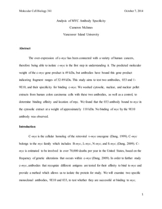
produced a western blot of the gel (Fig. 1)
6. Molecular Cell Biology 341 October 7, 2014
6
Fig. 1. Western blot of antibody 033, 9E10, and PBS/tween control. Each lane was loaded with
15 µl of extract, and SDS-page was run. After electrophoresis the gel was transblotted using a
Hoefer apparatus to produce the western blot. The only visible band was found using the
cytosolic extract washed with the 033 antibody. The approximate weight of the protein found is
110-120 kDa as determined by ColourPlus P7711S of New England Biolabs.
We expected to find myc in the nuclear extract with an approximate weight of 62 kDA.
According to Gibson et. al. (1991) this protein could have been bound by both the 033, and the
9E10 antibody. Considering Gibson et al. predicts that the human c-myc protein has a molecular
weight of about 49 kDa leads us to believe that myc is still not very well understood. The fact
that we did not have binding of the 9E10 antibody to myc may have been a result of human error.
It should have recognized p62, a 62 kDa proto-oncogene, which is a gene product of c-myc found
in the nucleus (Hilpert et. al., 2001).
150
100
033 Control 9E10
CE NE NP CE NE NP CE NE NP
25
7. Molecular Cell Biology 341 October 7, 2014
7
The overall conclusion of our experiment was that the 033 produced a clearly visible
band of about 110 kDA in the cytosolic extract. No bands were seen elsewhere on the western
blot. This allows us to assume that c-myc is not restricted to the nucleus. Hopefully in the future
myc will be better understood, as clearly there is still much to learn, as understanding myc is
crucial in the ongoing fight against cancer.
References
Dang, C.V. 1999. c-Myc target genes involved in cell growth, apoptosis, and metabolism.
Molecular Cell Biology. 19(1), 1-11
Gibson, A.W., Ye, R., Johnston, R.N., and L.W. Browder. 1992. Multiple antigens
recognized by anti-c-myc antibodies in human cells and Xenopus oocytes. Biochem Cell Biol.
70(10-11):998-1005.
Hilpert, K., Hansen, G., Wessner, H., Kuttner, G., Welfle, K., Seifert, M., and W. Hohne.
2001. Anti-c-myc antibody 9E10: epitope key positions and variability characterized using
peptide spot synthesis on cellulose. Protein Engineering. 14(10), 803-806.
Meyer, N., and Penn. L.Z. 2008. Reflecting on 25 years with myc. Nature Reviews
Cancer. 8, 976-990
Tansey, W.P. 2013. Mammalian MYC proteins and cancer. New Journal of Science.
2014, 1-27