
More Related Content
Similar to JC3article(2).pdf3 7 6 N A T U R E V O L 5 3 1 .docx
Similar to JC3article(2).pdf3 7 6 N A T U R E V O L 5 3 1 .docx (20)
More from christiandean12115
More from christiandean12115 (20)
Recently uploaded
Recently uploaded (20)
JC3article(2).pdf3 7 6 N A T U R E V O L 5 3 1 .docx
- 1. JC3article(2).pdf 3 7 6 | N A T U R E | V O L 5 3 1 | 1 7 M A R C H 2 0 1 6 LETTER doi:10.1038/nature17000 Co-ordinated ocular development from human iPS cells and recovery of corneal function Ryuhei Hayashi1,2, Yuki Ishikawa2, Yuzuru Sasamoto2, Ryosuke Katori2, Naoki Nomura2, Tatsuya Ichikawa2, Saori Araki2, Takeshi Soma2, Satoshi Kawasaki2, Kiyotoshi Sekiguchi3, Andrew J. Quantock4, Motokazu Tsujikawa2 & Kohji Nishida2 The eye is a complex organ with highly specialized constituent tissues derived from different primordial cell lineages. The retina, for example, develops from neuroectoderm via the optic vesicle, the corneal epithelium is descended from surface ectoderm, while the iris and collagen-rich stroma of the cornea have a neural crest origin. Recent work with pluripotent stem cells in culture has revealed a previously under-appreciated level of intrinsic cellular self-organization, with a focus on the retina and retinal cells1– 5. Moreover, we and others have demonstrated the in vitro induction of a corneal epithelial cell phenotype from pluripotent stem cells6–9.
- 2. These studies, however, have a single, tissue-specific focus and fail to reflect the complexity of whole eye development. Here we demonstrate the generation from human induced pluripotent stem cells of a self-formed ectodermal autonomous multi-zone (SEAM) of ocular cells. In some respects the concentric SEAM mimics whole-eye development because cell location within different zones is indicative of lineage, spanning the ocular surface ectoderm, lens, neuro-retina, and retinal pigment epithelium. It thus represents a promising resource for new and ongoing studies of ocular morphogenesis. The approach also has translational potential and to illustrate this we show that cells isolated from the ocular surface ectodermal zone of the SEAM can be sorted and expanded ex vivo to form a corneal epithelium that recovers function in an experimentally induced animal model of corneal blindness. To generate a SEAM of ocular cells, human induced pluripotent stem (iPS) cells were cultivated in differentiation medium in which they spontaneously and progressively formed a primordium compris- ing four identifiable concentric zones (Fig. 1a, Extended Data Fig. 1a). Cell morphology in each zone was distinctive, creating a visible delin- eation between zones (Extended Data Fig. 1b). The innermost central area (zone 1) formed first and this was followed by the emergence of three more radially distant concentric cell populations; zones
- 3. 2–4. (Fig. 1b, Supplementary Video). In our experiments 7.7 ± 1.8% of human iPS cells formed colonies and 67.9 ± 4.9% of these resulted in the generation of a SEAM (n = 5 technical replicates). Immunolabelling for the neural cell marker class III β-tubulin (TUBB3) was positive in zones 1 and 2, but not, more peripherally, in zones 3 or 4 (Fig. 1c). Cells in zones 1–3 expressed the ocular cell marker PAX6, while those in zones 3 and 4 were positive for the epithelial/surface ectodermal markers p63 and E-cadherin (Fig. 1d, e, f ). Thus, in a number of respects SEAM formation in two- dimensions mirrors whole eye development from the front of the ocular surface posteriorly to the retina (Fig. 1g). Cells in zone 1 expressed neural cell-specific markers TUBB3, SOX2 and SOX6, but no surface ectodermal markers (Fig. 2a). Accordingly, this central area was deemed to represent presumptive neuroectoderm. Zone 2 cells primarily expressed the optic vesicle marker RAX and the neural crest marker SOX10 (Fig. 2a). In eye development various cell types assume a tissue-specific arrangement at the juxtaposition of the embryonic anterior and posterior eye segments (Extended Data Fig. 1c), and perhaps this is reflected here. We also note that
- 4. zone 2 frequently contained retinal pigment epithelium (RPE)-like cells, and that SOX10+/p75+ neural crest cells were found to have emerged in satellite spheres in the presumptive zone 2 after two weeks in culture (Fig. 2b, Extended Data Fig. 1d). Differentiation of SEAMs by a pro- tocol which encourages retinal differentiation further demonstrated that CHX10+ neuro-retinal cells and MITF+ RPE cells were present in zone 2 towards its inner and outer margins, respectively (Fig. 2c, d). Collectively, the findings imply that zone 2 of the ocular SEAM is a developmental analogue of neural eye tissues comprising the neuro-retina, neural crest and RPE. At the margin of zones 2 and 3 α-crystallin+ lens cell clusters emerged after four weeks in culture and spread further through the SEAM by week six (Fig. 2e, Extended Data Fig. 1e). Cells in zone 3 did not display any neural features and this region was unique with its PAX6/p63-double-positive phenotype, representative of ocular surface ectoderm. Zone 3 cells also specifically expressed ocular surface ecto- derm markers such as PAX6, deltaN (DN)-p63, K18, and E- cadherin, but no neural cell markers (Fig. 2a). Thus, cells in zone 3 are consid- ered to be anlages of the ocular surface epithelium. Cells in
- 5. zone 4 expressed epithelial genes DN-p63 and E-cadherin, and did not express PAX6. This points to their identity as general surface ectodermal cells, which will probably differentiate into epidermal keratinocytes. The interactions of the different cell lineages complicit in whole eye for- mation in situ are thus mimicked in the research described here as illustrated schematically in Figs 1g and 2f. As mentioned, cells with retina-like characteristics have been generated from human iPS cells1–5, but functional ocular surface tissue has not. Thus, we sought to form a transplantable corneal epithelium; (i) as a conceptual example of the translational potential of the SEAM and (ii) to demonstrate that functional anterior eye tissue can indeed be fashioned from human iPS cells (Fig. 3a). At two and four weeks in culture cells in zone 3 co-expressed PAX6 and p63, first partially then fully. After isolation by the manual pipetting of cells in other zones (Extended Data Fig. 2a, b), zone 3 cells were also found to express K14 and cornea-specific keratin K1210 by 8–12 weeks (Fig. 3b). This was confirmed by qRT–PCR analysis (Extended Data Fig. 2c). Vimentin positive (VIM+) stroma-like cells were also present in the p63+ zone 3
- 6. at eight weeks (Extended Data Fig. 2d). Cellular differentiation in zone 3 of the SEAM thus follows that seen in the embryonic (mouse) eye (Extended Data Fig. 2e). BMP signalling influences the develop- ment of the surface ectoderm11,12, and we found that zone 3 of the SEAM was abolished following its treatment with the BMP4 inhib- itors Noggin and LDN-193189 (Extended Data Fig. 3). The TGFβ inhibitor, SB-431542, also disturbed the typical multi-zone forma- tion (Extended Data Fig. 3), suggesting that the suppression of ocular 1Department of Stem Cells and Applied Medicine, Osaka University Graduate School of Medicine, Suita, Osaka 565- 0871, Japan. 2Department of Ophthalmology, Osaka University Graduate School of Medicine, Suita, Osaka 565-0871, Japan. 3Laboratory of Extracellular Matrix Biochemistry, Institute for Protein Research, Osaka University, Suita, Osaka 565-0871, Japan. 4Structural Biophysics Group, School of Optometry and Vision Sciences, College of Biomedical and Life Sciences, Cardiff University, Cardiff CF24 4HQ, UK. © 2016 Macmillan Publishers Limited. All rights reserved http://www.nature.com/doifinder/10.1038/nature17000 1 7 M A R C H 2 0 1 6 | V O L 5 3 1 | N A T U R E | 3 7 7
- 7. LETTER RESEARCH surface ectodermal commitment is caused by the inhibition of early developmental events induced by endogenous BMP/TGFβ. As alluded to above, cells in SEAM zone 3 most closely resemble those of the presumptive ocular surface, and these were investigated for their translational, regenerative potential. After removal of zones 1 and 2 from the SEAM by manual pipetting, the cells that remained (that is, those in zone 3 and some in zone 4) were subjected to FACS to isolate a specific ocular surface lineage—corneal or conjunctival— and the results of this are shown in Extended Data Fig. 4a. SSEA-4 is a common marker of pluripotent stem cells and is specifically expressed in corneal epithelial cells in vivo13, including stem/progenitor cells (Extended Data Fig. 4b). Here, the use of SSEA-4 and the basal epithe- lial marker ITGB4 revealed that 14.1% of cells were SSEA- 4+/ITGB4+ (P3 cells), whereas 16.6% were SSEA-4−/ITGB4+ (P2 cells) (Fig. 3c). Thus, the population contains both corneal and non-corneal epithelial cells, with the indication that most colony-forming cells are derived from P2 and P3 fractions (Fig. 3d). Unlike P2, however, a
- 8. significant proportion of the P3 colonies expressed PAX6 (Extended Data Fig. 4c). P3 cells also had higher expression levels of corneal epithelial- specific markers PAX6, K12, CLU, and ALDH3A114, but lower levels of the epidermal marker K10 and the mucosal epithelial marker K13 (Extended Data Fig. 4d). P3 cell-derived colonies mostly consisted of PAX6+/K12+ corneal epithelial colonies along with a lower number of PAX6+/K12low limbal epithelial colonies, which are assumed to represent corneal epithelial stem/progenitor cells (Fig. 3e, Extended Data Fig. 5a). The limbus, at the edge of the cornea, is where corneal epithelial stem/progenitor cells are widely believed to reside. Stratified epithelia derived from 0d a 1st 1st 1st 1st 2nd 2nd 2nd
- 9. 2nd 1st 1st 1st2nd 2nd 3rd 3rd 3rd 3rd 4th 4th 4th 4th 5d 10d 15d 20d 25d b d PAX6 PAX6+PAX6+ c
- 11. E-cadherin Ed Ecad– p63+ f PAX6+ PAX6 PAX6– p63 Merged p63+ Ecad+ p63+ Ecad+ p63– PAX6– p63+ PAX6+ p63+ p63+ p63 Merged p63–
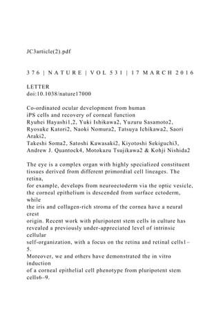
- 12. Ecad– g Embryonic eye 2nd 4th 3rd NC LE SE EK CE LEOSE SEAM of ocular cells 1st NE OC NR RPE CNS Figure 1 | Differentiation of multiple ocular cells in the SEAM.
- 13. a, A typical SEAM of differentiated human iPS cells after 40 days of culture. Representative of 19 independent experiments. Scale bar, 200 µm. b, Time-lapse microscopy of the differentiating human iPS cells during the first 25 days (d) of culture. Representative of six independent experiments. Scale bar, 200 µm. See also Supplementary Video 1. c–f, Immunostaining of TUBB3 (green) and p63 (red) (c, zones 1–3), PAX6 (green) and p63 (red) (d, zones 1–3), E-cadherin (Ecad, green) and p63 (red) (e, zones 1–3), and PAX6 (green) and p63 (red) (f, zones 2–4) in the SEAM after six to seven weeks of culture. Images are representative of three or four independent experiments. Nuclei, blue. Scale bar, 100 µm. g, The SEAM of human iPS cells induced different kinds of cells of ectodermal lineage, mimicking anterior and posterior eye development in vivo. CNS, central nervous system; NE, neuroectoderm; OC, optic cup; NR, neuroretina; NC, neural crest; LE; lens; OSE, ocular surface ectoderm; SE, surface ectoderm; CE, corneal epithelium; EK, epidermal keratinocyte. a 0 2 4 6
- 16. rd 4 th 1 st 2 n d 3 rd 4 th 1 st 2 n d 3 rd 4 th 1 st 2 n d 3 rd 4 th 1 st 2 n
- 17. d 3 rd 4 th 1 st 1 st 2 n d 3 rd 4 th 2 n d 3 rd 4 th 1 st 2 n d
- 18. 3 rd 4 th 1 st 2 n d 3 rd 4 th 1 st 2 n d 3 rd 4 th 0.0 2.0 4.0 6.0 8.0 0
- 20. ss io n r el at iv e to G A P D H (% ) PAX6 DN-p63 E-cadherin K18 c CHX10 MITFBF 3rd 2nd 1st
- 21. RPE cells 3Merged b 1st SOX10 2nd d 1st 2nd 3rd BF f 4 3rd 2nd 1st α-Crystallin2nd 3rd e 1st2nd RPE colony PAX6
- 22. CHX10 TUBB3 CRYAA p63/Ecad 1st 2nd 3rd 4th MITF Merged 1st p63 2nd 3rd 1st SOX10/p75 p63+ Lens Figure 2 | Characterization of cell zones in the SEAM. a, Gene expression analysis for ectoderm-related markers in each zone of the SEAM (zones 1 and 2, n = 12 colonies; zones 3 and 4, n = 7 colonies obtained from two independent experiments, respectively). Error bars are s.d. b, Phase-contrast image and SOX10 (red) expression around
- 23. SEAM zones 1 and 2 after two weeks of culture (representative of three independent experiments). Nuclei, blue. Scale bar, 200 µm. c, Bright- field (BF) appearance of neuroretinal cells (CHX10+, red) and RPE cells (MITF+, green) in zone 2 after seven weeks of culture using the protocol for retinal differentiation (representative of three independent experiments). Nuclei, blue. Scale bar, 200 µm. d, Cultivation of the isolated RPE cells (representative of three independent experiments). Scale bars, 200 µm (upper panel) and 50 µm (lower panel). e, Immunostaining of lens differentiation marker, α-crystallin (green) and p63 (red), after four weeks of culture (representative of three independent experiments). Nuclei, blue. Scale bar, 100 µm. f, Schematic of the expression pattern of ocular-cell-related genes in the SEAM. © 2016 Macmillan Publishers Limited. All rights reserved 3 7 8 | N A T U R E | V O L 5 3 1 | 1 7 M A R C H 2 0 1 6 LETTERRESEARCH P3 cells expressed K12 and PAX6, but not K13 (Fig. 3f ) or HOX genes (Extended Data Fig. 5b). P2 cells, on the other hand, expressed HOX
- 24. genes (Extended Data Fig. 5b), which are not expressed in cells of the ocular surface15,16. In the colonies of P2 cells, those expressing HOXB4 and PAX6 existed exclusively of each other (Extended Data Fig. 5c). Immunostaining of P2 cells further demonstrated that they consisted of PAX6+/K13+ conjunctival epithelial cells17 accompanied by a major- ity of PAX6−/K13+ non-ocular epithelial cells (Fig. 3e, f, Extended Data Fig. 5a). Long-term differentiation revealed that PAS- stained, MUC5AC+, K7+ conjunctival goblet-like cells appeared in presump- tive P2 cell regions (Extended Data Fig. 5d). Collectively, our data indicates that cells in zone 3 of the SEAM have characteristics of cor- neal, limbal, and conjunctival epithelial cells (Supplementary Table 1), raising the possibility that functional ocular surface epithelia could be developed for surgical use. To investigate this prospect we generated ex vivo expanded sheets of corneal epithelium from FACS-sorted cells acquired from zone 3 of the SEAM (Fig. 4a). The sheets expressed the corneal limbal stem- cell markers K15 and K19, along with the mucins MUC1, MUC4 and MUC16, tight junction protein ZO-1, the differentiation marker CX43, K3 and K12, all of which are characteristic for the
- 25. cornea (Fig. 4b, Extended Data Fig. 6a). The non-corneal keratins K10 and K18 were not expressed (Fig. 4b). Approximately 99% of the cells in the expanded sheets were stratified K14+ epithelial cells, 95% were of corneal epithelial lineage (SSEA-4+), and 70% were differentiated corneal epithelial cells (K12+) (Fig. 4c). Cells typically had a smooth apical surface with microvilli-like structures (Extended Data Fig. 6b). Analyses based on significantly changed genes revealed that cells in the expanded corneal epithelial sheet differed from human oral mucosal keratinocytes, dermal fibroblasts and, indeed, human iPS 4w Seeded on LN511E8-coated dish DM 0 iPS cells 4w 4w StemFit CDM 8w 10w Pipetting
- 27. c TRA-1-60– IT G B 4 TRA-1-60-negative cells P2 P3 14.1%16.6% 104 103 102 101 100 104 103 102 101 100 100 101 102 103 104
- 28. 100 101 102 103 104 100 101 102 103 104 104 103 102 101 100 9 10 d P2 2nd 3rd 2w 7–8w3–4w 12–13w1w Colony 1st + 2nd 1st + 2nd 1st + 2nd 1st + 2nd PAX6+ PAX6+ PAX6+ 3rd 3rd
- 29. PAX6+PAX6 + PAX6+ PAX6PAX6 PAX6PAX6PAX6 Pluripotent stem cells Ocular surface ectoderm Ocular surface epithelial cells Corneal/ conjunctival epithelial cells Y-27632 Head surface ectoderm b SSEA-4TRA-1-60 IT G B 4
- 31. Colony Colony p63+p63+ 3rd 3rd 3rd 3rd K14+ p63+ K14+ p63+ p63p63 p63p63 K14K14 K14K14 K12K12 K12K12 p63 K14 K12 0
- 32. 1 P1 P2 P3 P4 P3 cells P2 cells CECs (in vitro) Corneal epithelium Conjunctival epithelium (PAX6+ region) Non-ocular epithelium (PAX6– region) 6 f PAX6 PAX6 PAX6 PAX6 D o u b le s ta in in g
- 35. K13 K12 K13 K12 K13 K12 K13 p63 p63 p63 p63 1: Corneal epithelium ( P ro p o rt io n (% ) 1 2 3 80
- 36. 20 10 0 p 6 3 p p p p(PAX6+/K12+) 2: Conjunctival epithelium (PAX6+/K13+/K12–) 3: Other epithelium Figure 3 | Induction and isolation of ocular surface epithelial cells from the SEAM. a, Schematic representation of the strategy used for the generation of the ocular surface ectoderm and epithelium. CDM, corneal differentiation medium; CEM, corneal epithelium maintenance medium; CMM, corneal epithelium maturation medium; DM, differentiation medium; w, week(s). b, Immunostaining for ocular surface and corneal epithelial development-related markers, PAX6 (green), p63 (red), K14 (green), and K12 (red) during ocular surface epithelial differentiation culture (1–12 weeks, representative of three independent experiments). Nuclei, blue. Scale bar, 100 µm. c, A flow cytometric analysis
- 37. of SSEA-4, ITGB4 and TRA-1-60 in the differentiated human iPS cells after 10–15 weeks of culture revealed no undifferentiated human iPS cells (SSEA-4+/TRA-1-60+ cells) (left panels). TRA-1-60− cells were analysed by SSEA-4 and ITGB4 (right panel). The four populations are defined as P1–P4. The data shown here are representative images of 23 independent cell-sorting experiments. d, Colony-forming assay for the sorted P1–P4 cell fractions. Fixed colonies were stained with rhodamine (right upper panel, representative of seven independent cell-sorting experiments) and the colony-forming efficiency were calculated (graph: seven independent cell-sorting experiments). Error bars are s.d. Scale bar, 5 mm. e, The ratios of corneal epithelial (PAX6+/K12+ or PAX6+/K12low), conjunctival epithelial (PAX6+/K13+/K12−) and other epithelial cell types in the P2 and P3 colonies from five independent cell sorting experiments. Error bars are s.d. f, Triple-colour immunostaining for PAX6 (green), K12 (orange), K13(magenta), and p63 (green) expression in stratified P2 and P3 cells and cultivated human corneal limbal epithelial cells (CECs) (representative of three independent experiments, respectively). Nuclei, blue. Scale bar, 50 µm.
- 38. © 2016 Macmillan Publishers Limited. All rights reserved 1 7 M A R C H 2 0 1 6 | V O L 5 3 1 | N A T U R E | 3 7 9 LETTER RESEARCH cells, but were similar to cells harvested from the epithelium at the limbus of donated research human corneas (Fig. 4d, Extended Data Fig. 6c). Cells had the proliferative capability to be passaged 11 to 16 times (average population doubling, 35.1; Fig. 4e, Extended Data Fig. 6d) without any obvious karyotype aberration at each passage (Extended Data Fig. 6e). Holoclone analysis indicated that 15.9% of the corneal epithelial colony was composed of holoclones (Fig. 4f ). Together, the data show that corneal epithelial cells derived from our SEAM contain functional stem/progenitor cells equipped with high proliferative potential, which have the potential to reconstruct anterior eye epithelial tissue. Furthermore, all five different human iPS cell clones used were able to successfully induce corneal epithe- lial stem/progenitor cells, though each had a differential propensity for engendering corneal epithelial differentiation (Extended
- 39. Data Fig. 7). To investigate the translational potential of this approach, ex vivo expanded corneal epithelial cell sheets were cultivated as recoverable and translatable constructs, in which approximately 1.0% of cells maintained a colony-forming capability (Extended Data Fig. 8a, b). When the sheets were transplanted onto the eyes of rabbits in an experimental model of corneal epithelial stem-cell deficiency (Extended Data Fig. 8c–j), they successfully recovered a healthy corneal barrier function (Fig. 4g, Extended Data Fig. 8k) and continued to express cornea-specific proteins (Fig. 4h). This advance—that is, the generation of functional SEAM-derived ocular surface tissue with stem/progenitor cells, which can surgically repair the front of the eye—is noteworthy because, although somatic stem cells have been used to recover the ocular surface, the long-term clinical results have not been overly encouraging18–20. In resolving key purification steps for corneal epithelial stem/progenitor cells by applying a combination of specific antibodies to our human iPS cell-derived ocular SEAM, we are now in the position to initiate first- in-human clinical trials of anterior eye transplantation to restore visual function.
- 40. a b MUC1 MUC4 K3 PAX6 K12 K15 c e f H&E staining ZO-1 K18MUC16 K10 d 50 Holoclone Meroclone Paraclone DF OK 200 0 5 10 15
- 41. 20 25 30 35 40 45 0 10 20 30 40 50 60 70 80 90 iCEC1 iCEC2 iCEC3 C u m u la tiv e p o p u la tio n d o
- 42. u b lin g s 15.9% (10/63) 50.8% (32/63) 33.3% (21/63) CEC iCEC OSE (6w) iPS cell iPS cell OSE (6w) DF Stratified- epithelium iCEC CEC
- 44. hg Human nuclei p63 K14 MUC16 K12PAX6 iC E C 7d 14dBefore Days 40 50 60 70 80 90 100 110 iCEC * * Component 1 (30.97%) K14 MUC16 K3 K13
- 45. C o n tr o l ( sh am ) 0 10 20 30 40 0 2 4 6 8 10 12 14 Control Days after transplantation F lu o re sc ei n
- 46. -n eg at iv e ar ea (% ) K14 99.0% 68.4% 100 101 102 103 104 100 101 102 103 104 100 101 102 103 104 0 20 40 60 80 100 0 20 40
- 48. o u n t 95.0% Figure 4 | Characterization and surgical use of human iPS cell-derived corneal epithelial cells (iCECs). a, Phase-contrast microscopy and haematoxylin and eosin (H&E) staining of the human iCECs on cell culture inserts (representative of four independent experiments, respectively). Scale bars, 100 µm (phase), 50 µm (H&E). b, Immunostaining for corneal epithelial functional proteins and epithelial markers (green) in the stratified SEAM-derived human iCEC sheets (representative of three independent experiments). Nuclei, red. Scale bar, 50 µm. c, Results of flow cytometric analyses for K14, K12 and SSEA-4 expression in the stratified human iCECs (representative of three independent experiments). d, Results of a principle component analysis based on the global gene expression (examined by microarrays) comparing human iPS cells (n = 3 technical replicates), ocular surface ectoderm (OSE; that is, human iPS cell-derived cells after six weeks of differentiation, n = 3 technical replicates), human oral keratinocytes (OKs, n = 3 technical
- 49. replicates), human iCECs (n = 4 independent experiments), human dermal fibroblasts (DFs, n = 3 technical replicates) and human corneal epithelial cells from the limbus (CECs, n = 3 independent experiments). A total of 25,262 significantly changed genes (fold change >2.0, fasle discovery rate <0.05) were analysed. Compared to human DFs, human iPS cells, human OKs or the OSE, the gene expression in human iCECs was most similar to that of human CECs. e, Proliferation profiles of the human iCECs during serial passages (n = 3 independent experiments, average population doubling, 35.1). f, Result of a holoclone analysis for human iCEC colonies. Representative images of holoclone-, meroclone- and paraclone-derived colonies and their frequencies are shown (n = 63 single colonies from four independent cell-sorting experiments). Scale bar, 10 mm. g, Barrier function assay using fluorescein staining for transplanted and sham-operated control corneas on days 0, 7 and 14 post-surgery (left panels). A statistical analysis of the barrier function interpreted as the size of the fluorescein-negative area in the human iCEC-sheet-transplanted and control corneas (graph). *P < 0.05; n = 7 biological replicates, Steel’s test (Bonferroni corrected). Error
- 50. bars are s.d. h, Immunostaining for human nuclei and corneal epithelial markers (green) in the SEAM-derived human iCEC-sheet-transplanted corneas on postoperative day 14 (n = 6 animal transplantation experiments). Nuclei, red. Scale bar, 50 µm. © 2016 Macmillan Publishers Limited. All rights reserved 3 8 0 | N A T U R E | V O L 5 3 1 | 1 7 M A R C H 2 0 1 6 LETTERRESEARCH Online Content Methods, along with any additional Extended Data display items and Source Data, are available in the online version of the paper; references unique to these sections appear only in the online paper. Received 2 June 2015; accepted 14 January 2016. Published online 9 March 2016. 1. Eiraku, M. et al. Self-organizing optic-cup morphogenesis in three-dimensional culture. Nature 472, 51–56 (2011). 2. Nakano, T. et al. Self-formation of optic cups and storable stratified neural retina from human ESCs. Cell Stem Cell 10, 771–785 (2012). 3. Zhong, X. et al. Generation of three-dimensional retinal tissue with functional
- 51. photoreceptors from human iPSCs. Nature Commun. 5, 4047 (2014). 4. Reichman, S. et al. From confluent human iPS cells to self- forming neural retina and retinal pigmented epithelium. Proc. Natl Acad. Sci. USA 111, 8518–8523 (2014). 5. Mellough, C. B. et al. IGF-1 signaling plays an important role in the formation of three-dimensional laminated neural retina and other ocular structures from human embryonic stem cells. Stem Cells 33, 2416–2430 (2015). 6. Hayashi, R. et al. Generation of corneal epithelial cells from induced pluripotent stem cells derived from human dermal fibroblast and corneal limbal epithelium. PLoS ONE 7, e45435 (2012). 7. Shalom-Feuerstein, R. et al. Pluripotent stem cell model reveals essential roles for miR-450b-5p and miR-184 in embryonic corneal lineage specification. Stem Cells 30, 898–909 (2012). 8. Ahmad, S. et al. Differentiation of human embryonic stem cells into corneal epithelial-like cells by in vitro replication of the corneal epithelial stem cell niche. Stem Cells 25, 1145–1155 (2007). 9. Brzeszczynska, J. et al. Differentiation and molecular profiling of human embryonic stem cell-derived corneal epithelial cells. Int. J. Mol.
- 52. Med. 33, 1597–1606 (2014). 10. Lavker, R. M., Tseng, S. C. & Sun, T.-T. Corneal epithelial stem cells at the limbus: looking at some old problems from a new angle. Exp. Eye Res. 78, 433–446 (2004). 11. Liem, K. F. Jr, Tremml, G., Roelink, H. & Jessell, T. M. Dorsal differentiation of neural plate cells induced by BMP-mediated signals from epidermal ectoderm. Cell 82, 969–979 (1995). 12. McMahon, J. A. et al. Noggin-mediated antagonism of BMP signaling is required for growth and patterning of the neural tube and somite. Genes Dev. 12, 1438–1452 (1998). 13. Truong, T. T., Huynh, K., Nakatsu, M. N. & Deng, S. X. SSEA4 is a potential negative marker for the enrichment of human corneal epithelial stem/ progenitor cells. Invest. Ophthalmol. Vis. Sci. 52, 6315–6320 (2011). Supplementary Information is available in the online version of the paper. Acknowledgements We thank K. Baba, Y. Oie, H. Takayanagi, S. Hara, Y. Yasukawa, J. Toga and M. Yagi of Osaka University and M. Nakagawa of Kyoto University for technical assistance and scientific discussions.
- 53. This work was supported in part by the project for the realization of regenerative medicine of The Japan Agency for Medical Research and Development (AMED), The Japan Science and Technology Agency (JST) and The Ministry of Health, Labour, and Welfare of Japan and the Grants-in-Aid for Scientific Research from The Ministry of Education, Culture, Sports, Science and Technology of Japan. Author Contributions R.H., M.T. and K.N. designed the research; R.H, Y.I., R.K. and S.A. performed the in vitro experiments and acquired the data; Y.S., N.N., T.I. and T.S. performed animal experiments and acquired the data; K.S. provided reagents (LN511E8); R.H., Y.I. and R.K. analysed the data and wrote the respective methods and results; S.K., K.S. and A.J.Q. supervised the project; and R.H., M.T., A.J.Q. and K.N. wrote the paper. Author Information Reprints and permissions information is available at www.nature.com/reprints. The authors declare no competing financial interests. Readers are welcome to comment on the online version of the paper. Correspondence and requests for materials should be addressed to K.N. ([email protected]). 14. Estey, T., Piatigorsky, J., Lassen, N. & Vasiliou, V. ALDH3A1: a corneal crystallin
- 54. with diverse functions. Exp. Eye Res. 84, 3–12 (2007). 15. Pearson, J. C., Lemons, D. & McGinnis, W. Modulating Hox gene functions during animal body patterning. Nature Rev. Genet. 6, 893–904 (2005). 16. Mallo, M. & Alonso, C. R. The regulation of Hox gene expression during animal development. Development 140, 3951–3963 (2013). 17. Krenzer, K. L. & Freddo, T. F. Cytokeratin expression in normal human bulbar conjunctiva obtained by impression cytology. Invest. Ophthalmol. Vis. Sci. 38, 142–152 (1997). 18. Nishida, K. et al. Functional bioengineered corneal epithelial sheet grafts from corneal stem cells expanded ex vivo on a temperature- responsive cell culture surface. Transplantation 77, 379–385 (2004). 19. Pellegrini, G. et al. Long-term restoration of damaged corneal surfaces with autologous cultivated corneal epithelium. Lancet 349, 990– 993 (1997). 20. Nakamura, T. et al. Transplantation of cultivated autologous oral mucosal epithelial cells in patients with severe ocular surface disorders. Br. J. Ophthalmol. 88, 1280–1284 (2004). © 2016 Macmillan Publishers Limited. All rights reserved
- 55. http://www.nature.com/doifinder/10.1038/nature17000 http://www.nature.com/doifinder/10.1038/nature17000 http://www.nature.com/reprints http://www.nature.com/doifinder/10.1038/nature17000 mailto:[email protected] LETTER RESEARCH METHODS No statistical methods were used to predetermine sample size. The experiments were not randomized and investigators were not blinded to allocation during experiments and outcome assessment. Human iPS cell culture. The human iPS cell lines 201B7, 253G1, and 454E2 were obtained from the RIKEN Bio Resource Center (Tsukuba, Japan)21,22. The 1231A3 and 1383D2 human iPS cells were provided by the Center for iPS Cell Research and Application, Kyoto University23. All cells were cultured in StemFit medium (Ajinomoto, Tokyo, Japan) on LN511E8-coated (0.5 µg cm−2) dishes23,24. LN511E8, produced using cGMP-banked CHO-S cells (Life Technologies, Carlsbad, CA), was obtained from Nippi (Tokyo, Japan). In part, LN511E8 was produced using human 293-F cells as previously described12. The 201B7 and 454E2 human iPS cell lines were used in the in vitro experiments, while 201B7 and 1383D2 cells were used in the animal experiments; 253G1 and 1231A3 cells were used in the supplementary
- 56. experiments, the results of which are reported in Extended Data Fig. 7. All of the experiments using recombinant DNA were approved by the Recombinant DNA Committees of Osaka University and were performed according to our institu- tional guidelines. Ocular cell differentiation from human iPS cells. The differentiation culture for human iPS cells was performed as indicated in Fig. 3a. First, human iPS cells were seeded on LN511E8-coated dishes at 350–700 cells cm−2, after which they were cul- tivated in StemFit medium for 8–12 days. The culture medium was then changed to DM (differentiation medium; GMEM (Life Technologies) supplemented with 10% knockout serum replacement (KSR; Life Technologies), 1 mM sodium pyru- vate (Life Technologies), 0.1 mM non-essential amino acids (Life Technologies), 2 mM l-glutamine (Life Technologies), 1% penicillin- streptomycin solution (Life Technologies) and 55 µM 2-mercaptoethanol (Life Technologies) or mon- othioglycerol (Wako, Osaka, Japan))25. In some experiments, as indicated in the Results section, Noggin (R&D systems, Minneapolis, MN), LDN-193189 (Wako) or SB-431542 (Wako) were added for the first four days. BMP4 (R&D systems) was used in some early experiments at concentrations up to 0.125 nM. This had no discernible effect on SEAM formation, however, so its use was discontinued. After four weeks of culture in DM, the medium was changed to
- 57. corneal differentiation medium (CDM; DM and Cnt-20 or Cnt-PR (w/o; EGF and FGF2) (1:1, CELLnTEC Advanced Cell Systems, Bern, Switzerland) containing 5 ng ml−1 FGF2 (Wako), 20 ng ml−1 KGF (Wako) 10 µM Y-27632 (Wako) and 1% penicillin-streptomycin solution). FGF2 in CDM was not essential for corneal epithelial induction. During CDM culture (around six to eight weeks of differentiation), non-epithelial cells were removed by manual pipetting under microscopy (Extended Data Fig. 2a, b). After pipetting, the medium was changed to fresh CDM. After four weeks of culture in CDM, the medium was changed to corneal epithelium maintenance medium (CEM; DMEM/F12 (2:1), Life Technologies) containing 2% B27 supplement (Life Technologies), 1% penicillin-streptomycin solution, 20 ng ml−1 KGF and 10 µM Y-27632 for two to seven weeks. To achieve retinal differentiation (Fig. 2c) after four weeks of differentiation the medium was directly changed to CEM. Isolated RPE cell colonies were cultivated in CEM on separate dishes coated with LN511E8. Phase-contrast microscopic observations were performed with an Axio-observer. Z1, D1 (Carl Zeiss, Jena, Germany) and an EVOS FL Auto (Life Technologies). Flow cytometry and cell sorting. Differentiated human iPS cells in CEM were dissociated using Accutase (Life Technologies), and resuspended in ice-cold KCM medium (DMEM without glutamine and Nutrient Mixture F-12
- 58. Ham (3:1, Life Technologies) supplemented with 5% FBS (Japan Bio Serum, Hiroshima, Japan), 0.4 µg ml−1 hydrocortisone succinate (Wako), 2 nM 3,3′,5- Triiodo-l-thyronine sodium salt (MP biomedicals, Santa Ana, CA), 1 nM cholera toxin (List Biological Laboratory, Campbell, CA), 2.25 µg ml−1 bovine transferrin HOLO form (Life Technologies), 2 mM l-glutamine, 0.5% insulin transferrin selenium solution (Life Technologies) and 1% penicillin-streptomycin solution). The harvested cells were filtered with a cell strainer (40 µm, BD Biosciences, San Diego, CA) and then stained with anti-SSEA-4 (MC813-70, Biolegend, San Diego, CA), TRA-1-60 (TRA-1-60-R, Biolegend) and CD104 (ITGB4; 58XB4, Biolegend) antibodies for 1 h on ice. After being washed twice with PBS, stained cells underwent cell sorting with a FACSAria II instrument (BD Biosciences). For intracellular protein staining, a BD Cytofix/Cytoperm (BD Biosciences) kit was used. In all of the experiments, cells were stained with non-specific isotype IgG or IgM as controls (Biolegend). The data were analysed using the BD FACSDiva Software (BD Biosciences) and the FlowJo software program (TreeStar, San Carlos, CA). Fabrication and harvest of human iPS cell-derived corneal epithelial cell (human iCEC) Sheets. Sorted human iPS cell-derived epithelial cells obtained from zone 3 of the SEAM (human iCECs) were seeded on LN511E8 coated
- 59. (0.5 µg cm−2) cell culture inserts or temperature-responsive dishes (UpCell, CellSeed, Tokyo, Japan) without cell passaging, and were cultured in CEM until confluence26. To promote maturation, the epithelial cells were cultivated in CMM (corneal epithelium maturation medium; KCM medium containing 20 ng ml−1 KGF and 10 µM Y-27632) for an additional 3–14 days after CEM culture. The human iCECs cultivated on temperature-responsive dishes were released from their substrate by reducing the temperature to 20 °C. Quantitative real-time reverse-transcriptase PCR (qRT–PCR). Total RNA was obtained from differentiated human iPS cells after specific culture periods, from human epidermal keratinocytes (EKs (foreskin), Life Technologies and TaKaRa Bio, Otsu, Japan), and from human corneal limbal epithelial cells (CECs) using the RNeasy total RNA kit or the QIAzol reagent (Qiagen, Valencia, CA). Reverse tran- scription was performed using the SuperScript III First-Strand Synthesis System for qRT–PCR (Life Technologies) according to the manufacturer’s protocol, and cDNA was used as a template for PCR. qRT–PCR was performed using the ABI Prism 7500 Fast Sequence Detection System (Life Technologies) in accordance with the manufacturer’s instructions. The TaqMan MGB used in the present study are shown in Supplementary Table 2. The thermocycling program was performed
- 60. with an initial cycle at 95 °C for 20 s, followed by 45 cycles at 95 °C for 3 s and 60 °C for 30 s. Immunofluorescence staining. Research grade human skin tissue sections were obtained from US Biomax Inc. (MD, USA) and human oral mucosal tissue was obtained from Science Care (Phoenix, AZ). The cells were fixed in 4% paraform- aldehyde (PFA) or cold methanol, washed with Tris-buffered saline (TBS, TaKaRa Bio) three times for 10 min and incubated with TBS containing 5% donkey serum and 0.3% Triton X-100 for 1 h to block non-specific reactions. They were then incubated with the antibodies shown in Supplementary Table 3 at 4 °C overnight or at room temperature for 3 h. The cells were again washed twice with TBS for 10 min, and were incubated with a 1:200 dilution of Alexa Fluor 488-, 568-, 647- conjugated secondary antibodies (Life Technologies) for 1 h at room temperature. Counterstaining was performed with Hoechst 33342 (Molecular Probes) before fluorescence microscopy (Axio Observer.D1, Carl Zeiss). Haematoxylin and eosin staining. Fabricated human iCEC sheets were fixed with 10% formaldehyde neutral buffer solution (Nacalai Tesque, Kyoto, Japan). After washing with distilled water, the human iCEC sheets were embedded in paraffin from which 3-µm-thick sections were cut. These were stained with haematoxylin and eosin following deparaffinization and hydration. The sections were observed
- 61. with a NanoZoomer-XR C12000 (Hamamatsu Photonics, Hamamatsu, Japan), BZ-9000 (KEYENCE, Osaka, Japan) and an Axio Observer.D1. PAS staining. Differentiated human iPS cells (more than 12 weeks of differen- tiation) were fixed with 10% formaldehyde neutral buffer solution, after which PAS staining was performed with a PAS staining kit (MERCK KGaA, Darmstadt Germany) according to the manufacturer’s protocol. The sections were observed with an Axio Observer.D1. Colony-formation assay (CFA). Epithelial cells were seeded onto MMC-treated NIH-3T3 feeder layers at a density of 3,000–20,000 cells per well. These were cul- tivated in CMM for 7–14 days. The colonies were fixed with 10% formaldehyde neutral buffer solution and then stained with rhodamine B (Wako). Colony for- mation was then assessed using a dissecting microscope and the colony-forming efficiency (CFE) was calculated. For the holoclone analysis, a single human iCEC colony derived from the SEAM was cultivated on 3T3-J2 (provided by H. Green, Harvard Medical School, Boston, MA) in CMM for 7–11 days was picked up under a dissecting microscope and dissociated by TrypLE Select (Life Technologies). The dissociated human iCECs were again seeded on a MMC-treated 3T3-J2 feeder layer and cultivated in CMM for 10–13 days. The colonies were scored under a micro- scope and classified as holoclones, paraclones or meloclones based on previously
- 62. reported methods27. Microarray analysis. Human CECs were harvested from corneoscleral rims (Northwest Lions Eye Bank, Seattle, WA) as reported previously28. Human CECs and human oral keratinocytes (OKs; ScienCell, Carlsbad, CA) along with SEAM- derived human iCECs were cultivated on LN511E8 coated cell culture inserts in CEM until confluent. They were then cultivated in CMM. Human dermal fibro- blasts (DFs; ScienCell) were cultivated in DMEM/F12 (2:1) containing 10% FBS. Total RNA was obtained from human iPS cells, iCECs, CECs, OKs, DFs, and six- week differentiated iPS cells (that is, OSE) using the QIAzol reagent. A microarray analysis using Sure Print G3 human 8x60K slides (Agilent technologies, Palo Alto, CA) was performed at Takara Bio. The data were analysed using the GeneSpring GX software program (Agilent technologies). Microarray data used in this study are deposited in Gene Expression Omnibus under accession number GSE73971. Scanning electron microscopy (SEM). The cultivated epithelial cell sheets were fixed in 2.5% glutaraldehyde (Nacalai Tesque) at 4 °C overnight. Subsequently, the sheets were washed in buffer, dehydrated with ethanol and tert-butyl alcohol (Wako), and critical point dried (JFD-320, JEOL, Tokyo, Japan). After sputter-coating with platinum in an auto fine coater (JFCL-1600, JEOL), the samples were observed by scanning electron microscopy (JSM-6510LA,
- 63. JEOL) at 5 kV. © 2016 Macmillan Publishers Limited. All rights reserved LETTERRESEARCH Serial cell passaging and karyotype analysis. FACS-isolated human iCECs were cultivated on MMC-treated NIH-3T3 feeder layers in CMM up to 70–80% conflu- ence. The human iCECs were harvested using TrypLE Select following the removal of feeder cells by manual pipetting. The total cell numbers were counted, after which the cells were passaged at a 1:8 ratio onto newly prepared feeder layers. These were cultivated in CMM until sub-confluence was reached again. The G-band karyotype analysis for human iCECs was performed at Nihon Gene Research Laboratories (Sendai, Japan). Animal experiments. All animal experimentation was performed in accordance with the ARVO Statement for the Use of Animals in Ophthalmic and Vision Research, and was approved by the animal ethics committees of Osaka University. To examine embryonic mouse eyes, pregnant females (C57/BL6, E9.5–18.5) were acquired from SLC Japan (Shizuoka, Japan). For the transplantation experi- ments, Female New Zealand white rabbits (2.5–3.0 kg (approximately 12–14 weeks)) were obtained from Kitayama Labes (Nagano, Japan).
- 64. Harvested human iCEC sheets were grafted onto rabbit corneas, in which a total epithelial limbal stem-cell deficiency had been created following a corneal and limbal lamellar keratectomy (Extended Data Fig. 8c–j). After surgery, 0.3% ofloxacin ointment (Santen Pharmaceutical, Osaka, Japan), 0.1% betamethasone phosphate eye drops (Shionogi Pharmaceutical, Osaka, Japan) and 0.1% sodium hyaluronate eye drops (Santen Pharmaceutical) were applied three to four times per day. Triamcinolone acetonide (8 mg; Bristol Myers Squibb, Tokyo, Japan) was also administered by sub- conjunctival injection. Tacrolimus (0.05 mg kg−1 per day, Astellas Pharma, Tokyo, Japan) and Mizoribine (4.0 mg kg−1 per day Sawai Pharmaceutical, Osaka, Japan) were systemically administered using an osmotic pump (DURECT, Cupertino, CA). The corneal barrier function following surgery was assessed by 0.5% fluo- rescein eye drop instillation at day 7 and day 14 after surgery and the fluorescein negative area was calculated using the AxioVision software program (Carl Zeiss). Throughout the healing period, the cornea was observed with a digital slit-lamp camera (SL-7F, TOPCON, Tokyo, Japan) and 3D OCT1000 MARK II (TOPCON) or CASIA SS-1000 (TOMEY, Nagoya, Japan) machines. If an infection was found or if unexpected weight loss occurred, animals were excluded from the analysis.
- 65. The rabbits were euthanized by the intravenous administration of sodium pento- barbitone 14 days after transplantation, after which the eyes were immediately enu- cleated for the histological analyses. No blinding or randomization was conducted to allocate animals to each group. Statistical analyses. The data are expressed as means ± standard deviation (s.d.). The statistical analyses were performed using the Mann–Whitney rank sum test or Steel’s test. Bonferroni’s correction was applied to the data in animal experiments. All of the statistical analyses were performed using the JMP software program (SAS institute Inc., Cary, NC). No statistical methods were used to predetermine sample size. Comprehensive technical details can be found in Protocols Exchange, http:// dx.doi.org/10.1038/protex.2016.009. 21. Nakagawa, M. et al. Generation of induced pluripotent stem cells without Myc from mouse and human fibroblasts. Nature Biotechnol. 26, 101–106 (2008). 22. Takahashi, K. et al. Induction of pluripotent stem cells from adult human fibroblasts by defined factors. Cell 131, 861–872 (2007). 23. Nakagawa, M. et al. A novel efficient feeder-free culture system for the derivation of human induced pluripotent stem cells. Sci. Rep. 4, 3594 (2014).
- 66. 24. Miyazaki, T. et al. Laminin E8 fragments support efficient adhesion and expansion of dissociated human pluripotent stem cells. Nature Commun. 3, 1236 (2012). 25. Kawasaki, H. et al. Generation of dopaminergic neurons and pigmented epithelia from primate ES cells by stromal cell-derived inducing activity. Proc. Natl Acad. Sci. USA 99, 1580–1585 (2002). 26. Miyashita, H. et al. Long-term maintenance of limbal epithelial progenitor cells using Rho kinase inhibitor and keratinocyte growth factor. Stem Cells Transl. Med. 2, 758–765 (2013). 27. Barrandon, Y. & Green, H. Three clonal types of keratinocyte with different capacities for multiplication. Proc. Natl Acad. Sci. USA 84, 2302–2306 (1987). 28. Hayashi, R. et al. N-Cadherin is expressed by putative stem/progenitor cells and melanocytes in the human limbal epithelial stem cell niche. Stem Cells 25, 289–296 (2007). © 2016 Macmillan Publishers Limited. All rights reserved http://dx.doi.org/10.1038/protex.2016.009 http://dx.doi.org/10.1038/protex.2016.009
- 67. LETTER RESEARCH Extended Data Figure 1 | Differentiation of multiple ocular cells in the SEAM. a, A differentiated human iPS cell colony after 40 days of culture (macro photograph, representative of six independent experiments). Scale bar, 5 mm. b, Magnified views of each SEAM zone margin after six weeks in culture (phase-contrast and bright-field views, each representative of three independent experiments). Arrow heads indicate borders between each zone. Scale bars, 50 µm. c, Schematic for the development of the anterior eye. CE, corneal epithelium; CEnd, corneal endothelium; IS, iris stroma; NR, neuroretina; RPE, retinal pigment epithelium; LE, lens. d, Immunostaining for p75 (green) and SOX10 (red) in SEAM zones 1 and 2, two weeks (w) after the start of the differentiation culture (representative of three independent experiments). Asterisks indicate SOX10+/p75+ neural crest cells. Nuclei, blue. Scale bar, 100 µm. e, Immunostaining of lens differentiation marker α-crystallin (green) and epithelial marker p63 (red) in zones 2 and 3 of the SEAM after six weeks of culture (representative of three independent experiments). Nuclei, blue. Scale bar, 100 µm.
- 68. © 2016 Macmillan Publishers Limited. All rights reserved LETTERRESEARCH Extended Data Figure 2 | Enrichment and differentiation of a SEAM- derived ocular surface epithelium. a, The enrichment of zone 3 ocular surface epithelium by manual pipetting. Cells in the SEAM before (upper panel) and after (lower panel) pipetting are shown (n = 1). Scale bar, 200 µm. b, The expression of ocular-cell-related genes in removed and attached cells after pipetting (n = 1). c, The time course of PAX6, DN-p63, K14, K12 and LIN28A expression during human iPS cell differentiation culture (0, 2, 4, 6, 8 and 12 weeks, each n = 5 independent experiments). Error bars are s.d. d, Immunostaining for vimentin (VIM, green) and p63 (red) before and after FACS purification conducted at 11 weeks of culture (representative of three independent experiments). VIM+ stroma-like cells of zone 3 were removed by FACS. Nuclei, blue. Scale bar, 50 µm. e, Immunostaining for anterior eye development-related markers Pax6 (green), p63 (red), K14 (green) and K12 (red) during mouse eye development (E9.5–18.5, each representative
- 69. of three experiments). Nuclei, blue. Asterisks indicate p63- expressing cells. PCE, presumptive corneal epithelium; OSEpi, ocular surface epithelium; CE, corneal epithelium; CS, corneal stroma; LV, lens vesicle; LE, lens; AC, anterior chamber; EL, eyelid; OV, optic vesicle. Scale bar, 50 µm. © 2016 Macmillan Publishers Limited. All rights reserved LETTER RESEARCH Extended Data Figure 3 | The effect of BMP/TGFβ inhibitors on the development of ocular surface epithelium in the SEAM. a, Microscopic observation of the SEAM pre-treated with Noggin, LDN-193189 (LDN) or SB-431542 (SB) for four days at the start of differentiation culture (control (−), Noggin and LDN data are representative of six independent experiments, while SB data are representative of four independent experiments). Both BMP and TGFβ inhibitors resulted in the abolishment of zone 3. Scale bar, 200 µm. b, The effects of BMP or TGFβ inhibitors on the expression of ocular ectoderm-related genes at five to six weeks of differentiation culture (*P < 0.05, control; n = 13: Noggin, LDN and SB;
- 70. n = 8 independent experiments, respectively, Mann–Whitney test). Error bars are s.d. © 2016 Macmillan Publishers Limited. All rights reserved LETTERRESEARCH Extended Data Figure 4 | Isolation of corneal epithelial cells from the SEAM. a, Schematic for the induction of ocular surface epithelium from human iPS cells. The ocular surface ectoderm expressing PAX6 and p63 in zone 3 of the SEAM further differentiated into functional ocular surface epithelial stem cells that expressed K14. Among the ocular surface epithelial lineage cells, corneal epithelial progenitor cells were isolated by FACS as SSEA-4+/ITGB4+ cells. Conjunctival epithelial cells (PAX6+/K13+/K12−) were obtained as SSEA-4− cells. NE, neuroectoderm; NR, neuroretina; NC, neural crest; OSE, ocular surface ectoderm; SE, surface ectoderm. b, Immunostaining for SSEA-4 (green) and K12 (red) in stratified epithelial tissues, including human ocular surface epithelium (cornea, limbus and conjunctiva), epidermis and oral mucosa. A magnified view of the limbus (highlighted box) indicated that SSEA-4
- 71. was expressed in all layers of the limbal epithelium, including the basal layer, which had no K12 (asterisks). No other stratified epithelium expressed SSEA-4 and K12. Nuclei, blue. Scale bars, 50 µm. (Data for corneal, limbal and conjunctival tissue; n = 3; epidermis and oral mucosa; n = 1). c, Immunostaining for PAX6 (green) in colonies derived from P3 and P2 cells (representative of five independent cell-sorting experiments). Nuclei, blue. Scale bar, 100 µm. d, The expression of corneal epithelial- specific genes and non-corneal epithelial genes in the sorted P3 cells (that is, human iPS cell-derived corneal epithelial cells, iCECs) and P2 cells. *P < 0.05 (n = 7 independent cell sorting experiments, Mann– Whitney test). Error bars are s.d. © 2016 Macmillan Publishers Limited. All rights reserved LETTER RESEARCH Extended Data Figure 5 | Characterization of SEAM-derived ocular surface epithelial cells. a, Triple colour immunostaining for PAX6 (green), K12 (orange) and K13 (magenta) in the epithelial colonies from sorted P3 cells (that is, SEAM-derived human iCECs) and P2 cells
- 72. (representative of five independent cell-sorting experiments). Nuclei, blue. Scale bars, 100 µm. b, HOX gene expression in the sorted P3 cells, P2 cells, human epidermal keratinocytes (EKs) and human corneal limbal epithelial cells (CECs). (P3 and P2 cells; n = 7: human EKs and human CECs; n = 5 independent experiments). Error bars are s.d. c, The PAX6 (green) and HOXB4 (red) expression in the colonies of P2 cells (n = 1). Nuclei, blue. Scale bar, 100 µm. d, Goblet-cell-like differentiation in the SEAM-derived epithelium after long-term culture (more than 12 weeks of differentiation) without FACS in CEM (n = 1). Goblet-cell-like morphology was observed in presumptive P2 cell regions (left panel). These cells were PAS-positive and expressed the goblet cell markers MUC5AC (green) and K7 (red) in the superficial region and PAX6 in the basal region. Nuclei, blue. Scale bars, 50 µm. © 2016 Macmillan Publishers Limited. All rights reserved LETTERRESEARCH Extended Data Figure 6 | Characterization of the SEAM-derived corneal epithelium. a, Immunostaining for K19 and CX43 (green) in the
- 73. stratified SEAM-derived human iCECs (representative of n = 3 independent experiments). Magnified view of the dotted area is shown in the lower panel. Nuclei, red. Scale bars, 50 µm. b, Scanning electron microscopy of the apical surface of the stratified human iCECs (representative of two human iCEC sheets) and human CECs (n = 1). Scale bars, 10 µm (upper panels) and 1 µm (lower panels). c, Results of a hierarchical cluster analysis based on the global gene expression as examined by microarrays. Data are shown for human iPS cells (n = 3 technical replicates), human iPS cell- derived ocular surface ectoderm (OSE; that is, human iPS cell- derived cells after six weeks of differentiation, n = 3 technical replicates), human iCECs (n = 4 independent experiments), human oral keratinocytes (OKs, n = 3 technical replicates), human dermal fibroblasts (DFs, n = 3 technical replicates) and human corneal epithelial cells obtained from the limbus (CECs, n = 3 independent experiments). A total of 25,262 significantly changed genes (fold change >2.0, false discovery rate <0.05) were analysed. d, SEAM-derived human iCECs at passage (P) 1, 3, 9 and 15 during serial passages (representative of three independent experiments).
- 74. Scale bar, 200 µm. e, The G-band karyotype of human iCECs at P = 1, 6 and 12 (n = 1, respectively). © 2016 Macmillan Publishers Limited. All rights reserved LETTER RESEARCH Extended Data Figure 7 | Induction of corneal epithelial cells from different human iPS cell clones. a, SEAM formation patterns of five different human iPS cell clones (201B7 and 1383D2; representative of three independent experiments: 253G1, 454E2 and 1231A3; n = 1). PBMC, peripheral blood mononuclear cell; TF, transcription factor. Scale bar, 200 µm. b, Stratified human iCECs from 454E2, 253G1, 1231A3 and 1383D2 clones in vitro (454E2, 253G1 and 1231A3, n = 1; 1383D2, representative of three independent experiments). Phase- contrast microscopy (upper panel), haematoxylin and eosin (H&E) staining (middle panel) and immunostaining for K12 (lower panel, green) are shown. Nuclei, red. Scale bars, 200 µm (phase-contrast), 50 µm (H&E staining and immunostaining). c, Efficiency of corneal epithelial differentiation of the various human iPS cell clones (201B7, n =
- 75. 23; 253G1, n = 5; 454E2, n = 11; 1231A3, n = 9 and 1383D2, n = 6 independent experiments). Error bars are s.d. © 2016 Macmillan Publishers Limited. All rights reserved LETTERRESEARCH Extended Data Figure 8 | Transplantation of the human iCEC sheet to repair the ocular surface. a, Phase-contrast microscopy of SEAM-derived human iCECs cultivated on a temperature-responsive dish at 1, 5 and 14 days (representative of three independent experiments). A harvested human iCEC sheet is shown in the lower right panel. Scale bar, 200 µm (phase-contrast), 5 mm (macro photo). b, Immunostaining for PAX6 (green), K12 (orange) and K14 (magenta) in the human iCEC sheets after 21 days in culture (left panels, representative of three human iCEC sheets). The right panel shows an image used for a colony-forming assay (CFA) for the human iCEC sheets (15,000 cells per well, representative of eight independent experiments). The colony-forming efficiency (CFE) was 1.04 ± 0.43% (s.d., n = 8 independent experiments). Nuclei, blue. Scale bars, 100 µm (immunostaining) and 5 mm (CFA). c, Treatment
- 76. of rabbit ocular surface with a surgical swab soaked in 99% ethanol for 30–60 s. d, e, Elimination of corneal epithelial stem cells by the surgical removal of corneal and limbal epithelial tissue (that is, lamellar keratectomy). f, The ocular surface was invaded by conjunctival tissue and vessels at postoperative day 28. g, Removal of the conjunctival tissue that covered the ocular surface. h, Widespread fluorescein staining of the ocular surface after surgical removal of epithelial tissue. i, The harvest of a human iCEC sheet from a temperature-responsive dish after lowering the temperature. j, Transplantation of the human iCEC sheet onto the rabbit cornea with a surgically induced corneal epithelial stem-cell deficiency treated as described in c–h. k, H&E staining for human iCEC-sheet- transplanted and control corneas on postoperative day 14 (n = 6 animal transplantation experiments). Scale bar, 100 µm. © 2016 Macmillan Publishers Limited. All rights reserved Co-ordinated ocular development from human iPS cells and recovery of corneal function AuthorsAbstractReferencesAcknowledgementsAuthor ContributionsFigure 1 Differentiation of multiple ocular cells in the SEAM.Figure 2 Characterization of cell zones in the SEAM.Figure 3 Induction and isolation of ocular surface epithelial cells from the SEAM.Figure 4 Characterization and
- 77. surgical use of human iPS cell-derived corneal epithelial cells (iCECs).Extended Data Figure 1 Differentiation of multiple ocular cells in the SEAM.Extended Data Figure 2 Enrichment and differentiation of a SEAM-derived ocular surface epithelium.Extended Data Figure 3 The effect of BMP/TGFβ inhibitors on the development of ocular surface epithelium in the SEAM.Extended Data Figure 4 Isolation of corneal epithelial cells from the SEAM.Extended Data Figure 5 Characterization of SEAM-derived ocular surface epithelial cells.Extended Data Figure 6 Characterization of the SEAM- derived corneal epithelium.Extended Data Figure 7 Induction of corneal epithelial cells from different human iPS cell clones.Extended Data Figure 8 Transplantation of the human iCEC sheet to repair the ocular surface. __MACOSX/._JC3article(2).pdf