Our team has developed a prototype for a novel aortic cannula design with the goal of reducing embolic events during cardiopulmonary bypass procedures. We assessed the design using criteria such as peak flow velocities, pressure gradients, shear stress, and dispersion. Testing showed the prototype had superior performance to leading cannulas, with comparable pressure gradients and velocities. Certain hole designs and a small tapered region were shown to improve cannula performance by increasing surface area, dispersion, and flow while lowering pressure and velocities.
Miniaturized cpb in congenital heart surgeryDinesh Patil
Miniaturisation of cardiopulmonary bypass is an advanced perfusion technique need to be gain in use during cardiac surgery to minimize adverse effects of extracorporeal circulation. ie systemic inflammatory response syndrome, haemodilution.
this presentation will give you a brief idea about miniaturized CPB circuit
REVIEW OF FLOW DISTRIBUTION NETWORK ANALYSIS FOR DISCHARGE SIDE OF CENTRIFUGA...ijiert bestjournal
A computational fluid dynamics (CFD) analysis has been conducted to find the pressure losses for dividing and combining fluid flow through a junction of discharge system. Simulations are performed for a range of flow ratios and equations are developed for pressure loss coefficients at junctions. A mathematical model based on s uccessive approximations then would be employed to estimate the pressure losses. The proposed CFD based strategy can be used for the analysis of all the three pipe branches of s ome diameter are selected along with equal length so that only the effect of bend angle can be st udied. The effect of bend angle,pipe diameter,pipe length,reynolds number on the resistan ce coefficient is studied. The software used is CATIA for modeling and ANSYS fluent for analysis purpose.
EFFECT OF NEGATIVE ANGLE CANNULATION DURING CARDIOPULMONARY BYPASS – A COMPUT...ijbesjournal
Creation of emboli in the aortic root and changes in flow distribution between supra-aortic arteries and descending aorta can lead to stroke and perfusion related tissue damage during cardiopulmonary bypass. A thorough understanding of how the angle of cannulation affects the overall success of cardiopulmonary bypass during cannulation of the ascending aorta is needed. Previous simulation research has observed the effect of outflow cannula position by changing the location of the cannulation site to the subclavian artery and other vessels, as well as positions for innovative cannula designs. The purpose of this study is to evaluate the success of the procedure while using a straight cannula, modulating the angle of cannulation below horizontal in the frontal plane. A simplified geometry of the aorta was used. The success of the procedure was quantified by observing wall shear stress, normal stress, intra-fluid shear stress, and flow distribution. A numerical study was performed to solve the Reynolds Averaged Naiver Stokes governing equations, which were used in conjunction with a constant density fluid to simulate blood, and a realizable two-layer k-ε turbulence model
Lab 2 Fluid Flow Rate.pdf
MEE 491 Lab #2: Fluid Flow Rate
The goal of the fluid flow lab is to become familiar with measuring fluid pressure and flow rate
with orifice obstruction meters.
Reading: Beckwith pgs 489-576
Moran, Shapiro, Munson, and Dewitt (i.e. your thermofluids book): Ch 11, 12 & 14
Introduction
This experiment introduces you to orifice obstruction meters, which are a common tool used
to measure fluid flow rate. The experimental system includes two types of orifice obstruction
meters: flow nozzles and orifice plates. The differential pressure across the orifice obstruction
meter is needed to calculate flow rate, and so pressure measuring devices are included to
measure a) the differential pressure across the flow nozzle and b) the differential pressure across
the orifice plate. Figure 1 illustrates the experimental system and its relevant components.
Air from the room enters the plenum chamber through the nozzle. The air then flows through
flexible black tubing and into a transparent circular duct that is instrumented with the orifice
plate. Lastly the air flow enters the vacuum pump via more flexible black tubing and is returned
to the room via the vacuum pumps outlet. Variable air flow through the system can be achieved
by a rheostat knob that controls the vacuum pump. We will assume that any leaks in the system
are negligible. Since the obstruction meters are connected in series, both obstruction meters
measure the same mass flow rate (i.e. conservation of mass).
In the case of the flow nozzles, two different sizes are provided. Both nozzles are
standardized ASME long-radius flow nozzles with diameters of 1.265 cm and 2.530 cm for the
small and medium nozzles, respectively. The orifice plate has a diameter of 0.795 in and is
located in a pipe with a diameter of 2 in.
Figure 1. Photograph of the experimental system and relevant components for
part A of this lab
The discharge coefficient, CD, is a very important performance parameter for an orifice
obstruction meter. The discharge coefficient tells you the ratio of the actual orifice flow rate,
Qactual, to the ideal orifice flow rate, Qideal:
𝐶! =
!!"#$!%
!!"#$%
[1]
The ideal flow rate corresponds to the flow rate as derived from Bernoulli’s equation. Two of
the assumptions that Bernoulli’s equation makes are isentropic and incompressible flow. While
these are good approximations in many engineering situations, no real system is every truly
isentropic and incompressible. Hence the discharge coefficient is always less than 1. In this lab
you will determine the discharge coefficient for the nozzles as well as the orifice plate.
Procedure
• With the small nozzle measure at five different steady-state (i.e. make sure pressures are
not changing with time) flow rates measure:
o The differential pressure across the flow nozzle.
o The differential pressure across the orifice plate wi ..
A Bio-Inspired Pressure Compensating Emitter for Low-Cost Drip Irrigation Sys...Ruo-Qian (Roger) Wang
One of the major issues that prevent a largescale
dissemination of drip irrigation system is
the requirement of high pumping pressure,
which incurs a high cost of pumping and power
systems. The high pressure is used to maintain
the working condition of pressure-compensate
emitters, which are installed at the outlets of drip
irrigation system to compensate pressure loss
and evenly distribute flows for each crop.
A new architecture of the pressure-compensate
emitter is proposed using a flexible tube
enclosed in a pressurized chamber similar to the
design of a medical instrument called “Starling
resistor”. This design enables the external
pressure of the tube to correlate with the driving
pressure, such that a higher driving pressure
leads to a higher external pressure and
consequently collapses the tube. The desirable
feature that the flow rate is independent of the
upstream pressure variation can be achieved
with this new design at a lower driving pressure.
Hemodynamic assessment of partial mechanical circulatory support: data derive...Paul Schoenhagen
Partial mechanical circulatory support represents a new concept for the treatment of advanced heart failure. The Circulite Synergy Micro Pump®, where the inflow cannula is connected to the left atrium and the outflow cannula to the right subclavian artery, was one of the first devices to introduce this concept to the clinic. Using computational fluid dynamics (CFD) simulations, hemodynamics in the aortic tree was visualized and quantified from computed tomography angiographic (CTA) images in two patients. A realistic computational model was created by integrating flow information from the native heart and from the Circulite device. Diastolic flow augmentation in the descending aorta but competing/antagonizing flow patterns in the proximal innominate artery was observed. Velocity time curves in the ascending aorta correlated well with those in the left common carotid, the left subclavian and the descending aorta but poorly with the one in the innominate. Our results demonstrate that CFD may be useful in providing a better understanding of the main flow patterns in mechanical circulatory support devices.
Hybrid optimization of pumped hydro system and solar- Engr. Abdul-Azeez.pdffxintegritypublishin
Advancements in technology unveil a myriad of electrical and electronic breakthroughs geared towards efficiently harnessing limited resources to meet human energy demands. The optimization of hybrid solar PV panels and pumped hydro energy supply systems plays a pivotal role in utilizing natural resources effectively. This initiative not only benefits humanity but also fosters environmental sustainability. The study investigated the design optimization of these hybrid systems, focusing on understanding solar radiation patterns, identifying geographical influences on solar radiation, formulating a mathematical model for system optimization, and determining the optimal configuration of PV panels and pumped hydro storage. Through a comparative analysis approach and eight weeks of data collection, the study addressed key research questions related to solar radiation patterns and optimal system design. The findings highlighted regions with heightened solar radiation levels, showcasing substantial potential for power generation and emphasizing the system's efficiency. Optimizing system design significantly boosted power generation, promoted renewable energy utilization, and enhanced energy storage capacity. The study underscored the benefits of optimizing hybrid solar PV panels and pumped hydro energy supply systems for sustainable energy usage. Optimizing the design of solar PV panels and pumped hydro energy supply systems as examined across diverse climatic conditions in a developing country, not only enhances power generation but also improves the integration of renewable energy sources and boosts energy storage capacities, particularly beneficial for less economically prosperous regions. Additionally, the study provides valuable insights for advancing energy research in economically viable areas. Recommendations included conducting site-specific assessments, utilizing advanced modeling tools, implementing regular maintenance protocols, and enhancing communication among system components.
Overview of the fundamental roles in Hydropower generation and the components involved in wider Electrical Engineering.
This paper presents the design and construction of hydroelectric dams from the hydrologist’s survey of the valley before construction, all aspects and involved disciplines, fluid dynamics, structural engineering, generation and mains frequency regulation to the very transmission of power through the network in the United Kingdom.
Author: Robbie Edward Sayers
Collaborators and co editors: Charlie Sims and Connor Healey.
(C) 2024 Robbie E. Sayers
Hierarchical Digital Twin of a Naval Power SystemKerry Sado
A hierarchical digital twin of a Naval DC power system has been developed and experimentally verified. Similar to other state-of-the-art digital twins, this technology creates a digital replica of the physical system executed in real-time or faster, which can modify hardware controls. However, its advantage stems from distributing computational efforts by utilizing a hierarchical structure composed of lower-level digital twin blocks and a higher-level system digital twin. Each digital twin block is associated with a physical subsystem of the hardware and communicates with a singular system digital twin, which creates a system-level response. By extracting information from each level of the hierarchy, power system controls of the hardware were reconfigured autonomously. This hierarchical digital twin development offers several advantages over other digital twins, particularly in the field of naval power systems. The hierarchical structure allows for greater computational efficiency and scalability while the ability to autonomously reconfigure hardware controls offers increased flexibility and responsiveness. The hierarchical decomposition and models utilized were well aligned with the physical twin, as indicated by the maximum deviations between the developed digital twin hierarchy and the hardware.
Welcome to WIPAC Monthly the magazine brought to you by the LinkedIn Group Water Industry Process Automation & Control.
In this month's edition, along with this month's industry news to celebrate the 13 years since the group was created we have articles including
A case study of the used of Advanced Process Control at the Wastewater Treatment works at Lleida in Spain
A look back on an article on smart wastewater networks in order to see how the industry has measured up in the interim around the adoption of Digital Transformation in the Water Industry.
Water Industry Process Automation and Control Monthly - May 2024.pdf
James F. Lincoln Gold Award Winner 2010
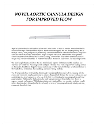
1. NOVEL AORTIC CANNULA DESIGN
FOR IMPROVED FLOW
___________________________________
High incidences of stroke and embolic events have been known to occur in patients with atherosclerotic
disease following a cardiopulmonary bypass. Recent research suggests that this may be partially due to
the high velocity flow jetting effects produced by conventional end-hole arterial cannula which may cause
aortic dissection or dislodge atheromatous plaque along the aortic wall. Our team has redesigned the
cannula in order to reduce these effects from occurring. Our team has assessed the performance of our
design using conventional criteria of peak flow velocities, dispersion, shear stress, and pressure gradients.
Our team has produced a prototype that has demonstrated superior performance under analytical and
empirical test conditions. Pressure gradients and peak velocities have been comparable to leading cannula
on the market. In addition, the performance relationships of other marketed designs have been identical to
the results found in literature, thus validating our team’s testing methods.
The development of our prototype has illuminated which design features may help in reducing embolic
events and which ones may be detrimental to patients. Selected hole designs that increase surface area and
create dispersion have been proven to be one of the best ways to reduce the pressure gradient and lower
peak velocities. Additionally, the location of a small tapered region on the aorta has been shown to
improve cannula performance. While features that create rotational flow can provide a moderate benefit
in flow direction, they ultimately offer lower benefit in terms of pressure gradient and velocity and may
even create thrombotic risk.
2. Page 2 of 14
I Background and Need
Clinical Background
During cardiopulmonary bypass (CPB), arterial cannulae are used to transfer and deliver oxygenated
blood from the heart lung machine to the body during open heart surgery to sustain systemic circulation.
However, traditional end-hole cannulae produce high velocity jets, due to their narrowed tapered bodies,
that can cause arterial dissection or may disrupt vulnerable atherosclerotic plaque in the aorta resulting in
embolic stroke. In order to minimize these jets, diffuser tips can be used to reduce the risk of embolisms
[2]. However, diffuser tips create high shear stresses that can cause detrimental changes to the blood, such
as hemolysis, the rupture of red blood cells, and platelet activation. Besides high shear stress, low shear
stress also poses a problem in that local stagnation can lead to thrombus formation. Aortic dissection, air
embolization, aortic wall tear, injury to the back wall, and excessive bleeding are all complications
associated arterial cannulation [7]. Many of these complications can lead to embolic stroke and even
death. Studies have shown that patients who undergo CPB display a 2.5% frequency of stroke related to
atheroemboli. Those patients with severe atherosclerosis display a 37% incidence of embolic events [14].
During cannulation, a hole is cut in the ascending aorta, the cannula is inserted into the tissue, and purse-
string sutures are tied around the hole to keep the cannula fixed in position. The cannula must be properly
chosen and positioned to ensure complications are reduced. The tip and size of the cannula, usually the
narrowest part of the high flow perfusion circuit, is one element that can be modified based on the patient
[1]. The performances of arterial cannulae are conventionally based on flow and pressure gradient across
the cannula. The optimal design criteria has been shown to be cannulae with lower pressure gradients,
lower peak velocities, optimal shear rates, and wider dispersion with minimal turbulence.
Clinical Need
A way to reduce the incidence of embolic events due to aortic cannulation in cardiopulmonary bypass
procedures through a novel arterial cannula design as demonstrated by lower pressure gradients, wider
dispersion, and lower peak velocities.
II Project Scope
The scope of our project is limited to exploring ways of improving blood flow in a cannula specifically
indicated for use in the aorta during CPB. This will be shown by presenting novel, manufacturable
designs that will lead to a reduction in atheroemboli in the cerebral circulation and thus leading to a
reduced risk of stroke. The following deliverables can be expected from our report:
1. Prototype of Improved Aortic Cannula: A working, physical model that has the ability to sustain at
least 5 L/min of flow against 70-80 mmHg back pressure, which is the typical back pressure seen in the
systemic circulation during diastole and during CPB. A high performing cannula will have reduced
maximum velocities in the cannula itself and in the aorta and have reduced pressure gradients across
varying flow rates compared to the measured in other marketed products.
3. Page 3 of 14
2. Numerical Simulation Model of the Final Design: A computational fluid dynamics (CFD) model of
various cannula designs using proper boundary conditions. This simulation will serve as the main method
by which we will iterate our designs and assess their performance in pressure gradients, shear stresses,
and velocity profiles. Strong design candidates will be prototyped and later assessed in the testing
apparatus.
3. In Vitro Testing Apparatus: This setup will provide (1) a way of empirically validating the scientific
rationale and some of the modeling analysis of the novel cannula design and (2) a standard method for
comparing the design with other competitive cannula products. In order to accomplish this, a thorough
regiment of tests will be made to verify instrument calibration and acceptable levels of testing
repeatability. These tests will monitor the back pressure, or the pressure exerted back on the cannula
itself. This parameter is important for monitoring the consistency of the flow conditions for each
experiment. The apparatus will also generate data on the pressure gradient vs. flow relationship for each
cannula. This will show the pressure drop from the proximal end of the cannula to the distal tip. This
relationship is significant since it is the primary criteria by which arterial cannula are assessed. Increased
pressure gradients are associated with higher jetting effects and those gradients greater than 100 mmHg
are associated with accelerated hemolysis and protein denaturation rates.
4. High Speed Camera Images: Video and images of air bubbles flowing out of the tips of different
marketed products as well as our final prototype. This will provide a visual representation of the
dispersion and impact the fluid is having within a mock aorta pressurized tube.
III Methods for Cannula Design Evaluation
Computational Fluid Dynamic Methodology
The CFD model consists of a CAD design of a cannula and a representation of the aorta, which in our
model is a 2.5cm inner diameter tube, with no slip conditions on an impermeable wall. An assembly of
these two objects are made and then imported into SolidWorks’ FloWorks fluid dynamics package.
The fluid flow for our analysis is governed by a set of 3-D, non-linear, partial differential equations
known as the Navier-Stokes equations [9]. These equations are based on conservation of mass and
momentum in the fluid. The fluid dynamics flow assumptions used to model the blood in our CFD
analysis were as a homogenous, incompressible, Newtonian fluid. Flow is also assumed to be steady and
laminar, and gravitational effects were assumed to be negligible. The boundary conditions used for our
model were an average inlet volume flow rate and aortic pressure for cardiopulmonary bypass.
The CFD analysis will measure three parameters that will allow the engineers to judge the designs and
prototype accordingly [10]. First, pressures at the entrance and exit of the cannula were measured. These
values produced a pressure gradient across the cannula. Second, regions of velocity were visualized to
assess the direction and magnitude of the jetting. Third, the shear rates in the cannula have been measured
as well [11]. The higher the shear stress, the more likely hemolysis will occur. Ideal cannula designs will
also minimize points of extremely low shear due to the likelihood of generating stagnant flow or potential
eddies, which can promote thrombosis. Regions of low velocity, not low shear, by the cannula tip and in
the aortic lumen are sought.
4. Page 4 of 14
This model contains an aorta with an opening for a 21 French cannula, the standard sized cannula tested.
Each early stage concept was modeled in CAD and imported it into the aorta assembly, shown here. At
this point boundary conditions were added to the assembly. The specific boundary conditions used for our
simulations are 5 L/min volumetric flow rate for the inlet, the expected flow rate in typical CPB
procedures, 80 mmHg for the outlet, representing the systemic back pressure in our system, and fluid
properties of water. The mesh was consistently sized across the flow area and was in the order of 800
elements.
There are several figures that can be produced that are useful in prototyping and designing. These include,
velocity magnitude (m/s), velocity streamlines (m/s), pressure gradients (Pa), and shear stress (Pa) in the
cannula. As seen below in Figure 1, the sample simulation appears with a colored reference bar,
maximum and minimum values, and units for each variable.
Figure 1: Simulation figure. Blue arrow shows color reference bar. Green arrow shows minimum value,
and red arrow shows maximum value. Black arrow shows units.
Testing Apparatus Methodology
Shown here is the empirical testing apparatus schematic:
5. Page 5 of 14
Our team decided that the best approach is to construct a test apparatus that can generate reliable pressure
and flow data for each cannula and rely on the high speed camera to capture a qualitative understanding
of the flow dispersion at the tip. If the empirical data of the pressure and flow is validated by the data
generated by the CFD model, then our team can use the modeling data to analyze the peak velocities
within the system. The following photograph shows our final test apparatus that is only intended to
measure the flow and pressure gradients of a cannula within a simulated cardiopulmonary bypass system.
This closed circuit system begins with a 500 GPH bilge pump. The pump must be submerged in a
reservoir of room temperature water (blue arrow). The pump drives the water 40 cm upward through a
½”ID Tygon PVC tube through a water strainer to remove debris (white arrow). The fluid then flows
through a liquid sensor flow meter (Omega Engineering, FLR1013) that is surrounded by two check
valves (black arrows). The check valves prevent backflow from the high pressure aortic system into the
flow meter and out through the pump when the pump is not operating. The flow meter is a pre-calibrated
liquid sensor capable of detecting flows from 1-10 L/min (orange arrow). It operates through a patented
Pelton-turbine wheel that rotates at a rate that is linear over the dynamic range of the device.
Two stopcocks are used to relieve air bubbles and pockets that may form in the tubing or within the aorta
(green arrows). They are also used to drain fluid into an overflow reservoir (pink bucket) when new
cannulae are being replaced for testing.
There are two pressure transducers in the apparatus (red arrows). Each of the transducers (Omega® 0-5
psi silicon gauge sensor) receives 10V of power through a DC Regulated Power Supply that outputs
50mV for 5 psi ±0.25% FS linearity. The first transducer is embedded within a 3/8” ID Tygon PVC tube
approximately 8 cm proximal to the opening of the cannula. This 3/8” tube is used to closely resemble the
6. Page 6 of 14
largest inner diameter of most cannula on the market. The second pressure transducer is embedded within
wall of the aorta directly opposed to the cannulation site. This distal pressure transducer measures the
backpressure of the aorta during stagnant flow as well as the pressure at the tip of the cannula during flow
experiments. The pressure gradient is calculated by the difference between the proximal and distal
pressure transducer at a prescribed flow.
The signals from the pressure and flow transducers are acquired using a 24 bit NI-DAQ 9219. A data
acquisition and recording program, using LabView 8.5, has been written to acquire continuous signals at a
10 Hz sampling rate with 10 samples to read. The signals are acquired with a high speed ADC timing
mode at a differential terminal configuration. The program acquires DC signals ranging from -10 to
+10V. Each DC signal is measured using an averaging window that averages the DC signal every 1
second.
IV Analytic Results and Final Prototype
Market Models
In order to assess how effective our designs would perform on the market, our team compared our
cannula to several leading brands through both numeric modeling and on the physical testing apparatus.
These included the 21 Fr 3M Sarns cannula, 21 Fr Edwards EZ Glide (curved and straight tip), 21 Fr
Edwards Embol-X, and the 22 Fr RMI cannula. The following photos illustrate some of the cannulae
benchmarked against our own design:
Figure 2: Edwards Curved EZ Glide (left), RMI End-Hole Cannula (middle), 3M Sarns Soft-Flow (right)
Final Prototype Design
After iterating through many cannula designs, we arrived at our top choice for a prototype. This design
has an end-hole, four holes on the cannula face, and a 40 mm pitch rectangular spiral internal feature. The
following CAD images in Figure 3 illustrate our final cannula design. The internal spring is meant to
induce the optimal rotational flow through the cannula and reduce jetting and high pressures at the aortic
wall. The rectangular spring was made to increase the efficiency and effectiveness of the spring since
more surface area is essential to induce high axial rotation of flow. While most of the body is straight,
there is a tapered region near the distal end of the cannula. Also at the tip, there are four holes to disperse
the flow into four different flow directions. This will reduce the high impact of the flow on the aortic
wall.
7. Page 7 of 14
Figure 3: CAD Model of Final Prototype
CFD Final Prototype Comparison Tests
In summary, our prototype design performed better than a 21 Fr 3M Sarns and a straight 21 Fr end-hole
cannula in each simulation, except for shear stress. In all of the following simulations, water was used as
the fluid in order to compare the results of the CFD model with those of the testing apparatus. In the table
below a side-by-side comparison of the velocity, pressure and shear stresses numbers can be seen.
Cannula
pressure gradient
(mmHg)
max wall velocity
(m/s)
max velocity
(m/s)
max shear
stress (Pa)
Prototype 38.66342956 0.68 2.9 338.6*
Sarns 44.84453787 0.91 3 63
Endhole 85.95159339 2.5 4.58 43.2
*This shear stress is on one individual node while the rest of the shear stresses are comparable to the other cannulae.
This is most likely due to a meshing issue. Due to the fine geometric features, there is a good chance a large element
was placed beside a very small element, causing an ill-conditioned matrix.
Velocity Profiles on the Wall
These plots show the velocity of the fluid against the aorta wall and illustrate the superiority of the Sarns
and our prototype designs over the end-hole cannula. They are all set to the same scale, and have the max
velocities labeled. The best cannula was the team’s prototype, which had a max wall velocity 25% better
than the Sarns with 0.68 m/s vs. 0.91 m/s, respectively. The end-hole cannula produced an expected jet
and showed a maximum wall velocity of 2.5 m/s over a large area.
8. Page 8 of 14
Velocity Profiles Through the Cannulae
The plots below show the velocity of the fluid through
the cannula. They are all set to the same scale [0 m/s –
2.71437 m/s], and have the max velocities labeled.
Again, the best cannula was our team’s prototype. Its
overall max velocity was 3.3% better than the Sarns
with 2.9 m/s vs 3.0m/s, respectively. The end-hole
cannula’s performance was far inferior to the Sarns
and the prototype, with a maximum wall velocity of
4.58m/s.
Prototype
3M Sarns
End-Hole
End-Hole
3M Sarns
Prototype
9. Page 9 of 14
Pressure Gradients of the Cannulae
These plots show the differences between the pressure gradients at 5 L/min for the end-hole cannula and
our prototoype. The Sarns model is not shown since it had a nearly identical pressure distribution as the
prototype. Each cannula is set to its own scale, so its total pressure gradient can be seen. Figure 4 shows
the flow vs. pressure gradient curves for each of the cannulae. Again, out of these three cannulae the best
cannula was the team’s prototype. Its pressure gradient at 5 L/min is 38.66 mmHg compared to 44.84
mmHg for the Sarns. The end-hole cannula was far above the Sarns and prototype, with a pressure
gradient of 85.95 mmHg.
The following plot summarizes these pressure gradients for these three models based on the CFD
simulations only.
Figure 4: Pressure gradient curves
based on CFD analysis
Final Prototype
Based on these promising findings, a prototype was rapidly made out
of a photopolymer resin using a stereolithography apparatus (SLA).
End-Hole Prototype
10. Page 10 of 14
V Empirical Test and Video Results and Analysis
Here are the pressure gradients of across varying cannula designs, measured by the test apparatus, using
room temperature water flow rates from 1-5 L/min, with 1 L/min intervals:
Figure 5: Pressure gradient curves based on test apparatus experiments
Our test results showed that the cannulae rank from best (lowest pressure gradient) to worst as follows:
3M Sarns, Final Prototype, Endhole Prototype, RMI Endhole, Edwards EZGuide Curved Tip, Edwards
Embol-X, Edwards EZGuide Straight Tip, and a 10 mm pitch spring coil “BlackSpring Prototype".
The CFD simulations showed nearly identical rankings, except that the CFD had a slightly lower pressure
gradient at 5 L/min on our final prototype (38.66mmHg) compared to the Sarns (44.84mmHg). On the
testing apparatus, the Sarns had a slightly lower pressure gradient of 18.30mmHg compared to our final
prototype’s pressure gradient of 19.94mmHg. However, since the pressure gradient has an inherent error
of ±4 mmHg, the pressure gradients of these designs may be considered statistically identical.
In our analysis, one of our main goals was to examine how our cannulae prototypes and the competing
cannulae compared to the standard end-hole cannula, represented here by the RMI model. One of the
design criteria our new dispersion cannula design needed to meet was to have a pressure gradient lower
than a standard end-hole cannula. In order to test for the comparability between the testing apparatus data
and our CFD data, we measured the percent difference between the measured pressure gradient of each of
the cannulae and the RMI end-hole cannula based on the equation: (Cannula’s Pressure gradient-RMI
End-hole Pressure Gradient)/RMI End-hole Pressure Gradient. Based on the table below, the testing
11. Page 11 of 14
apparatus showed that our final prototype was 56% better than the RMI end-hole cannula, while the CFD
showed that our final prototype was 55% better than the RMI end-hole cannula.
Cannula
Measured
Pressure
Gradient
(mmHg)
Theoretica
l CFD
Pressure
Gradient
(mmHg)
Measured
Percent
Difference of
Cannula vs.
Endhole
CFD Percent
Difference of
Cannula vs.
Endhole (O-E)^2/E
Final Prototype 19.9398829 38.66 0.564560969 0.550203607 0.00037465
Sarns 18.30648129 44.84 0.600230527 0.478301338 0.031082345
RMI Endhole 45.79259435 85.95 0 0 0
60mmPitch
BlackSpring
Prototype 90.85070071 127.80 -0.983960551 -0.49 0.507399225
Edwards
EZGuide
Straight Tip 91.63448816 400.00 -1.001076582 -3.65 1.9259875
We performed a Chi squared test to test for significance between our measured and CFD percent
difference data. Since our X2
statistic (2.46) is less than the critical value for 0.05 probability level (9.49)
we can accept the null hypothesis that the difference between the measured and theoretical data is not
statistically significant. This establishes that the results from our CFD models are comparable to the
testing apparatus’s results. Thus, since our main goal was to perform comparative testing, our methods
can be considered valid since both the testing apparatus and CFD simulations lead to similar cannula
rankings with equivalent percentage differences among models. The differences between the magnitude
of the pressure gradients themselves can be explained by a variation in boundary conditions and
geometrical differences of the tips in the CFD model and the cannulae themselves.
Images of Video Recording
High speed camera testing was used to provide a high quality visualization technique that will allow our
team and surgeons to observe the flow patterns that emerge from the tips of different cannula designs. In
our tests, we operated the camera at 250 frames per second with each frame image captured at 1
megapixel in size. To trace the flow, we injected air at the upstream end of our cannula through a syringe.
Shown below are images from the RMI End-Hole (left), Sarns (middle), and our team’s prototype (right).
12. Page 12 of 14
The images confirm the velocity distribution profiles seen in the earlier CFD models. The RMI end-hole
cannula has minimal dispersion and generates high velocity jets along the aortic wall. The Sarns design,
however, generates a much wider dispersive profile with lower jetting impact regions along the wall.
Although our prototype has a low to moderate dispersive effect, confirmed by the localized dispersion in
the CFD model, one of the interesting findings seen in the recording had been the presence of rotational
flow within the cannula body. This suggests that, while the dispersion profile may be narrower than the
Sarns, there is evidence that the fluid in flowing out of the prototype is rotating and possibly leading to
the lower velocities seen in the CFD models.
Comparison of Results with Literature
The results of the CFD modeling and the testing
apparatus are further validated by existing literature
that has also assessed the performance of the same
arterial cannula models. The relationships of the
pressure gradients among many of the marketed
models we have tested are confirmed by the graphs
shown. First, the inferiority of the Edwards Embol-X
to the RMI end-hole cannula is shown in Figure 6
[17].
Additionally, Figure 7 from Edwards Lifesciences [18]
demonstrates the curved tipped cannula’s (EZC21)
superiority over the straight tipped cannula (EZS21),
both of which show pressure gradients above the RMI end-hole cannula (70 mmHg@5L/min). All of
these relationships are also seen in the results of our testing in Figure 5.
Finally, Figure 8 [20] shows that the Sarns Soft-Flow cannula is superior to the RMI end-hole cannula
with pressure gradients at 5 L/min at 54 mmHg and 80 mmHg, respectively. Both the CFD and the test
apparatus confirm this relationship as well. This analysis demonstrates that, while there may be some
discrepancies with the magnitude of the pressure gradients measured in the literature, CFD and test
apparatus environments, the performance relationships among the cannula tested remains validated by all
these methods. Discrepancies are expected due to variations in testing methods and variations in the sizes
Figure 6
Figure 7
Figure 8
13. Page 13 of 14
of the cannulae tested. In any case, the equivalence of our results with existing literature allows our team
to confidently predict where our prototype’s performance will rank among other arterial cannula.
VI Design Conclusions and Recommendations
In summary, our team has developed an arterial cannula prototype design that is comparable to the
superior performance of a leading model on the market, the 3M Sarns Soft Flow. This design has also
been shown to be superior to a typical end-hole cannula and various models from Edwards Lifesciences
based on pressure gradient and peak velocity data. Our CFD analysis has further shown that this design
may even project lower peak velocities than the leading Sarns model. The pressure gradients of our
prototype fall within the acceptable ranges of our functional requirements. Its pressure gradient of 19
mmHg is well below 100 mmHg that can cause hemolysis and little to no regions of stagnant flow that
may induce clotting. However, one drawback of the prototype’s spiral design may be the increased
chance of forming thromboses within the grooves of the internal features. Overall, the low pressure
gradients and low peak velocities have a lower likelihood of inducing embolic events after CPB in
patients with atheroma within their aorta.
There have been many design considerations to learn from this project that may be used in the future
development of any arterial cannula. First, dispersive features, such as holes on the tip, can aid in
lowering pressure gradients. However, not all hole designs may help, as demonstrated by several of our
team’s early design concepts and the inferiority of the Edwards EZGlide cannula, which also have
dispersive hole features on the tip. Designs should seek to maximize the surface area of the holes, as
shown in our prototype and the Sarns models. Although our prototype has an end-hole feature, it did not
prove to increase the overall peak velocity since the other holes have allowed dispersion to take place. On
the contrary, this end-hole feature, which is not found on the Sarns model, has helped reduce peak
velocities.
Second, the location of a tapered feature on the cannula is crucial. Current end-hole designs that are
tapered serve to accelerate the fluid and produce high velocity jetting effects at the opening. Tapered
features at the proximal end of the cannula, like those in the Edwards models, perform the same way and
accelerate the fluid down the body of the cannula. Our model and the Sarns model both have a smaller
tapered region located at the distal end of the cannula body next to the tip. The rest of the cannula body is
straight. As shown by the velocity models in the CFD, this important feature promotes lower velocity in
the cannula body and only brief acceleration near the distal end. Due to this shortened acceleration time,
the peak velocities at the tip of the cannula and within the lumen of the aorta are significantly lower. Any
future cannula designs should examine the location and draft of this feature carefully.
Other design elements that were shown not to be advantageous in performance were many. Rotational
flow through spirals, hexagonal patterns, or fins, while may be helpful, provide only a small improvement
in pressure and velocity at the risk of creating thromboses within the cannula body. Although circular
flow has been shown to occur in the high speed recording, it is fairly minimal at such high flow rates.
Future work should examine flow using a fluid that more closely mimics blood, possibly a
glycerol mixture to better simulate blood viscosity. Other tests include measuring peak velocity
directly in the apparatus or placing solid material along the walls of the aorta and observing any
dislodgement that may take place.
14. Page 14 of 14
VII References
[1] Jegger, D, Horisberger J, et. al. Vascular access for Cardiopulmonary Bypass Procedures. Artificial
Organs. 28 (7). 649-654.
[2] Tovar F, Escobedo C, et al. Structural Performance and Hydrodynamic Resistance of a new silicon
Auricula cannula tip, 2006. Engineering in Medicine and Biology Society. 5396-5399.
[3] Brodman R, Siegel H, Lesser M, Frater R. 1985, A comparison of flow gradients across disposable
arterial perfusion cannulas. Ann. Thorac. Surgery, 29, 225-233
[4] Segadal, L and Matre, K. “Blood velocity distribution in the human ascending aorta.” Circulation.
1987;76:90-100.
[5] Fogel, Mark A. “Effect of Surgical Reconstruction on Flow Profiles in the Aorta Using Magnetic
Resonance Blood Tagging.” Annals of Thoracic Surgery 1997;63:1691-1700.
[6] Leibaschoff, Gustavo H. “Efficacy of the Different Types of Reciprocating Cannulas: A Comparative
Clinical and Surgical Research Study.” International Journal of Cosmetic Surgery and Aesthetic
Dermatology 2001; 3: 13-16.
[7] Swaminathan, Madhav. “The “Sandblasting’ effect of Aortic cannula on arch atheroma during
cardiopulmonary bypass.” International Anesthesia Research Society 2007; 104: 1350-1351.
[8] Bedingham, W., Neavin, T.D., 1991. Application of finite element analysis for assessing
biocompatibility of intra-arterial catheters and probes. Asaio Transactions 37, M179–M180.
[9] Cuvelier, C., Segal, A., van Steenhoven, A., 1986. Finite element methods and Navier-Stokes
equations. D. Reidel, Dordrecht, Netherlands.
[10] Riley, J.R., Hardin, S.B., Winn, B.A., Hurdle, M.B., 1986. In vitro comparison of cavoatrial (dual
stage) cannulae for use during cardiopulmonary bypass. Perfusion 1, 197.
[11] Grigioni M, Daniele C, Morbiducci U, D'Avenio G, Di Benedetto G, Del Gaudio C, Barbaro V.
Computational model of the fluid dynamics of a cannula inserted in a vessel: incidence of the presence of
side holes in blood flow. J Biomech. 2002 Dec;35(12):1599-612.
[12] United States Patent Office. www.uspto.gov
[13] Joubert-Houebnert E, Gerdes A, Sievers H-H. An in vitro evaluation of new cannula tip design
compared with two clinically established cannula tip designs regarding aortic arch vessel perfusion
characteristics. Perfusion 2000; 15; 69
[14] Minakawa, M et al. Effect of Cannula Shape on Aortic Wall and Flow Turbulence: Hydrodynamic
Study During Extracorporeal Circulation in Mock Thoracic Aorta. Artificial Organs (31) 12: 880-886
[15] MAQUET Website: www.maquet.com
[16] FDA CFR. www.fda.gov
[17] Gerdes, A., Hanke, T., Sievers H. In vitro hydrodynamics of the Embol-X Cannula. Perfusion, 2002;
17; 153.
[18] Edwards Lifesciences Product Catalog
[19] Sarns Soft-Flow Arterial Cannula Brochure. Tarum Cardiovascular Systems.
[20] Muehreke D, et al. Flow Characteristics of Aortic Cannula. J. Card. Surg. 1995; 10: 514-519
![Page 2 of 14
I Background and Need
Clinical Background
During cardiopulmonary bypass (CPB), arterial cannulae are used to transfer and deliver oxygenated
blood from the heart lung machine to the body during open heart surgery to sustain systemic circulation.
However, traditional end-hole cannulae produce high velocity jets, due to their narrowed tapered bodies,
that can cause arterial dissection or may disrupt vulnerable atherosclerotic plaque in the aorta resulting in
embolic stroke. In order to minimize these jets, diffuser tips can be used to reduce the risk of embolisms
[2]. However, diffuser tips create high shear stresses that can cause detrimental changes to the blood, such
as hemolysis, the rupture of red blood cells, and platelet activation. Besides high shear stress, low shear
stress also poses a problem in that local stagnation can lead to thrombus formation. Aortic dissection, air
embolization, aortic wall tear, injury to the back wall, and excessive bleeding are all complications
associated arterial cannulation [7]. Many of these complications can lead to embolic stroke and even
death. Studies have shown that patients who undergo CPB display a 2.5% frequency of stroke related to
atheroemboli. Those patients with severe atherosclerosis display a 37% incidence of embolic events [14].
During cannulation, a hole is cut in the ascending aorta, the cannula is inserted into the tissue, and purse-
string sutures are tied around the hole to keep the cannula fixed in position. The cannula must be properly
chosen and positioned to ensure complications are reduced. The tip and size of the cannula, usually the
narrowest part of the high flow perfusion circuit, is one element that can be modified based on the patient
[1]. The performances of arterial cannulae are conventionally based on flow and pressure gradient across
the cannula. The optimal design criteria has been shown to be cannulae with lower pressure gradients,
lower peak velocities, optimal shear rates, and wider dispersion with minimal turbulence.
Clinical Need
A way to reduce the incidence of embolic events due to aortic cannulation in cardiopulmonary bypass
procedures through a novel arterial cannula design as demonstrated by lower pressure gradients, wider
dispersion, and lower peak velocities.
II Project Scope
The scope of our project is limited to exploring ways of improving blood flow in a cannula specifically
indicated for use in the aorta during CPB. This will be shown by presenting novel, manufacturable
designs that will lead to a reduction in atheroemboli in the cerebral circulation and thus leading to a
reduced risk of stroke. The following deliverables can be expected from our report:
1. Prototype of Improved Aortic Cannula: A working, physical model that has the ability to sustain at
least 5 L/min of flow against 70-80 mmHg back pressure, which is the typical back pressure seen in the
systemic circulation during diastole and during CPB. A high performing cannula will have reduced
maximum velocities in the cannula itself and in the aorta and have reduced pressure gradients across
varying flow rates compared to the measured in other marketed products.](data:image/gif;base64,R0lGODlhAQABAIAAAAAAAP///yH5BAEAAAAALAAAAAABAAEAAAIBRAA7)