
Recommended
More Related Content
Similar to Introduction to the Respiratory system and basic anatomy and physiology for preclinical years
Similar to Introduction to the Respiratory system and basic anatomy and physiology for preclinical years (20)
Recently uploaded
Recently uploaded (20)
Introduction to the Respiratory system and basic anatomy and physiology for preclinical years
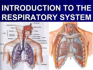
- 1. 1 INTRODUCTION TO THE RESPIRATORY SYSTEM
- 2. GROSS ANATOMY OF THE RESPIRATORY SYSTEM (OVERVIEW)
- 3. THREE COMPONENTS OF THE RESPIRATORY SYSTEM: RESPIRATORY TRACT – Conducting portion – Respiratory portion LUNGS & PULMONARY CIRCULATION ADDITIONAL STRUCTURES INVOLVED IN PULMONARY VENTILATION
- 5. RESPIRATORY TRACT (AIRWAY) Its conducting portion is composed of interconnected hollow organs, thus forming a branching out passageway, which has hard elements (bones, cartilages, ligaments) in its walls. Those elements maintain to keep lumina of hollow organs patent, because they prevent collapsing of walls of air passages.
- 6. Two Major Subdivisions of Conducting Portion of Airway: a) Upper part/airway (nasal cavity with paranasal air sinuses, oral cavity, nasopharynx, oropharynx and upper portion of laryngopharynx); b) Lower part/airway (larynx, trachea, extrapulmonary bronchi, intrapulmonary bronchi, regular and terminal bronchioles). It includes bronchial trees.
- 7. Subdivisions of Respiratory Tract (Airway) Upper Lower
- 8. Upper & Lower Airway 4 5 6 Nasal Cavity Pharynx Oral Cavity Larynx Trachea
- 10. RESPIRATORY TRACT (AIRWAY) Its respiratory portion – “Respiratory Tree”: consists of functional units “Acini”, which include respiratory bronchioles, alveolar ducts, alveolar atria or vestibules and alveolar sacs with alveoli. Acini are attached to the smallest elements of the bronchial tree – the terminal bronchioles.
- 12. Lowest part of Conducting Portion & Respiratory Portion
- 13. Outside air: Varies in temperature. At the alveolar surface it must be at body temperature Varies from very dry to very humid. At the alveolar surface it must be saturated with water vapour Contains dust and debris. These must not reach the alveolar wall Contains micro-organisms, which must be filtered out of the inspired air and disposed, before they reach the alveoli, enter the blood and cause possible problems. It is easy to see that the temperature and humidity of inspired air will increase as it passes down a long series of tubes lined with a moist mucosa at body temperature. The mechanisms for filtering are not so obvious though the turbulence of inspired air could play some role in it.
- 14. Functions of Nasal Cavity It is also filtered & cleaned there.
- 15. LUNGS Lungs are paired parenchymatous organs, which consist of lobes, bronchopulmonary segments and lobules. They also include intrapulmonary bronchi, bronchioles and numerous Respiratory Trees or functional units: Acini, which contain Respiratory Membranes (air-blood barriers). Latter serve for gas exchange between atmospheric air and blood. Transport of gases with the blood to and from lungs is provided via the pulmonary circulation.
- 16. Components of Respiratory Membrane
- 17. ADDITIONAL STRUCTURES INVOLVED IN PULMONARY VENTILATION They include the: –Visceral and parietal pleurae –Two plural cavities (right & left) –Bones and joints of the thoracic cage –Muscles of respiration with their blood and nerve supply)
- 18. Mechanism of breathing. In order to grasp the way in which we breathe we have to grasp the following facts: – Each lung is surrounded by a pleural cavity or sac, except where the plumbing joins it to the rest of the body, rather like a hand in a boxing glove. The glove has an outer and inner surface, separated by a layer of padding. The pleura, similarly, has two surfaces, but the padding is replaced by a thin layer of fluid. – Each lung is enclosed in a rib cage bounded below by the diaphragm and at the sides by the chest wall and the mediastinum. – Breathing works by making the cage bigger: the pleural layers slide over each other and the pressure in the lung is decreased, so air is sucked in. Breathing out does the reverse, the cage collapses and air is expelled.
- 20. Ribs, Respiratory Muscles, Lungs & Pleurae Parietal Pleura Visceral Pleura
- 21. THANK YOU!!!