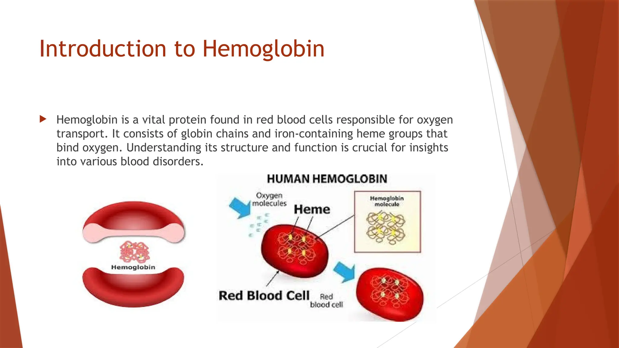
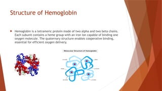
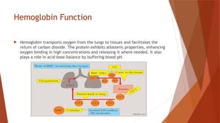
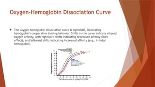
Introduction to Hemoglobin
 Hemoglobin is a vital protein found in red blood cells responsible for oxygen
transport. It consists of globin chains and iron-containing heme groups that
bind oxygen. Understanding its structure and function is crucial for insights
into various blood disorders.
Structure of Hemoglobin
 Hemoglobin is a tetrameric protein made of two alpha and two beta chains.
Each subunit contains a heme group with an iron ion capable of binding one
oxygen molecule. The quaternary structure enables cooperative binding,
essential for efficient oxygen delivery.
Hemoglobin Function
 Hemoglobin transports oxygen from the lungs to tissues and facilitates the
return of carbon dioxide. The protein exhibits allosteric properties, enhancing
oxygen binding in high concentrations and releasing it where needed. It also
plays a role in acid-base balance by buffering blood pH.
Oxygen-Hemoglobin Dissociation Curve
 The oxygen-hemoglobin dissociation curve is sigmoidal, illustrating
hemoglobin's cooperative binding behavior. Shifts in the curve indicate altered
oxygen affinity, with rightward shifts indicating decreased affinity (Bohr
effect), and leftward shifts indicating increased affinity (e.g., in fetal
hemoglobin).
Hemoglobin Variants
 Different forms of hemoglobin exist, including Hemoglobin A (normal adult
form), Hemoglobin F (fetal form), and variants like Hemoglobin S and
Hemoglobin C, associated with sickle cell disease and hemoglobinopathies.
Table: Hemoglobin Types and Properties
Hemoglobin Type Description Clinical Significance
Hemoglobin A Normal adult hemoglobin Main oxygen transporter in
adults
Hemoglobin F Fetal hemoglobin Higher oxygen affinity; replaced
after birth
Hemoglobin S Sickle cell variant Causes sickle cell anemia under
hypoxic conditions
Hemoglobinopathies
 Hemoglobinopathies are genetic disorders affecting hemoglobin structure and
function. Common hemoglobinopathies include sickle cell anemia,
thalassemia, and other variants that lead to impaired oxygen transport and
related clinical symptoms.
Sickle Cell Disease
 Sickle cell disease is caused by a mutation in the beta-globin gene, leading to
abnormal hemoglobin S. Under low oxygen conditions, hemoglobin S
polymerizes, causing red blood cells to become sickle-shaped and leading to
vaso-occlusive crises and hemolysis.
Thalassemia
 Thalassemia are a group of inherited blood disorders characterized by
reduced production of one of the globin chains (alpha or beta). This results in
imbalanced hemoglobin production and leads to anemia, ineffective
erythropoiesis, and iron overload.
Hemoglobin and Iron Metabolism
 Iron is essential for hemoglobin synthesis. Iron deficiency leads to microcytic
anemia, while iron overload can damage organs. Proper regulation of iron
absorption, storage, and recycling is crucial for maintaining hemoglobin
levels.
Hemoglobin Degradation
 Hemoglobin degradation occurs in macrophages, where the heme is broken
down into biliverdin, bilirubin, and iron. Bilirubin is processed by the liver,
and iron is recycled for new hemoglobin synthesis. Imbalances in degradation
lead to jaundice and other conditions.
Clinical Tests for Hemoglobin Levels
 Common tests to assess hemoglobin levels include the complete blood count
(CBC), hemoglobin electrophoresis, and blood smear analysis. These tests
help diagnose anemia, hemoglobinopathies, and other blood disorders.
THANK YOU

Introduction to Hemoglobin, synthesis, working, Bohr's effect, types.

  • 1.
    Introduction to Hemoglobin Hemoglobin is a vital protein found in red blood cells responsible for oxygen transport. It consists of globin chains and iron-containing heme groups that bind oxygen. Understanding its structure and function is crucial for insights into various blood disorders.
  • 2.
    Structure of Hemoglobin Hemoglobin is a tetrameric protein made of two alpha and two beta chains. Each subunit contains a heme group with an iron ion capable of binding one oxygen molecule. The quaternary structure enables cooperative binding, essential for efficient oxygen delivery.
  • 3.
    Hemoglobin Function  Hemoglobintransports oxygen from the lungs to tissues and facilitates the return of carbon dioxide. The protein exhibits allosteric properties, enhancing oxygen binding in high concentrations and releasing it where needed. It also plays a role in acid-base balance by buffering blood pH.
  • 4.
    Oxygen-Hemoglobin Dissociation Curve The oxygen-hemoglobin dissociation curve is sigmoidal, illustrating hemoglobin's cooperative binding behavior. Shifts in the curve indicate altered oxygen affinity, with rightward shifts indicating decreased affinity (Bohr effect), and leftward shifts indicating increased affinity (e.g., in fetal hemoglobin).
  • 5.
    Hemoglobin Variants  Differentforms of hemoglobin exist, including Hemoglobin A (normal adult form), Hemoglobin F (fetal form), and variants like Hemoglobin S and Hemoglobin C, associated with sickle cell disease and hemoglobinopathies.
  • 6.
    Table: Hemoglobin Typesand Properties Hemoglobin Type Description Clinical Significance Hemoglobin A Normal adult hemoglobin Main oxygen transporter in adults Hemoglobin F Fetal hemoglobin Higher oxygen affinity; replaced after birth Hemoglobin S Sickle cell variant Causes sickle cell anemia under hypoxic conditions
  • 7.
    Hemoglobinopathies  Hemoglobinopathies aregenetic disorders affecting hemoglobin structure and function. Common hemoglobinopathies include sickle cell anemia, thalassemia, and other variants that lead to impaired oxygen transport and related clinical symptoms.
  • 8.
    Sickle Cell Disease Sickle cell disease is caused by a mutation in the beta-globin gene, leading to abnormal hemoglobin S. Under low oxygen conditions, hemoglobin S polymerizes, causing red blood cells to become sickle-shaped and leading to vaso-occlusive crises and hemolysis.
  • 9.
    Thalassemia  Thalassemia area group of inherited blood disorders characterized by reduced production of one of the globin chains (alpha or beta). This results in imbalanced hemoglobin production and leads to anemia, ineffective erythropoiesis, and iron overload.
  • 10.
    Hemoglobin and IronMetabolism  Iron is essential for hemoglobin synthesis. Iron deficiency leads to microcytic anemia, while iron overload can damage organs. Proper regulation of iron absorption, storage, and recycling is crucial for maintaining hemoglobin levels.
  • 11.
    Hemoglobin Degradation  Hemoglobindegradation occurs in macrophages, where the heme is broken down into biliverdin, bilirubin, and iron. Bilirubin is processed by the liver, and iron is recycled for new hemoglobin synthesis. Imbalances in degradation lead to jaundice and other conditions.
  • 12.
    Clinical Tests forHemoglobin Levels  Common tests to assess hemoglobin levels include the complete blood count (CBC), hemoglobin electrophoresis, and blood smear analysis. These tests help diagnose anemia, hemoglobinopathies, and other blood disorders.
  • 13.