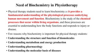
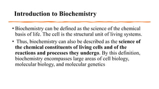
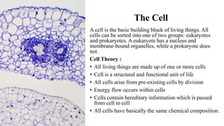
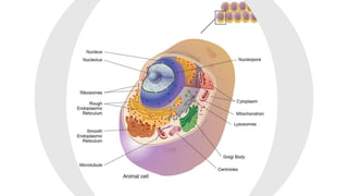
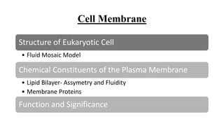
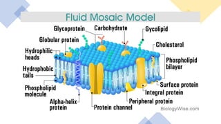
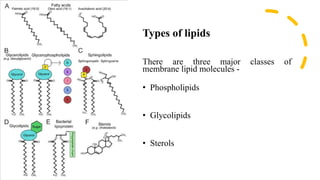
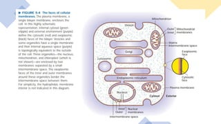
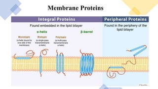
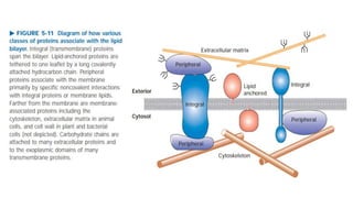
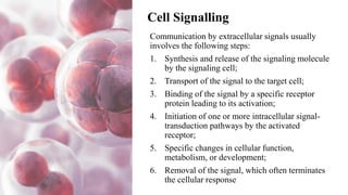
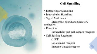
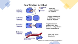
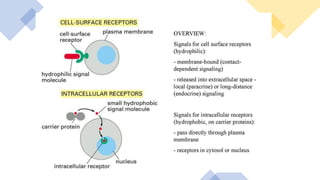
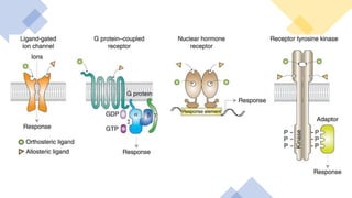
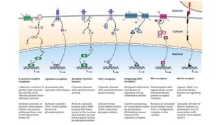
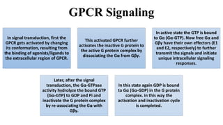
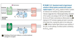
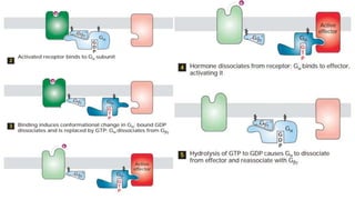
Unit 1. CELL
Doctor of Physiotherapy
Sanobar Ashoora
04/03/2023
Why biochemistry is
important for physical
therapy?
Need of Biochemistry in Physiotherapy
• Physical therapy students need to learn biochemistry as it provides a
fundamental understanding of the physiological processes underlying
human movement and function. Biochemistry is the study of the chemical
processes that occur within living organisms, and these processes are
essential to understanding how the body functions and responds to injury or
disease.
• Few reasons why biochemistry is important for physical therapy students:
• Understanding the structure and function of biomolecules
• Understanding metabolism and energy production
• Understanding pharmacology
• Understanding the molecular basis of diseases
Introduction to Biochemistry
• Biochemistry can be defined as the science of the chemical
basis of life. The cell is the structural unit of living systems.
• Thus, biochemistry can also be described as the science of
the chemical constituents of living cells and of the
reactions and processes they undergo. By this definition,
biochemistry encompasses large areas of cell biology,
molecular biology, and molecular genetics
The Cell
A cell is the basic building block of living things. All
cells can be sorted into one of two groups: eukaryotes
and prokaryotes. A eukaryote has a nucleus and
membrane-bound organelles, while a prokaryote does
not.
Cell Theory :
• All living things are made up of one or more cells
• Cell is a structural and functional unit of life
• All cells arise from pre-existing cells by division
• Energy flow occurs within cells
• Cells contain hereditary information which is passed
from cell to cell
• All cells have basically the same chemical composition.
Cell Membrane
Structure of Eukaryotic Cell
• Fluid Mosaic Model
Chemical Constituents of the Plasma Membrane
• Lipid Bilayer- Assymetry and Fluidity
• Membrane Proteins
Function and Significance
Types of lipids
There are three major classes of
membrane lipid molecules -
• Phospholipids
• Glycolipids
• Sterols
Asymmetry and Fluidity of CM
• A common feature of all eukaryotic membranes is the non-random distribution of different lipid
species in the lipid bilayer (lipid asymmetry).
• Lipid asymmetry in membranes is a consequence of multiple factors, including the biophysical
properties of lipids that dictate their ability to spontaneously “flip” their polar headgroups through
the hydrophobic membrane interior, and the presence of transporters (enzymes) that assist in active
lipid translocation across the bilayer
• Fluidity is a term used to describe the ease of movement of molecules in the membrane and is an
important characteristic of cell function. Fluidity depends on the temperature (increased temperatures
make it more fluid and decreased temperatures make it more solid), saturated fatty acids and
unsaturated fatty acids. Saturated fatty acids make the membrane less fluid while unsaturated fatty
acids make it more fluid.
Lipid Rafts
• The van der Waals attractive forces between
neighboring fatty acid tails are not selective
enough to hold groups of molecules
together.
• For some lipid molecules, however, such as
the sphingolipids which tend to have long
and saturated fatty hydrocarbon chains, the
attractive forces can be just strong enough to
hold the adjacent molecules together
transiently in small microdomains.
• Such microdomains, or lipid rafts, can be
thought of as transient phase separations in
the fluid lipid bilayer where sphingolipids
become concentrated.
Membrane Proteins
Integral and Peripheral Proteins
Lipid Anchored proteins
Transport Proteins
Cell coat/Glycocalyx
Blood Group Antigens
Membrane Proteins
Blood Group Antigens
• Blood group antigens are
surface markers on the
outside of the red blood cell
(RBC) membrane. They are
proteins and carbohydrates
attached to lipid or protein.
• The carbohydrate chains of
the blood group substances
are built by sequential
assembly of the respective
monosaccharide residues
through the action of specific
glycosyltransferases
Cell Signalling
Communication by extracellular signals usually
involves the following steps:
1. Synthesis and release of the signaling molecule
by the signaling cell;
2. Transport of the signal to the target cell;
3. Binding of the signal by a specific receptor
protein leading to its activation;
4. Initiation of one or more intracellular signal-
transduction pathways by the activated
receptor;
5. Specific changes in cellular function,
metabolism, or development;
6. Removal of the signal, which often terminates
the cellular response
Cell Signalling
• Extracellular Signalling
• Intracellular Signalling
• Signal Molecules
Membrane-bound and Secretory
molecules
• Receptors
Intracellular and cell-surface receptors
• Cell Surface Receptors
GPCR
Ion-channel receptor
Enzyme-Linked receptor
GPCR Signaling
In signal transduction, first the
GPCR gets activated by changing
its conformation, resulting from
the binding of agonists/ligands to
the extracellular region of GPCR.
This activated GPCR further
activates the inactive G protein to
the active G protein complex by
dissociating the Gα from Gβγ.
In active state the GTP is bound
to Gα (Gα-GTP). Now free Gα and
Gβγ have their own effectors (E1
and E2, respectively) to further
transmit the signals and initiate
unique intracellular signaling
responses.
Later, after the signal
transduction, the Gα-GTPase
activity hydrolyze the bound GTP
(Gα-GTP) to GDP and Pi and
inactivate the G protein complex
by re-associating the Gα with
Gβγ.
In this state again GDP is bound
to Gα (Gα-GDP) in the G protein
complex. in this way the
activation and inactivation cycle
is completed.
Enzyme Linked Receptors
• Receptor tyrosine kinases mediate responses to a large number of signals, including peptide hormones like insulin
and growth factors like epidermal growth factor.
• Binding of signal molecules to the EC domains of receptor tyrosine kinase molecules causes two receptor
molecules to dimerize. This brings the cytoplasmic tails of the receptors close to each other and causes the
tyrosine kinase activity of these tails to be turned on. The activated tails then phosphorylate each other on several
tyrosine residues. This is called autophosphorylation.
• The phosphorylation of tyrosines on the receptor tails triggers the assembly of an IC signaling complex on the
tails. The newly phosphorylated tyrosines serve as binding sites for signaling proteins that then pass the message
on to yet other proteins. An important protein that is subsequently activated by the signaling complexes on the
receptor tyrosine kinases is called Ras.
• The Ras protein is a monomeric guanine nucleotide binding protein that is associated with the cytosolic face of
the plasma membrane. Ras is active when GTP is bound to it and inactive when GDP is bound to it. Ras can
hydrolyze the GTP to GDP.
• When a signal arrives at the receptor tyrosine kinase, the receptor monomers come together and phosphorylate
each others' tyrosines, triggering the assembly of a complex of proteins on the cytoplasmic tail of the receptor.
One of the proteins in this complex interacts with Ras and stimulates the exchange of the GDP bound to the
inactive Ras for a GTP. This activates the Ras.
• Activated Ras triggers a phosphorylation cascade of three protein kinases, which relay and distribute the signal.
These protein kinases are members of a group called the MAP kinases (Mitogen Activated Protein Kinases). The
final kinase in this cascade phosphorylates various target proteins, including enzymes and transcriptional
activators that regulate gene expression.
• Harpers Illustrated Biochemistry
• Cell and Molecular Biology, Concepts and experiments-
Gerald Karp
• Life Science Fundamentals and Practice Part I by Pranav
Kumar
• https://www.ncbi.nlm.nih.gov/books/NBK26871/#:~:text
=There%20are%20three%20major%20classes,phospholipi
ds%2C%20cholesterol%2C%20and%20glycolipids.
Thank you

Intro to Biochem Class1.pptx

  • 1.
    Unit 1. CELL Doctorof Physiotherapy Sanobar Ashoora 04/03/2023
  • 2.
    Why biochemistry is importantfor physical therapy?
  • 3.
    Need of Biochemistryin Physiotherapy • Physical therapy students need to learn biochemistry as it provides a fundamental understanding of the physiological processes underlying human movement and function. Biochemistry is the study of the chemical processes that occur within living organisms, and these processes are essential to understanding how the body functions and responds to injury or disease. • Few reasons why biochemistry is important for physical therapy students: • Understanding the structure and function of biomolecules • Understanding metabolism and energy production • Understanding pharmacology • Understanding the molecular basis of diseases
  • 4.
    Introduction to Biochemistry •Biochemistry can be defined as the science of the chemical basis of life. The cell is the structural unit of living systems. • Thus, biochemistry can also be described as the science of the chemical constituents of living cells and of the reactions and processes they undergo. By this definition, biochemistry encompasses large areas of cell biology, molecular biology, and molecular genetics
  • 5.
    The Cell A cellis the basic building block of living things. All cells can be sorted into one of two groups: eukaryotes and prokaryotes. A eukaryote has a nucleus and membrane-bound organelles, while a prokaryote does not. Cell Theory : • All living things are made up of one or more cells • Cell is a structural and functional unit of life • All cells arise from pre-existing cells by division • Energy flow occurs within cells • Cells contain hereditary information which is passed from cell to cell • All cells have basically the same chemical composition.
  • 7.
    Cell Membrane Structure ofEukaryotic Cell • Fluid Mosaic Model Chemical Constituents of the Plasma Membrane • Lipid Bilayer- Assymetry and Fluidity • Membrane Proteins Function and Significance
  • 10.
    Types of lipids Thereare three major classes of membrane lipid molecules - • Phospholipids • Glycolipids • Sterols
  • 11.
    Asymmetry and Fluidityof CM • A common feature of all eukaryotic membranes is the non-random distribution of different lipid species in the lipid bilayer (lipid asymmetry). • Lipid asymmetry in membranes is a consequence of multiple factors, including the biophysical properties of lipids that dictate their ability to spontaneously “flip” their polar headgroups through the hydrophobic membrane interior, and the presence of transporters (enzymes) that assist in active lipid translocation across the bilayer • Fluidity is a term used to describe the ease of movement of molecules in the membrane and is an important characteristic of cell function. Fluidity depends on the temperature (increased temperatures make it more fluid and decreased temperatures make it more solid), saturated fatty acids and unsaturated fatty acids. Saturated fatty acids make the membrane less fluid while unsaturated fatty acids make it more fluid.
  • 12.
    Lipid Rafts • Thevan der Waals attractive forces between neighboring fatty acid tails are not selective enough to hold groups of molecules together. • For some lipid molecules, however, such as the sphingolipids which tend to have long and saturated fatty hydrocarbon chains, the attractive forces can be just strong enough to hold the adjacent molecules together transiently in small microdomains. • Such microdomains, or lipid rafts, can be thought of as transient phase separations in the fluid lipid bilayer where sphingolipids become concentrated.
  • 15.
    Membrane Proteins Integral andPeripheral Proteins Lipid Anchored proteins Transport Proteins Cell coat/Glycocalyx Blood Group Antigens
  • 16.
  • 20.
    Blood Group Antigens •Blood group antigens are surface markers on the outside of the red blood cell (RBC) membrane. They are proteins and carbohydrates attached to lipid or protein. • The carbohydrate chains of the blood group substances are built by sequential assembly of the respective monosaccharide residues through the action of specific glycosyltransferases
  • 21.
    Cell Signalling Communication byextracellular signals usually involves the following steps: 1. Synthesis and release of the signaling molecule by the signaling cell; 2. Transport of the signal to the target cell; 3. Binding of the signal by a specific receptor protein leading to its activation; 4. Initiation of one or more intracellular signal- transduction pathways by the activated receptor; 5. Specific changes in cellular function, metabolism, or development; 6. Removal of the signal, which often terminates the cellular response
  • 22.
    Cell Signalling • ExtracellularSignalling • Intracellular Signalling • Signal Molecules Membrane-bound and Secretory molecules • Receptors Intracellular and cell-surface receptors • Cell Surface Receptors GPCR Ion-channel receptor Enzyme-Linked receptor
  • 27.
    GPCR Signaling In signaltransduction, first the GPCR gets activated by changing its conformation, resulting from the binding of agonists/ligands to the extracellular region of GPCR. This activated GPCR further activates the inactive G protein to the active G protein complex by dissociating the Gα from Gβγ. In active state the GTP is bound to Gα (Gα-GTP). Now free Gα and Gβγ have their own effectors (E1 and E2, respectively) to further transmit the signals and initiate unique intracellular signaling responses. Later, after the signal transduction, the Gα-GTPase activity hydrolyze the bound GTP (Gα-GTP) to GDP and Pi and inactivate the G protein complex by re-associating the Gα with Gβγ. In this state again GDP is bound to Gα (Gα-GDP) in the G protein complex. in this way the activation and inactivation cycle is completed.
  • 31.
    Enzyme Linked Receptors •Receptor tyrosine kinases mediate responses to a large number of signals, including peptide hormones like insulin and growth factors like epidermal growth factor. • Binding of signal molecules to the EC domains of receptor tyrosine kinase molecules causes two receptor molecules to dimerize. This brings the cytoplasmic tails of the receptors close to each other and causes the tyrosine kinase activity of these tails to be turned on. The activated tails then phosphorylate each other on several tyrosine residues. This is called autophosphorylation. • The phosphorylation of tyrosines on the receptor tails triggers the assembly of an IC signaling complex on the tails. The newly phosphorylated tyrosines serve as binding sites for signaling proteins that then pass the message on to yet other proteins. An important protein that is subsequently activated by the signaling complexes on the receptor tyrosine kinases is called Ras. • The Ras protein is a monomeric guanine nucleotide binding protein that is associated with the cytosolic face of the plasma membrane. Ras is active when GTP is bound to it and inactive when GDP is bound to it. Ras can hydrolyze the GTP to GDP. • When a signal arrives at the receptor tyrosine kinase, the receptor monomers come together and phosphorylate each others' tyrosines, triggering the assembly of a complex of proteins on the cytoplasmic tail of the receptor. One of the proteins in this complex interacts with Ras and stimulates the exchange of the GDP bound to the inactive Ras for a GTP. This activates the Ras. • Activated Ras triggers a phosphorylation cascade of three protein kinases, which relay and distribute the signal. These protein kinases are members of a group called the MAP kinases (Mitogen Activated Protein Kinases). The final kinase in this cascade phosphorylates various target proteins, including enzymes and transcriptional activators that regulate gene expression.
  • 34.
    • Harpers IllustratedBiochemistry • Cell and Molecular Biology, Concepts and experiments- Gerald Karp • Life Science Fundamentals and Practice Part I by Pranav Kumar • https://www.ncbi.nlm.nih.gov/books/NBK26871/#:~:text =There%20are%20three%20major%20classes,phospholipi ds%2C%20cholesterol%2C%20and%20glycolipids.
  • 35.