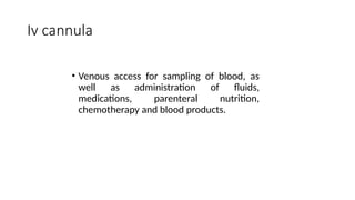
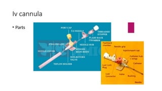
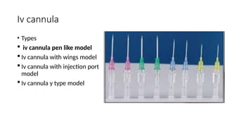
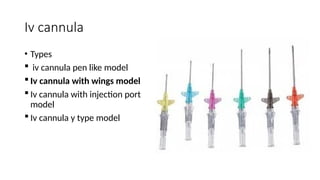
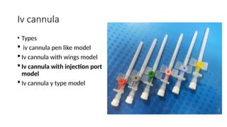
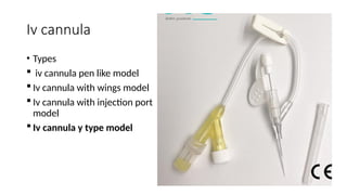
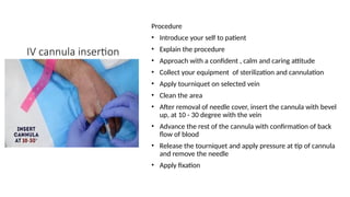
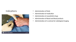
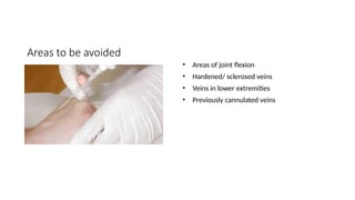
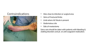
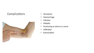
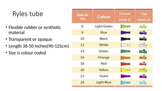
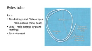
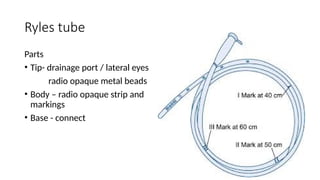
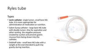
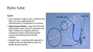
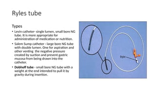
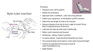
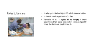
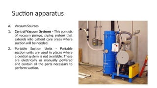
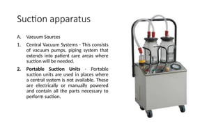
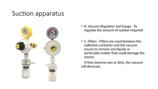
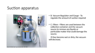
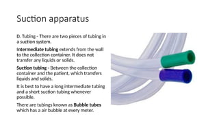
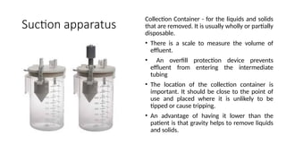
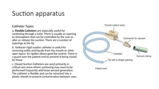
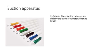
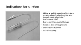
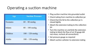
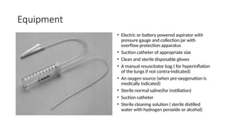
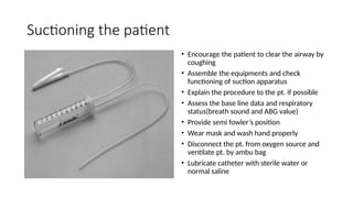
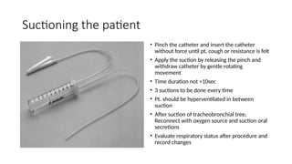
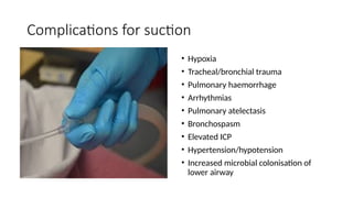
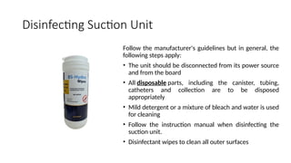
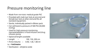
Topics
• Iv cannula
• Ryles tube
• Suction apparatus
• Pressure monitoring line(PM-line)
Presenter- M. Imran
Moderator – Dr. Rafat
Iv cannula
• Venous access for sampling of blood, as
well as administration of fluids,
medications, parenteral nutrition,
chemotherapy and blood products.
Iv cannula
• Parts
Iv cannula
• Colour coding
Iv cannula
• Types
 iv cannula pen like model
 Iv cannula with wings model
 Iv cannula with injection port
model
 Iv cannula y type model
Iv cannula
• Types
 iv cannula pen like model
 Iv cannula with wings model
 Iv cannula with injection port
model
 Iv cannula y type model
Iv cannula
• Types
 iv cannula pen like model
 Iv cannula with wings model
 Iv cannula with injection port
model
 Iv cannula y type model
Iv cannula
• Types
 iv cannula pen like model
 Iv cannula with wings model
 Iv cannula with injection port
model
 Iv cannula y type model
Iv cannula
• Types
 iv cannula pen like model
 Iv cannula with wings model
 Iv cannula with injection port
model
 Iv cannula y type model
IV cannula insertion
Procedure
• Introduce your self to patient
• Explain the procedure
• Approach with a confident , calm and caring attitude
• Collect your equipment of sterilization and cannulation
• Apply tourniquet on selected vein
• Clean the area
• After removal of needle cover, insert the cannula with bevel
up, at 10 - 30 degree with the vein
• Advance the rest of the cannula with confirmation of back
flow of blood
• Release the tourniquet and apply pressure at tip of cannula
and remove the needle
• Apply fixation
Indications • Administration of fluids
• Administration of medication
• Administration of anaesthetic drugs
• Administration of blood and blood products
• Administration of iv contrast for radiological imaging
Areas to be avoided
• Areas of joint flexion
• Hardened/ sclerosed veins
• Veins in lower extremities
• Previously cannulated veins
Contraindications • Sites close to infection or surgical area
• Veins of fractured limbs
• Limb where AV fistula is present
• Oedematous site
• Side of mastectomy
Extra care should be taken with patients with bleeding &
clotting disorders and pt. on anti-coagulant medication.
Complications • Hematoma
• Haemorrhage
• Infection
• Phlebitis
• Puncturing an artery or a nerve
• Infiltration
• Extravasation
Ryles tube
Ryles tube
• Flexible rubber or synthetic
material
• Transparent or opaque
• Length 36-50 inches(90-125cm)
• Size is colour coded
Ryles tube
Parts
• Tip- drainage port / lateral eyes
radio opaque metal beads
• Body – radio opaque strip and
markings
• Base – connect
Ryles tube
Parts
• Tip- drainage port / lateral eyes
radio opaque metal beads
• Body – radio opaque strip and
markings
• Base - connect
Ryles tube
Types
• Levin catheter- single lumen, small bore NG
tube. It is more appropriate for
administration of medication or nutrition.
• Salem Sump catheter - large bore NG tube
with double lumen. One for aspiration and
other venting the negative pressure
created by suction and prevent gastric
mucosa from being drawn into the
catheter.
• Dobhoff tube - small bore NG tube with a
weight at the end intended to pull it by
gravity during insertion.
Ryles tube sterilization • Gamma radiation
• Ethylene oxide gas sterilization
• Boiling
Ryles tube insertion
Procedure
• Introduce your self to patient
• Explain the procedure
• Approach with a confident , calm and caring attitude
• Collect your equipment of sterilization and RT insertion
• Determine the length of tube to be inserted
• Measure distance from tip of nose/ angle of mouth TO ear
lobe To xyphoid process of sternum
• Lubricate the tube tip with water soluble jelly
• Wider nostril selected and cleaned
• Positioning –sitting / fowler’s position
• In supine patient , head should be flexed by one hand
• Tip is passed along nasal septum, insert upto measured length
• Confirm by aspiration/Immersing in bowl of
water/Ascultation/x-ray
• Apply fixation
Ryles tube care • If tube gets blocked inject 10 ml air/normal saline
• It should be changed every 3rd
day
• Removal of RT – inject air to empty it from
secretions then close the end of tube and gently
bring the tube out by pinching it.
Ryles tube indications • To feed the pt. when oral intake is not possible
(comatose pt.)
• Pt. who refuses to eat (disoriented pt.)
• To dilute and remove consumed poison (gastric
lavage)
• To instill ice cold solution to control gastric bleed
• To prevent stress on operated site by decompression
• To relieve vomiting and distension
• To collect gastric juice for diagnostic purpose
Ryles tube
contraindications
• Acute corrosive poisoning
• Stricture esophagus, Tracheoesophageal fistula
• Faciomaxillary trauma, , recent nasal surgery and
facial surgery
• Gastric and Oesophageal surgery
• Polyps in nose
• Deviated nasal septum
• Patient on anticoagulants
Ryles tube
complications
• Sinusitis
• Epistaxis
• Esophageal perforation
• Esophageal ulceration
• URTI
Suction apparatus
Principles
• Negative pressure
• Flow
A. Negative Pressure - Vacuum is the difference between the measured
negative pressure and ambient atmospheric pressure
B. Flow - All suction equipment from the source to the patient connection
acts a tube of varying diameter.
• The rate of flow will depend on the pressure difference between the
ends of the tube and the resistance to flow.
• To ensure good flow, the maximum vacuum and the shortest possible
length of tubing should be used
• The internal diameter of tubings , connectors, and the suction catheter
should be as large as possible, and the vacuum system should not leak.
• Increasing the number of intakes will result in a decrease of vacuum
and flow at each intake.
• Flow through suction apparatus is also affected by the physical
characteristics (elasticity, viscosity, adhesion, and cohesion) of the
material being suctioned.
• After material enters the collection container, it no longer causes
resistance to flow.
Suction apparatus
A. Vacuum Sources
1. Central Vacuum Systems - This consists
of vacuum pumps, piping system that
extends into patient care areas where
suction will be needed.
2. Portable Suction Units - Portable
suction units are used in places where
a central system is not available. These
are electrically or manually powered
and contain all the parts necessary to
perform suction.
Suction apparatus
• B. Vacuum Regulator and Gauge - To
regulate the amount of suction required
• C. Filters - Filters are used between the
collection container and the vacuum
source to remove any liquids or
particulate matter that could damage the
source.
If they become wet or dirty, the vacuum
will decrease.
Suction apparatus
D. Tubing - There are two pieces of tubing in
a suction system.
Intermediate tubing extends from the wall
to the collection container. It does not
transfer any liquids or solids.
Suction tubing - Between the collection
container and the patient, which transfers
liquids and solids.
It is best to have a long intermediate tubing
and a short suction tubing whenever
possible.
There are tubings known as Bubble tubes
which has a air bubble at every meter.
Suction apparatus
Collection Container - for the liquids and solids
that are removed. It is usually wholly or partially
disposable.
• There is a scale to measure the volume of
effluent.
• An overfill protection device prevents
effluent from entering the intermediate
tubing
• The location of the collection container is
important. It should be close to the point of
use and placed where it is unlikely to be
tipped or cause tripping.
• An advantage of having it lower than the
patient is that gravity helps to remove liquids
and solids.
Suction apparatus
Catheter Types
a. Flexible Catheters are especially useful for
suctioning through a tube. There is usually an opening
to atmosphere that can be controlled by the user to
alter or release the suction. There are a number of
openings at the tip
b. Yankauer rigid suction catheter is useful for
removing solids and liquids from the mouth or other
open space. Its rigidity allows good tip control. There is
a guard over the patient end to prevent it being closed
by tissue
c. Closed Suction Catheters are used primarily in
critical care areas where suctioning may need to be
performed frequently with least aerosol generation.
The catheter is flexible and can be retracted into a
plastic sheath to prevent contamination between uses.
Suction apparatus
2. Catheter Sizes- Suction catheters are
sized by the external diameter and shaft
length
Indications for suction
• Visible or audible secretions (Removal of
secretions from Tracheobronchial tree,
through endotracheal tube /
tracheostomy tube)
• Decreased O2 sat. due to blockage
• Increased peak airway pressure
• Decreased tidal volume
• Sputum sampling
Operating a suction machine
• Plug suction machine into grounded outlet
• Check tubing from machine to collection jar
• Check that the lid to the collection jar is
closed tightly
• Attach the extension tubing to the collection
jar.
• Turn the machine on and kink the extension
tubing to block the flow of air (if gauge did
not move, recheck all connections)
• Set pressure gauge as required
• Attach suction catheter to extension tubing
Equipment
• Electric or battery powered aspirator with
pressure gauge and collection jar with
overflow protection apparatus
• Suction catheter of appropriate size
• Clean and sterile disposable gloves
• A manual resuscitator bag ( for hyperinflation
of the lungs if not contra-indicated)
• An oxygen source (when pre-oxygenation is
medically indicated)
• Sterile normal saline(for instillation)
• Suction catheter
• Sterile cleaning solution ( sterile distilled
water with hydrogen peroxide or alcohol)
Suctioning the patient
• Encourage the patient to clear the airway by
coughing
• Assemble the equipments and check
functioning of suction apparatus
• Explain the procedure to the pt. if possible
• Assess the base line data and respiratory
status(breath sound and ABG value)
• Provide semi fowler’s position
• Wear mask and wash hand properly
• Disconnect the pt. from oxygen source and
ventilate pt. by ambu bag
• Lubricate catheter with sterile water or
normal saline
Suctioning the patient
• Pinch the catheter and insert the catheter
without force until pt. cough or resistance is felt
• Apply the suction by releasing the pinch and
withdraw catheter by gentle rotating
movement
• Time duration not >10sec
• 3 suctions to be done every time
• Pt. should be hyperventilated in between
suction
• After suction of tracheobronchial tree,
Reconnect with oxygen source and suction oral
secretions
• Evaluate respiratory status after procedure and
record changes
Complications for suction
• Hypoxia
• Tracheal/bronchial trauma
• Pulmonary haemorrhage
• Arrhythmias
• Pulmonary atelectasis
• Bronchospasm
• Elevated ICP
• Hypertension/hypotension
• Increased microbial colonisation of
lower airway
Disinfecting Suction Unit
Follow the manufacturer's guidelines but in general, the
following steps apply:
• The unit should be disconnected from its power source
and from the board
• All disposable parts, including the canister, tubing,
catheters and collection are to be disposed
appropriately
• Mild detergent or a mixture of bleach and water is used
for cleaning
• Follow the instruction manual when disinfecting the
suction unit.
• Disinfectant wipes to clean all outer surfaces
Pressure monitoring line
• Made from non-toxic medical grade PVC
• Provided with male luer lock at one end and
female luer lock at other end and fits all
standard equipment.
• Sterile, individually packed in Blister pack.
• Can withstand a pressure of 580 PSI (30,000
mmhg)
• Used for high pressure monitoring,
neonatal/pediatric iv fluid infusion and drug
infusion pump
• Optional lengths available
• Length – 100, 150, 200 cm
• Prime Volume – 0.90, 1.40, 1.80 ml
i.e. <1ml/metre
• Sterilization - ethylene oxide
Thank you

important_equipments_in_anaesthesia.pptx

  • 1.
    Topics • Iv cannula •Ryles tube • Suction apparatus • Pressure monitoring line(PM-line) Presenter- M. Imran Moderator – Dr. Rafat
  • 2.
    Iv cannula • Venousaccess for sampling of blood, as well as administration of fluids, medications, parenteral nutrition, chemotherapy and blood products.
  • 3.
  • 4.
  • 5.
    Iv cannula • Types iv cannula pen like model  Iv cannula with wings model  Iv cannula with injection port model  Iv cannula y type model
  • 6.
    Iv cannula • Types iv cannula pen like model  Iv cannula with wings model  Iv cannula with injection port model  Iv cannula y type model
  • 7.
    Iv cannula • Types iv cannula pen like model  Iv cannula with wings model  Iv cannula with injection port model  Iv cannula y type model
  • 8.
    Iv cannula • Types iv cannula pen like model  Iv cannula with wings model  Iv cannula with injection port model  Iv cannula y type model
  • 9.
    Iv cannula • Types iv cannula pen like model  Iv cannula with wings model  Iv cannula with injection port model  Iv cannula y type model
  • 10.
    IV cannula insertion Procedure •Introduce your self to patient • Explain the procedure • Approach with a confident , calm and caring attitude • Collect your equipment of sterilization and cannulation • Apply tourniquet on selected vein • Clean the area • After removal of needle cover, insert the cannula with bevel up, at 10 - 30 degree with the vein • Advance the rest of the cannula with confirmation of back flow of blood • Release the tourniquet and apply pressure at tip of cannula and remove the needle • Apply fixation
  • 11.
    Indications • Administrationof fluids • Administration of medication • Administration of anaesthetic drugs • Administration of blood and blood products • Administration of iv contrast for radiological imaging
  • 12.
    Areas to beavoided • Areas of joint flexion • Hardened/ sclerosed veins • Veins in lower extremities • Previously cannulated veins
  • 13.
    Contraindications • Sitesclose to infection or surgical area • Veins of fractured limbs • Limb where AV fistula is present • Oedematous site • Side of mastectomy Extra care should be taken with patients with bleeding & clotting disorders and pt. on anti-coagulant medication.
  • 14.
    Complications • Hematoma •Haemorrhage • Infection • Phlebitis • Puncturing an artery or a nerve • Infiltration • Extravasation
  • 15.
  • 16.
    Ryles tube • Flexiblerubber or synthetic material • Transparent or opaque • Length 36-50 inches(90-125cm) • Size is colour coded
  • 17.
    Ryles tube Parts • Tip-drainage port / lateral eyes radio opaque metal beads • Body – radio opaque strip and markings • Base – connect
  • 18.
    Ryles tube Parts • Tip-drainage port / lateral eyes radio opaque metal beads • Body – radio opaque strip and markings • Base - connect
  • 19.
    Ryles tube Types • Levincatheter- single lumen, small bore NG tube. It is more appropriate for administration of medication or nutrition. • Salem Sump catheter - large bore NG tube with double lumen. One for aspiration and other venting the negative pressure created by suction and prevent gastric mucosa from being drawn into the catheter. • Dobhoff tube - small bore NG tube with a weight at the end intended to pull it by gravity during insertion.
  • 20.
    Ryles tube Types • Levincatheter- single lumen, small bore NG tube. It is more appropriate for administration of medication or nutrition. • Salem Sump catheter - large bore NG tube with double lumen. One for aspiration and other venting the negative pressure created by suction and prevent gastric mucosa from being drawn into the catheter. • Dobhoff tube - small bore NG tube with a weight at the end intended to pull it by gravity during insertion.
  • 21.
    Ryles tube Types • Levincatheter- single lumen, small bore NG tube. It is more appropriate for administration of medication or nutrition. • Salem Sump catheter - large bore NG tube with double lumen. One for aspiration and other venting the negative pressure created by suction and prevent gastric mucosa from being drawn into the catheter. • Dobhoff tube - small bore NG tube with a weight at the end intended to pull it by gravity during insertion.
  • 22.
    Ryles tube sterilization• Gamma radiation • Ethylene oxide gas sterilization • Boiling
  • 23.
    Ryles tube insertion Procedure •Introduce your self to patient • Explain the procedure • Approach with a confident , calm and caring attitude • Collect your equipment of sterilization and RT insertion • Determine the length of tube to be inserted • Measure distance from tip of nose/ angle of mouth TO ear lobe To xyphoid process of sternum • Lubricate the tube tip with water soluble jelly • Wider nostril selected and cleaned • Positioning –sitting / fowler’s position • In supine patient , head should be flexed by one hand • Tip is passed along nasal septum, insert upto measured length • Confirm by aspiration/Immersing in bowl of water/Ascultation/x-ray • Apply fixation
  • 24.
    Ryles tube care• If tube gets blocked inject 10 ml air/normal saline • It should be changed every 3rd day • Removal of RT – inject air to empty it from secretions then close the end of tube and gently bring the tube out by pinching it.
  • 25.
    Ryles tube indications• To feed the pt. when oral intake is not possible (comatose pt.) • Pt. who refuses to eat (disoriented pt.) • To dilute and remove consumed poison (gastric lavage) • To instill ice cold solution to control gastric bleed • To prevent stress on operated site by decompression • To relieve vomiting and distension • To collect gastric juice for diagnostic purpose
  • 26.
    Ryles tube contraindications • Acutecorrosive poisoning • Stricture esophagus, Tracheoesophageal fistula • Faciomaxillary trauma, , recent nasal surgery and facial surgery • Gastric and Oesophageal surgery • Polyps in nose • Deviated nasal septum • Patient on anticoagulants
  • 27.
    Ryles tube complications • Sinusitis •Epistaxis • Esophageal perforation • Esophageal ulceration • URTI
  • 28.
  • 29.
    Principles • Negative pressure •Flow A. Negative Pressure - Vacuum is the difference between the measured negative pressure and ambient atmospheric pressure B. Flow - All suction equipment from the source to the patient connection acts a tube of varying diameter. • The rate of flow will depend on the pressure difference between the ends of the tube and the resistance to flow. • To ensure good flow, the maximum vacuum and the shortest possible length of tubing should be used • The internal diameter of tubings , connectors, and the suction catheter should be as large as possible, and the vacuum system should not leak. • Increasing the number of intakes will result in a decrease of vacuum and flow at each intake. • Flow through suction apparatus is also affected by the physical characteristics (elasticity, viscosity, adhesion, and cohesion) of the material being suctioned. • After material enters the collection container, it no longer causes resistance to flow.
  • 30.
    Suction apparatus A. VacuumSources 1. Central Vacuum Systems - This consists of vacuum pumps, piping system that extends into patient care areas where suction will be needed. 2. Portable Suction Units - Portable suction units are used in places where a central system is not available. These are electrically or manually powered and contain all the parts necessary to perform suction.
  • 31.
    Suction apparatus A. VacuumSources 1. Central Vacuum Systems - This consists of vacuum pumps, piping system that extends into patient care areas where suction will be needed. 2. Portable Suction Units - Portable suction units are used in places where a central system is not available. These are electrically or manually powered and contain all the parts necessary to perform suction.
  • 32.
    Suction apparatus • B.Vacuum Regulator and Gauge - To regulate the amount of suction required • C. Filters - Filters are used between the collection container and the vacuum source to remove any liquids or particulate matter that could damage the source. If they become wet or dirty, the vacuum will decrease.
  • 33.
    Suction apparatus • B.Vacuum Regulator and Gauge - To regulate the amount of suction required • C. Filters - Filters are used between the collection container and the vacuum source to remove any liquids or particulate matter that could damage the source. If they become wet or dirty, the vacuum will decrease.
  • 34.
    Suction apparatus D. Tubing- There are two pieces of tubing in a suction system. Intermediate tubing extends from the wall to the collection container. It does not transfer any liquids or solids. Suction tubing - Between the collection container and the patient, which transfers liquids and solids. It is best to have a long intermediate tubing and a short suction tubing whenever possible. There are tubings known as Bubble tubes which has a air bubble at every meter.
  • 35.
    Suction apparatus Collection Container- for the liquids and solids that are removed. It is usually wholly or partially disposable. • There is a scale to measure the volume of effluent. • An overfill protection device prevents effluent from entering the intermediate tubing • The location of the collection container is important. It should be close to the point of use and placed where it is unlikely to be tipped or cause tripping. • An advantage of having it lower than the patient is that gravity helps to remove liquids and solids.
  • 36.
    Suction apparatus Catheter Types a.Flexible Catheters are especially useful for suctioning through a tube. There is usually an opening to atmosphere that can be controlled by the user to alter or release the suction. There are a number of openings at the tip b. Yankauer rigid suction catheter is useful for removing solids and liquids from the mouth or other open space. Its rigidity allows good tip control. There is a guard over the patient end to prevent it being closed by tissue c. Closed Suction Catheters are used primarily in critical care areas where suctioning may need to be performed frequently with least aerosol generation. The catheter is flexible and can be retracted into a plastic sheath to prevent contamination between uses.
  • 37.
    Suction apparatus Catheter Types a.Flexible Catheters are especially useful for suctioning through a tube. There is usually an opening to atmosphere that can be controlled by the user to alter or release the suction. There are a number of openings at the tip b. Yankauer rigid suction catheter is useful for removing solids and liquids from the mouth or other open space. Its rigidity allows good tip control. There is a guard over the patient end to prevent it being closed by tissue c. Closed Suction Catheters are used primarily in critical care areas where suctioning may need to be performed frequently with least aerosol generation. The catheter is flexible and can be retracted into a plastic sheath to prevent contamination between uses.
  • 38.
    Suction apparatus Catheter Types a.Flexible Catheters are especially useful for suctioning through a tube. There is usually an opening to atmosphere that can be controlled by the user to alter or release the suction. There are a number of openings at the tip b. Yankauer rigid suction catheter is useful for removing solids and liquids from the mouth or other open space. Its rigidity allows good tip control. There is a guard over the patient end to prevent it being closed by tissue c. Closed Suction Catheters are used primarily in critical care areas where suctioning may need to be performed frequently with least aerosol generation. The catheter is flexible and can be retracted into a plastic sheath to prevent contamination between uses.
  • 39.
    Suction apparatus 2. CatheterSizes- Suction catheters are sized by the external diameter and shaft length
  • 40.
    Indications for suction •Visible or audible secretions (Removal of secretions from Tracheobronchial tree, through endotracheal tube / tracheostomy tube) • Decreased O2 sat. due to blockage • Increased peak airway pressure • Decreased tidal volume • Sputum sampling
  • 41.
    Operating a suctionmachine • Plug suction machine into grounded outlet • Check tubing from machine to collection jar • Check that the lid to the collection jar is closed tightly • Attach the extension tubing to the collection jar. • Turn the machine on and kink the extension tubing to block the flow of air (if gauge did not move, recheck all connections) • Set pressure gauge as required • Attach suction catheter to extension tubing
  • 42.
    Equipment • Electric orbattery powered aspirator with pressure gauge and collection jar with overflow protection apparatus • Suction catheter of appropriate size • Clean and sterile disposable gloves • A manual resuscitator bag ( for hyperinflation of the lungs if not contra-indicated) • An oxygen source (when pre-oxygenation is medically indicated) • Sterile normal saline(for instillation) • Suction catheter • Sterile cleaning solution ( sterile distilled water with hydrogen peroxide or alcohol)
  • 43.
    Suctioning the patient •Encourage the patient to clear the airway by coughing • Assemble the equipments and check functioning of suction apparatus • Explain the procedure to the pt. if possible • Assess the base line data and respiratory status(breath sound and ABG value) • Provide semi fowler’s position • Wear mask and wash hand properly • Disconnect the pt. from oxygen source and ventilate pt. by ambu bag • Lubricate catheter with sterile water or normal saline
  • 44.
    Suctioning the patient •Pinch the catheter and insert the catheter without force until pt. cough or resistance is felt • Apply the suction by releasing the pinch and withdraw catheter by gentle rotating movement • Time duration not >10sec • 3 suctions to be done every time • Pt. should be hyperventilated in between suction • After suction of tracheobronchial tree, Reconnect with oxygen source and suction oral secretions • Evaluate respiratory status after procedure and record changes
  • 45.
    Complications for suction •Hypoxia • Tracheal/bronchial trauma • Pulmonary haemorrhage • Arrhythmias • Pulmonary atelectasis • Bronchospasm • Elevated ICP • Hypertension/hypotension • Increased microbial colonisation of lower airway
  • 46.
    Disinfecting Suction Unit Followthe manufacturer's guidelines but in general, the following steps apply: • The unit should be disconnected from its power source and from the board • All disposable parts, including the canister, tubing, catheters and collection are to be disposed appropriately • Mild detergent or a mixture of bleach and water is used for cleaning • Follow the instruction manual when disinfecting the suction unit. • Disinfectant wipes to clean all outer surfaces
  • 47.
    Pressure monitoring line •Made from non-toxic medical grade PVC • Provided with male luer lock at one end and female luer lock at other end and fits all standard equipment. • Sterile, individually packed in Blister pack. • Can withstand a pressure of 580 PSI (30,000 mmhg) • Used for high pressure monitoring, neonatal/pediatric iv fluid infusion and drug infusion pump • Optional lengths available • Length – 100, 150, 200 cm • Prime Volume – 0.90, 1.40, 1.80 ml i.e. <1ml/metre • Sterilization - ethylene oxide
  • 48.