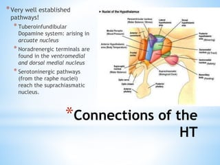
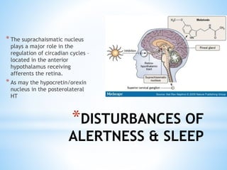
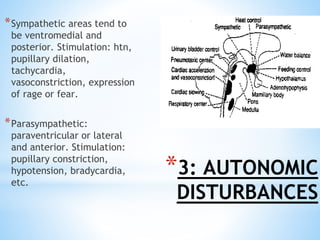
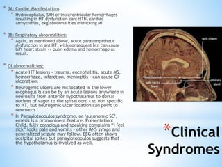
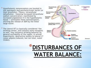
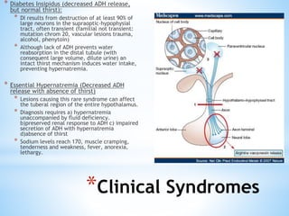
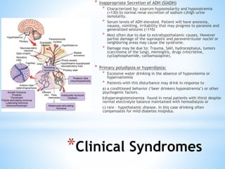
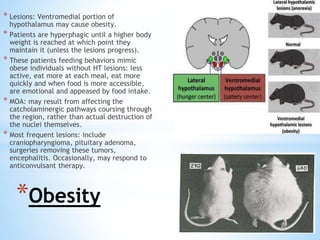
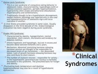
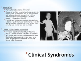
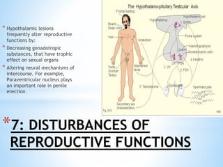
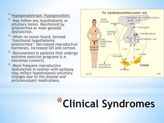
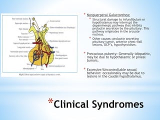
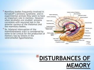
2. *Anatomy
* The hypothalamus constitutes the lateral wall of
the third ventricle.
* Walls of 3rd ventricle merge forming lamina
terminalis anteriorly.
* Lateroposteriorly borders: GP, basal forebrain,
internal capsule, crus cerebri.
* Inferior prolongation of the floor of 3rd ventricle →
pituitary stalk/infundibulum. Which joins the HT
with the pituitary gland.
3.
4. *Connections of the HT
*Still there needs to be a lot of
work done to clarify/define the
NT’s identified in the HT in
animals/humans… But what we
know (in addition to
hypophysiotropic hormones
regulating anterior pit
secretions):
* Body temperature → Bombesin,
Neurotensin
* Alertness → orexin, somatostatin
* Cardiopulmonary fxn →
thyrotropin-releasing hormone
* Reproductive fxn → oxytocin, VIP
5. *Connections of the
HT
*Very well established
pathways!
* Tuberoinfundibular
Dopamine system: arising in
arcuate nucleus
* Noradrenergic terminals are
found in the ventromedial
and dorsal medial nucleus
* Serotoninergic pathways
(from the raphe nuclei)
reach the suprachiasmatic
nucleus.
6. *DISTURBANCES OF
TEMPERATURE
REGULATION
*Hypothalamic ‘thermostat’ for
normal temp regulation:
Anterior preoptic HT area.
*However physiologic responses
for heating (vasoconstriction,
shivering, eating) and cooling
(sweating, vasodilation)
traverse post HT
*In general:
*Ant HT lesion:
hyperthermia
*Post HT lesion:
hypothermia
7. *Clinical Syndromes
*Hypothermia
*Chronic:
*posterior or entire HT
*MCC: Wernickes encephalopathy, Head
trauma, Craniopharyngioma, GBM, HCP
*Paroxysmal:
* Lesion in arcuate nucleus
* Rare syndrome; ‘spontaneous paroxysmal
hypothermia’ ‘diencephalic epilepsy’.
* Episodic abrupt decrease in temp (low as 30 c)
with sweating, dullness, n/v, decreased HR and
BP.
* May have assoc. w/ agenesis of corpus collusom
(very rarely)
8.
9. *Hyperthermia
* Pyogen Induced: Bacteria/Virus can directly
stimulate HT & also release IL-1-> acts in the
preptic, paraventricular and anterior HT.
* Acute Hyperthermia: from acute process such as
trauma, bleeding, craniotomy – anterior HT. <2
weeks
*Poikilothermia
* Fluctuation of more than 2’C, following ambient
temperature.
* Most common. Most unaware – no discomfort.
Posterior Hypothalamic lesion
*Clinical Syndromes
10. * The suprachaismatic nucleus
plays a major role in the
regulation of circadian cycles –
located in the anterior
hypothalamus receiving
afferents the retina.
* As may the hypocretin/orexin
nucleus in the posterolateral
HT
*DISTURBANCES OF
ALERTNESS & SLEEP
11. *Clinical Syndromes
*Circadian Abnormalities
*Loss of neurons in the
suprchaismatic nucleus -
Increased variability of
the sleep cycle phases
and of stability of the
circardiac rhythmicity.
*Seen in Alzheimers, and
lesions in that area,
optic glioma
* Narcolepsy
* Perifornical area of the
postero lateral HT: cluster of
cells that secrete hypcretin or
orexin - a peptide that
mediates wakefulness and
feeding behavior. A lack of
this substance causes
narcolepsy.
* speculated autoimmune
nature, patients have similar
HLA type
12. * Coma, Hypersomnia or Akinetic
Mutism: (weak)
* occasionally related to posterior
hypothalamus or larger lesions. MCC
tumors and wernicke’s
encephalopathy.
* Hypersomnia and coma result more
commonly from midbrain lesions rather
than HT lesions.
* Insomnia (weak)
* -<10 patient cases implicate
anterior HT, however some
insomniacs may have increased
production of stress-related
hormones.
*Clinical Syndromes
13. *3: AUTONOMIC
DISTURBANCES
*Sympathetic areas tend to
be ventromedial and
posterior. Stimulation: htn,
pupillary dilation,
tachycardia,
vasoconstriction, expression
of rage or fear.
*Parasympathetic:
paraventricular or lateral
and anterior. Stimulation:
pupillary constriction,
hypotension, bradycardia,
etc.
14. * 3A: Cardiac Manifestations
* Hydrocephalus, SAH or intraventricular hemorrhages
resulting in HT dysfunction can: HTN, cardiac
arrhythmias, ekg abnormalities mimicking MI.
* 3B: Respiratory abnormalities:
* Again, as mentioned above, acute parasympathetic
dysfunction in ant HT, with consequent htn can cause
left heart strain -> pulm edema and hemorrhage as
result.
* GI abnormalities:
* Acute HT lesions - trauma, encephalitis, acute MS,
hemorrhage, infarction, meningitis - can cause GI
ulceration.
* Neurogenic ulcers are mc located in the lower
esophagus & can be by an acute lesions anywhere in
neuroaxis from anterior hypothalamus to dorsal
nucleus of vagus to the spinal cord - so non specific
to HT, but neurogenic ulcer location can point to
neuroaxis
* In Panayiotopoulos syndrome, or ‘autonomic SE’,
emesis is a promoninent feature. Presentation:
Child, fully conscious and speaking complains “i feel
sick” looks pale and vomits - other ANS symps and
generalized seizure may follow. EEG often shows
occipital spikes but panayiotopoulos suggests that
the hypothalamus is involved as well.
*Clinical
Syndromes
15. * Diencephalic Epilepsy
episodes of htn, tachycardia, flushing,
salivation, sweating and fluctuations of
temperature with preserved alertness. And
though autonomic disturbances are common
in many seizure types, this clinical picture
has been seen in 3rd ventricle tumors or
dilation.
EEG may be abnormal with slowing in half
the cases, but most do not show paroxysmal
dysrhythmias characteristic of most forms of
epilepsy.
Half patients do respond to AEDs.
* Unilateral anhidrosis or hyperhidrosis
Unilateral hypothalamic lesions may cause
ipsilateral anhidrosis of the body, which is
generally incomplete. An ipsilateral Horner
syndrome is often present. Dysfunction of
the sympathetic fibers in the posterior HT
may be responsible.
*Clinical Syndromes
16. *DISTURBANCES OF
WATER BALANCE:
* Hypothalamic osmoreceptors are located in
the supraoptic and paraventricular nuclei (or
their proximity). Theory: Intracellular
dehydration - meaning increased intracellular
sodium concentration, manifested by
increased angiotensin II in the hypothalamic
blood -> stimulates these osmoreceptors,
eliciting the release of ADH by the these
large nuclei.
* The lateral HT is classically considered ‘the
drinking center’ and contains osmo receptors
as well, may influence drinking behavior by
general excitability of the region. In animal
experiments, destructive lesions to lateral LT
cause adipsia (reduced, but not total, water
intake).
17. * Diabetes Insipidus (decreased ADH release,
but normal thirst):
* DI results from destruction of at least 90% of
large neurons in the supraoptic-hypophysial
tract, often transient (familial not transient:
mutation chrom 20, vascular lesions trauma,
alcohol, phenytoin)
* Although lack of ADH prevents water
reabsorption in the distal tubule (with
consequent large volume, dilute urine) an
intact thirst mechanism induces water intake,
preventing hypernatremia.
* Essential Hypernatremia (Decreased ADH
release with absence of thirst)
* Lesions causing this rare syndrome can affect
the tuberal region of the entire hypothalamus.
* Diagnosis requires a) hypernatremia
unaccompanied by fluid deficiency.
b)preserved renal response to ADH c) impaired
secretion of ADH with hypernatremia
d)absence of thirst
* Sodium levels reach 170, muscle cramping,
tenderness and weakness, fever, anorexia,
lethargy.
*Clinical Syndromes
18. * Inappropriate Secretion of ADH (SIADH)
* Characterized by: a)serum hyposmolarity and hyponatremia
(<130) b) normal renal excretion of sodium c)high urine
osmolality.
* Serum levels of ADH elevated. Patient will have anorexia,
nausea, vomiting, irritability that may progress to paranoia and
generalized seizures (<110)
* Most often due to due to extrahypothalamic causes. However
partial damage of the supraoptic and paraventricular nuclei or
neighboring areas may cause the syndrome.
* Damage may be due to: Trauma, SAH, hydrocephalus, tumors
(carcinoma of the lung), meningitis, drugs (vincristine,
cyclophosphamide, carbamazapine).
* Primary polydipsia or hyperdipsia:
* Excessive water drinking in the absence of hypovolemia or
hypernatremia
* Patients with this disturbance may drink in response to
a) a conditioned behavior (‘beer drinkers hyponatremia’) or other
psychogenic factors.
b)hyperangiotensinemia –found in renal patients with thirst despite
normal electrolyte balance maintained with hemodialysis or
c) rare – hypothalamic disease. In this case drinking often
compensates for mild diabetes insipidus.
*Clinical Syndromes
19. * Lesions: Ventromedial portion of
hypothalamus may cause obesity.
* Patients are hyperphagic until a higher body
weight is reached at which point they
maintain it (unless the lesions progress).
* These patients feeding behaviors mimic
obese individuals without HT lesions: less
active, eat more at each meal, eat more
quickly and when food is more accessible,
are emotional and appeased by food intake.
* MOA: may result from affecting the
catcholaminergic pathways coursing through
the region, rather than actual destruction of
the nuclei themselves.
* Most frequent lesions: include
craniopharyngioma, pituitary adenoma,
surgeries removing these tumors,
encephalitis. Occasionally, may respond to
anticonvulsant therapy.
*Obesity
20. * Kleine-Levin Syndrome:
* This is a rare syndrome of compulsive eating behavior in
adolescent males, characterized by: hyperphagia (with or
without excessive appetite), periodic hypersomnolence,
hyperactivity when awake and behavioral disturbances
(hypersexuality and exhibitionism).
* Traditionally thought to be a hypothalamic derangement,
medial thalamic pathology was reported only in one case
and hypopigmentation of substantia nigra and locus
coeruleus in another.
* -A viral illness preceded syndrome in some cases. Usually
disappears in the third decade of life. Endocrinological
evaluation has been negative during acute syndrome.
* Prader-Wili Syndrome:
* Characterized by obesity, hypogenitalism, mental
retardation, short stature, micromelia and tendency for
diabetes mellitus.
* Infants hypotonic and hypophagic, but eat in excess and
become obese between 6months and 2 years.
* Mechanism: Abnormal luteinizing hormone-releasing
hormone neurons are thought to be responsible for
decreased levels of sex horomones → non descended
testes. Lack of growth hormone-releasing hormone may
also contribute to short stature.
* The number of oxytocin neurons - responsible for satiety -
in the hypothalamic paraventricular nucleus is markedly
decreased in this syndrome. The presumed basis for hunger
and obesity.
* -Fluctuating body temperature and daytime
hypersomnolense may also be a result of hypothalamic
dysfunction.
*Clinical
Syndromes
21. * Emaciation
* Diencephalic Syndrome of Infancy
* Characterized by: emaciation (progessive with
normal food intake) with loss of subcutaneous
fat, pallor and motor overactivity with an
inappropriately jovial behavior. Usually
appears in boys aged 3 to 12.
* Caused by a slow growing astrocytoma of the
anterior hypothalamus or optic nerve. Growth
hormone levels may be high. Nystagmus, optic
atrophy and tremor are less common.
* Lateral Hypothalamic Syndrome:
* Few case reports of lateral hypothalamic
lesions causing aphagia, cachexia, and death.
* MS, tumors and trauma have been implicated.
Huntington’s chorea and weight losee have
been associated with neuronal los sin the
lateral tuberal nucleus.
*Clinical Syndromes
22. * Hypothalamic lesions
frequently alter reproductive
functions by:
* Decreasing gonadotropic
substances, that have trophic
effect on sexual organs
* Altering neural mechanisms of
intercourse. For example,
Paraventricular nucleus plays
an important role in penile
erection.
*7: DISTURBANCES OF
REPRODUCTIVE FUNCTIONS
23. * Hypogonadotropic Hypogonadism:
* -May follow any hypothalamic or
pituitary lesion. Manifested by
amenorrhea or male gonadal
dysfunction.
* -When no lesion found, termed
‘functional hypothalamic
amenorrhea’. Decreased reproductive
hormones, increased GH and cortisol.
* -Documented in patients that undergo
extreme exercise programs (i.e.
marathon runners).
* -More frequent reproductive
dysfunction in women with epilepsy
may reflect hypothalamic-pituitary
changes due to the disease and
anticonvulsant medications.
*Clinical Syndromes
24. * Nonpuerperal Galactorrhea:
* Structural damage to infundibulum or
hypothalamus may interrupt the
dopaminergic pathway that inhibits
prolactin secretion by the pituitary. This
pathway originates in the arcuate
nucleus.
* Other causes: prolactin secreting
pituitary tumor, anterior chest wall
lesions, OCP’s, hypothyroidism.
* Precocious puberty: Generally idiopathic,
may be due to hypothalamic or pineal
tumors.
* Excessive/Uncontrollable sexual
behavior: occasionally may be due to
lesions in the caudal hypothalamus.
*Clinical Syndromes
25. *DISTURBANCES OF
MEMORY
* Mamillary bodies frequently involved in
Korsakoff’s amnestic syndrome, and in
experimental animals they seem to have
an important role in memory. However
when alcoholics are studied, amnesia
correlates with neuronal loss in the
anterior nucleus of the thalamus (not
the mamillary bodies.
* So, bilateral interruption of the
mamillothalamic tract is considered by
some to be critical for the production of
memory loss by lesions in the
ventromedial hypothalamus.
27. * Rage and fear: inappropriately
disinhibited behavior
* When caused by hypothalamic
lesions, usually ventromedial
region (as opposed to posterior
‘sympathetic’ area → fear and
horror).
* episodic outbursts of rage and fear,
triggered by delayed or frustrating
event (ex: feeding), and
coordinated with an intense
autonomic component.
* (delete disinhibited rage, similar to
orbitofrontal cortex lesions)
* Behavior normal between outbursts.
* May realize behavior is
inappropriate (will apologize)
*Apathy, Chronic Fatigue
*May follow a lesion of
posterior or lateral
hypothalamaus.
*Bilateral stereotactic
lesions in this area can
treat unmanageable
aggressive behavior
caused by brain
pathology
*DISTURBANCES OF EMOTIONAL
BEHAVIOR AND AFFECT
28. * Depression
* -No evidence of hypothalamic
dysfunction causes depression -
however, depressed patient have
excessively active Hypothalamic-
Pituitary-Adrenal axis.
* -Post-mortem studies after suicides:
shown increased corticotropin
releasing hormone neurons and
increased vasporessin expression in
the paraventricular nucleus. As well
as increased CRH in csf and
decreased receptors in the frontal
cortex.
* Gelastic Seizures
* -seizures characterized by involuntary
laughter.
* The patient may stare and giggle briefly
with or without any other motor
manifestations. Crying/sobbing may
alternate with gelastic spells.
* The patient may also have an internal
pressure or feeling to laugh without overt
manifestation.
* -Hypothalamic hamartomas are most
common cause and origin. Though cortical
displasia in cingulate gyrus has been seen.
* -Associated with prococious puberty
*DISTURBANCES OF
EMOTIONAL BEHAVIOR
AND AFFECT
29. *HEADACHE
* 1/7 patients with pituitary tumor
present with headache. Usually
bitemporal, bifrontal or behind the
eyes. Due to compression of the
diagphragma sella.
* -More frequent with pituitary
abscesses rather than with tumors.
* Episodic Headache:
* -Increased perfusion (PET) in
posterior-medial hypothalamus, dorsal
to mamillary bodies, has some
association with cluster HA’s,
paroxysmal hemicrania or SUNCT
(short-lasting unilateral headache
attacks with conjunctival injection
and tearing).
* -Deep brain stimulation shown some
improvement in these patients
30. *IMPAIRED VISUAL
ACUITY; VISUAL
FIELD DEFECTS
* Pituitary Adenoma: Some patients
present with visual complaints, but 15%
of patients on formal testing have visual
field defects or decreased acuity.
* Bitemporal defects are most common -
related to compression of the inferior
aspect of the chiasm.
* However asymmetric tumor growth may
result in unilateral blindness, or
complete homymous hemianopia
(compression of the optic tract)
* Position of the chiasm in relation to the
sella also determines the visual field
defect.
* -80% over the sella, 9% over tuberculum
sellae, 11% dosrsum sellae