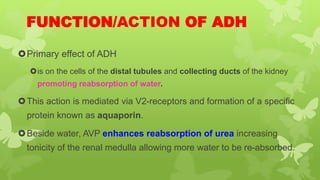
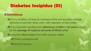
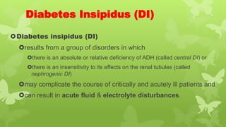
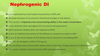
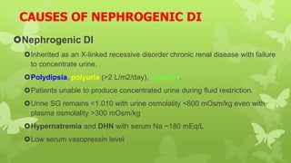
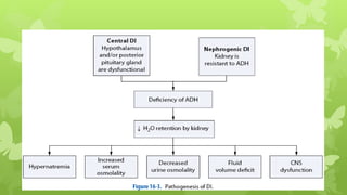
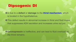
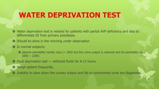
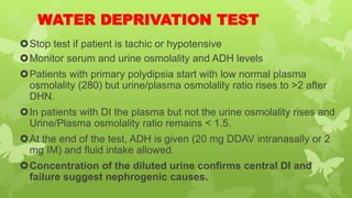
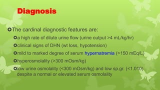
Emergency Lecture Series
Disorders of the Pituitary
By Ame M. (BSc, MSc in EMCC)
Hypothalamus and Pituitary
The hypothalamus-pituitary unit
is the most dominant portion of the entire endocrine system.
The output of the hypothalamus – pituitary unit:
regulates the function of the
thyroid,
adrenal and
reproductive glands
controls
somatic growth,
lactation,
milk secretion and
water metabolism.
Hypothalamus and Pituitary…..
 Pituitary function depends on the hypothalamus
The pituitary gland
lies in a pocket of bone at the base of the brain, just below the hypothalamus to
which it is connected by a stalk containing nerve fibers and blood vessels.
lies at the base of the skull in the sellaturcica, within the sphenoid bone.
weighs 500-900 mg
composed of two lobes
Anterior pituitary
Posterior Pituitary
Anatomically and functionally anterior and posterior lobes are distinct structures.
Pituitary Development
The pituitary originate from different source.
The anterior pituitary from Rathke´s pouch (which is an
embryonic invagination of the pharyngeal epithelium).
The posterior pituitary from an outgrow of the hypothalamus.
Pituitary Gland
1. The anterior lobe
consists 2/3 of the gland
originate from Rathke´s pouch (which is an
embryonic invagination of the pharyngeal epithelium).
Secretes
A) Adrenocortico Tropic Hormone (ACTH)
B) Growth hormone (GH)
C) Prolactin (Prl)
D) Thyroid stimulating hormone (TSH)
E) Follicle stimulating hormone (FSH)
F) Luteinizing Hormone(LH)
Posterior Pituitary/Lobe
2. Posterior Pituitary/Lobe
consists of neural tissue &
is an extension of hypothalamus.
originate from an outgrow of the hypothalamus
is composed of nerve fibers that have their cell bodies in the supraoptic and
paraventricular nuclei of the hypothalamus.
The neurosecretory cells in these nuclei synthesize Oxytocin and Vasopressin
which pass down the nerve fibers to be stored in and released from the
posterior pituitary.
Posterior Pituitary/Lobe
secretes –
Antidiuretic hormone (ADH) also called arginine vasopressin
(AVP) &
regulates water metabolism
Oxytocin
controls
lactation or milk ejection from breasts
uterine contraction
ADH
 The supraoptic nucleus (SON) is responsible predominantly for the synthesis of
vasopressin which is the ADH.
 The close structural similarity of vasopressin and oxytocin explains the overlap of
their biological actions.
 ADH is an octapeptide like oxytocin.
 The Arginine Vasopressin (AVP) is ADH in man and other mammals apart from the
pig and the hippopotamus where Lysine Vasopressin is the ADH.
FUNCTION/ACTION OF ADH
Primary effect of ADH
is on the cells of the distal tubules and collecting ducts of the kidney
promoting reabsorption of water.
This action is mediated via V2-receptors and formation of a specific
protein known as aquaporin.
Beside water, AVP enhances reabsorption of urea increasing
tonicity of the renal medulla allowing more water to be re-absorbed.
FUNCTION……
 Acting on V1-receptors in peripheral vessels AVP causes
vaso-constriction and increase in BP.
 Normally this is balanced by its inhibitory effect on sympathetic cardiac stimuli
causing bradycardia.
 During hypovolemia high plasma levels of AVP help maintain tissue perfusion.
 A lesser 2ndry effect that is mediated via V2 non-renal receptors is
stimulation of synthesis and release of factor VIII & Von Willebrand Factor.
REGULATION OF ADH SECRETION
ADH release is stimulated by:
A plasma osmolality >280 mosm/L
A fall in plasma volume
Emotional factors & stress
Sleep
Other factors
 Other ADH Stimulants
 Cholinergic stimulation
 -adrenergic stimulation
 Angiotensin II
 Prostaglandin E
 Opiates
 Nicotine
 Histamine
 Ether
 Phenobarbitone
ADH SECRETION IS INHIBITED BY:
Alcohol
Oropharyngeal water reflex
β-adrenergic stimulants
Atrial natriuretic factor (ANF)
Phenytoin
Disorders of the Posterior Pituitary
Outline
Diabetes Insipidus
Definitions
Causes
Types
Ass’t and Dx
Mgt
SIADH
Definitions
Causes
Dx
Mgt
Diabetes Insipidus (DI)
Definitions
DI is a condition ch’rized by excessive thirst and excretion of large
amounts of severely dilute urine, with reduction of fluid intake.
DI is a disorder resulting from deficiency of ADH or its action ch’zed
by the passage of copious amounts of dilute urine
It must be differentiated from other polyuric states
Primary polydipsia and
Osmotic diuresis.
Diabetes Insipidus (DI)
Diabetes insipidus (DI)
results from a group of disorders in which
there is an absolute or relative deficiency of ADH (called central DI) or
there is an insensitivity to its effects on the renal tubules (called
nephrogenic DI)
may complicate the course of critically and acutely ill patients and
can result in acute fluid & electrolyte disturbances.
DI …
Classification
Different types of DI, each with a different cause.
Neurogenic/Central DI: most common
Nephrogenic DI: second most common
Dipsogenic DI
Gestational DI
Neurogenic DI
more commonly known as central DI,
caused by a deficiency of Arginine vasopressin(AVP)
AVP also known as ADH.
ADH-deficient
is due to a lack of vasopressin production in the brain
is due to failure of the pituitary gland to secrete adequate
ADH.
secondary to Panhypopituitarism
Etiology of Central DI
 Idiopathic (30% of cases)
 Suprasellar tumours (30% of cases)
 Hypothalamic or pituitary tumors —
craniopharyngioma, germinoma,
histiocytosis, glioma,
lymphocytic hypophysitis.
 Autoimmune
Associated with thyroiditis
Antibodies target ADH-producing cells.
Familial (hereditary): 2 types AD and X-linked inheritance
Genetic (autosomal dominant inheritance)
Genetic — mutations in vasopressin gene and WFS1 gene (Wolfram syndrome)
 Midline brain defects — septo-optic dysplasia, holoprosencephaly
Etiology of Central DI…..
Neoplasms
Infections
Tuberculosis
Cryptococcosis
Syphilis
CNS infections
 Granulomatous diseases
Infectious Granulomatous Ds (e.g., sarcoidosis, TB)
Non-infectious granuloma e.g. Sarcoid, hand-schuller Christian disease
(histiocytosis)
 Langerhans cell histiocytosis – in which 25% of pts develop DI.
Etiology of Central DI……
 Brain injury, infection, or surgical resection
 Infections (Encephalitis, meningitis, TB, etc)
 Trauma and hypoxic ischemic brain injury
 Trauma or skull surgery
basilar skull fractures,
neurosurgical complications
 Leukemia
Vascular Ds (e.g., aneurysms, lesions)
 Cerebrovascular hemorrhage
 Aneurysm (circle of Willis)
 Cerebral thrombosis
 Wolfram syndrome (also known as DIDMOAD syndrome) ch’zed by
 DI, DM, Nerve Deafness and Optic Atrophy.
Nephrogenic DI
 is characterized by renal tubule insensitivity to ADH and
 develops because of structural or functional changes in the kidney.
 This results in impaired urine-concentrating ability & free water conservation.
 is less dramatic than neurogenic DI in its onset and appearance.
 ADH-resistance (kidney does not respond to ADH)
 is due to inability/insensitivity of the kidneys to respond normally to ADH
 is due to the renal tubules of the kidneys fail to respond to circulating ADH.
 can also an iatrogenic artifact of drug use
 The resulting renal concentration defect leads to the loss of large volumes of dilute urine.
 This causes cellular and extracellular DHN and hypernatremia.
Causes of Nephrogenic DI
 Primary Familial (hereditary):
 X-linked recessive that is severe in boys and
mild in girls.
 Secondary to:
Renal disease
chronic pyelonephritis
chronic renal failure,
obstructive uropathy,
polycystic disease
Electrolyte disorders
Hypokalemia
Hypercalcemia
Sickle cell disease
Protein deprivation
Amyloidosis
Drugs:
Lithium, Colchicine, Fluoride,
Rifampin, Cidofovir, Demeclocycline
Methoyflurane, Cisplatin, Methicillin
Amphotericin B, Gentamicin,
Furosemide
CAUSES OF NEPHROGENIC DI
Nephrogenic DI
Inherited as an X-linked recessive disorder chronic renal disease with failure
to concentrate urine.
Polydipsia, polyuria (>2 L/m2/day), nocturia.
Patients unable to produce concentrated urine during fluid restriction.
Urine SG remains <1.010 with urine osmolality <600 mOsm/kg even with
plasma osmolality >300 mOsm/kg
Hypernatremia and DHN with serum Na ~180 mEq/L
Low serum vasopressin level
Etiology …
Excessive Water Intake (Secondary DI)
Excessive IV fluid administration
Psychogenic polydipsia (lesion in thirst center)
Dipsogenic DI
is due to a defect or damage to the thirst mechanism, which
is located in the hypothalamus.
This defect results in abnormal increase in thirst and fluid intake
that suppresses ADH secretion and increases urine out put.
Desmopressin is ineffective, and can lead to fluid overload as
the thirst remains.
Gestational DI
only occurs during pregnancy.
While all pregnant women produce vasopressinase in
the placenta, which breaks down ADH, this can assume
extreme forms in GDI.
CLINICAL FEATURES
 Polyuria, polydipsia & thirst
 Nocturia or nocturnal enuresis
 Hypernatremic DHN
 Anorexia, constipation and FTT
 Hyperthermia and lack of sweating
 Symptoms of underlying cause
Abnormalities of the CNS such as:
irritability, altered consciousness, increased muscle tone, convulsions, and coma occur
secondary to hypernatremia.
these findings correlate with the degree and rapidity of the rise in serum Na.
COMPLICATIONS
Hypernatremic DHN and its neurological sequelea
Growth retardation
Hydronephrosis (due to excessive urine output)
Differential Diagnosis
Psychogenic polydipsia
Osmotic diuresis
Diabetes mellitus
Beer potomania
Hypertonic DHN secondary to diarrhea
Exogenous salt administration
DIAGNOSTIC WORKUP
Careful history and examination
U/A and microscopy together with plasma electrolytes help
exclude most of the causes of polyuria.
Water deprivation test
Measuring glucose, sodium, and osmolality in urine and
serum can quickly distinguish DM, hypertonic DHN,
psychogenic polydipsia, and exogenous salt intake from
DI.
DIAGNOSTIC WORKUP
Polyuria >2000ml/day
 Document presence of polyuria (usually 4-15 L/24hrs)
Urinary sp. gr. of 1.001-1.005
Increased serum osmolality-Na
In a normal well hydrated subject:
plasma osmolality is <290 mosml/L and
urine osmolality is 300-450 mosmol/L.
In patients with DI and free excess to water:
plasma osmolality is >295 mosmol/L &
urine osmolality is 50-150 mosmol/L.
WATER DEPRIVATION TEST
 Water deprivation test is needed for patients with partial AVP deficiency and also to
differentiate DI from primary polydipsia.
 Should be done in the morning under observation
 In normal subjects:
 plasma osmolality hardly rises (< 300) but the urine output is reduced and its osmolality rises
(800 – 1200)
 Fluid deprivation test — withhold fluids for 8-12 hours.
 Weigh patient frequently.
 Inability to slow down the urinary output and fail to concentrate urine are diagnostic.
WATER DEPRIVATION TEST
Stop test if patient is tachic or hypotensive
Monitor serum and urine osmolality and ADH levels
Patients with primary polydipsia start with low normal plasma
osmolality (280) but urine/plasma osmolality ratio rises to >2 after
DHN.
In patients with DI the plasma but not the urine osmolality rises and
Urine/Plasma osmolality ratio remains < 1.5.
At the end of the test, ADH is given (20 mg DDAV intranasally or 2
mg IM) and fluid intake allowed.
Concentration of the diluted urine confirms central DI and
failure suggest nephrogenic causes.
Diagnosis
The cardinal diagnostic features are:
a high rate of dilute urine flow (urine output >4 mL/kg/hr)
clinical signs of DHN (wt loss, hypotension)
mild to marked degree of serum hypernatremia (>150 mEq/L)
hyperosmolality (>300 mOsm/kg)
low urine osmolality (<300 mOsm/kg) and low sp.gr. (<1.010)
despite a normal or elevated serum osmolality
Management
Identify treatable causes of DI
The DOC for central DI is DDAVP (synthetic ADH)
Desmopressin acetate (DDAVP), an ADH analogue,
can be given intranasally, SQ, IV, IM or po to stimulate
the kidneys to retain water and reverse the polyuria,
polydipsia, and hypernatremia.
Management…….
 Desmopressin (DDAVP) a synthetic analog is superior to native AVP b/c:
it has longer duration of action (8-10 h vs 2-3 h)
more potent
its antidiuretic activity is 3000 times greater than its presser activity.
If renal in origin,—thiazide diuretics, NSAIDs (prostaglandin
inhibition) and salt depletion may help.
Educate patient about actions of medications, how to administer
meds, wear medical alert bracelet.
TREATMENT OF NEPHROGENIC DI
Provision of adequate fluids and calorie
Low sodium diet
Diuretics
High dose of DDAVP
Correction of underlying cause
DRUGS:
Indomethacin,
Chlorpropramide,
Clofibrate and
Carbamazepine.
Demeclocycline and other tetracyclines cause permanent if subtle nephrogenic DI; be
cautious using them for acne during puberty.
?
SIADH
Excessive ADH secretion
Retain fluids and develop a dilutional hyponatremia known as
syndrome of hyponatremia with inappropriate increased
secretion of Vasopressin (SIADH)
can be identified in an individuals with encephalitis, brain tumors,
head trauma, or psychiatric disease.
SIADH
Causes:
Disorders of the CNS like head injury, brain surgery, tumor or infections.
Many drugs are capable of interfering with free water clearance
(lisinopril, vincristine, phenothiazine's, TCAs, carbamazepine, thiazide
diuretics and others).
can either affect the pituitary or increase sensitivity to renal tubules
to ADH.
Often non-endocrine in origin — such as bronchogenic carcinoma.
SIADH
Clinical Manifestations
Patients present with normovolemic hyponatremia, relatively
concentrated urine, and normal renal, thyroid, and adrenal
function.
Symptoms are related to the degree of hyponatremia and how
rapid the hyponatremia progressed.
symptoms unlikely with a Na >125 mEq/L.
Headache, nausea, lethargy, and other CNS findings may occur
when sodium falls ≤125 mEq/L.
SIADH
Management:
eliminate cause,
give diuretics (Lasix),
fluid restriction,
monitor in put and out put
daily wt.,
lab chemistries
Restoration of electrolytes must be gradual
May use 3% NaCl in conjunction with Lasix
SIADH
Management
SIADH is a diagnosis of exclusion.
Other causes of hyponatremia must be ruled out (hyperglycemia,
increased serum lipids or protein).
A serum osmolality <280mOsm/kg combined with urine osmolality
>200mOsm and urine Na+ concentration >20mEq/L are consistent with
SIADH.
The patient should appear euvolemic.
Most cases of SIADH are self-limited, and the mode of mgt is fluid
restriction.
The END

Diabetes Insipidus

  • 1.
    Emergency Lecture Series Disordersof the Pituitary By Ame M. (BSc, MSc in EMCC)
  • 2.
    Hypothalamus and Pituitary Thehypothalamus-pituitary unit is the most dominant portion of the entire endocrine system. The output of the hypothalamus – pituitary unit: regulates the function of the thyroid, adrenal and reproductive glands controls somatic growth, lactation, milk secretion and water metabolism.
  • 3.
    Hypothalamus and Pituitary….. Pituitary function depends on the hypothalamus The pituitary gland lies in a pocket of bone at the base of the brain, just below the hypothalamus to which it is connected by a stalk containing nerve fibers and blood vessels. lies at the base of the skull in the sellaturcica, within the sphenoid bone. weighs 500-900 mg composed of two lobes Anterior pituitary Posterior Pituitary Anatomically and functionally anterior and posterior lobes are distinct structures.
  • 4.
    Pituitary Development The pituitaryoriginate from different source. The anterior pituitary from Rathke´s pouch (which is an embryonic invagination of the pharyngeal epithelium). The posterior pituitary from an outgrow of the hypothalamus.
  • 5.
    Pituitary Gland 1. Theanterior lobe consists 2/3 of the gland originate from Rathke´s pouch (which is an embryonic invagination of the pharyngeal epithelium). Secretes A) Adrenocortico Tropic Hormone (ACTH) B) Growth hormone (GH) C) Prolactin (Prl) D) Thyroid stimulating hormone (TSH) E) Follicle stimulating hormone (FSH) F) Luteinizing Hormone(LH)
  • 6.
    Posterior Pituitary/Lobe 2. PosteriorPituitary/Lobe consists of neural tissue & is an extension of hypothalamus. originate from an outgrow of the hypothalamus is composed of nerve fibers that have their cell bodies in the supraoptic and paraventricular nuclei of the hypothalamus. The neurosecretory cells in these nuclei synthesize Oxytocin and Vasopressin which pass down the nerve fibers to be stored in and released from the posterior pituitary.
  • 7.
    Posterior Pituitary/Lobe secretes – Antidiuretichormone (ADH) also called arginine vasopressin (AVP) & regulates water metabolism Oxytocin controls lactation or milk ejection from breasts uterine contraction
  • 8.
    ADH  The supraopticnucleus (SON) is responsible predominantly for the synthesis of vasopressin which is the ADH.  The close structural similarity of vasopressin and oxytocin explains the overlap of their biological actions.  ADH is an octapeptide like oxytocin.  The Arginine Vasopressin (AVP) is ADH in man and other mammals apart from the pig and the hippopotamus where Lysine Vasopressin is the ADH.
  • 9.
    FUNCTION/ACTION OF ADH Primaryeffect of ADH is on the cells of the distal tubules and collecting ducts of the kidney promoting reabsorption of water. This action is mediated via V2-receptors and formation of a specific protein known as aquaporin. Beside water, AVP enhances reabsorption of urea increasing tonicity of the renal medulla allowing more water to be re-absorbed.
  • 10.
    FUNCTION……  Acting onV1-receptors in peripheral vessels AVP causes vaso-constriction and increase in BP.  Normally this is balanced by its inhibitory effect on sympathetic cardiac stimuli causing bradycardia.  During hypovolemia high plasma levels of AVP help maintain tissue perfusion.  A lesser 2ndry effect that is mediated via V2 non-renal receptors is stimulation of synthesis and release of factor VIII & Von Willebrand Factor.
  • 11.
    REGULATION OF ADHSECRETION ADH release is stimulated by: A plasma osmolality >280 mosm/L A fall in plasma volume Emotional factors & stress Sleep Other factors  Other ADH Stimulants  Cholinergic stimulation  -adrenergic stimulation  Angiotensin II  Prostaglandin E  Opiates  Nicotine  Histamine  Ether  Phenobarbitone
  • 12.
    ADH SECRETION ISINHIBITED BY: Alcohol Oropharyngeal water reflex β-adrenergic stimulants Atrial natriuretic factor (ANF) Phenytoin
  • 13.
    Disorders of thePosterior Pituitary
  • 14.
    Outline Diabetes Insipidus Definitions Causes Types Ass’t andDx Mgt SIADH Definitions Causes Dx Mgt
  • 15.
    Diabetes Insipidus (DI) Definitions DIis a condition ch’rized by excessive thirst and excretion of large amounts of severely dilute urine, with reduction of fluid intake. DI is a disorder resulting from deficiency of ADH or its action ch’zed by the passage of copious amounts of dilute urine It must be differentiated from other polyuric states Primary polydipsia and Osmotic diuresis.
  • 16.
    Diabetes Insipidus (DI) Diabetesinsipidus (DI) results from a group of disorders in which there is an absolute or relative deficiency of ADH (called central DI) or there is an insensitivity to its effects on the renal tubules (called nephrogenic DI) may complicate the course of critically and acutely ill patients and can result in acute fluid & electrolyte disturbances.
  • 17.
    DI … Classification Different typesof DI, each with a different cause. Neurogenic/Central DI: most common Nephrogenic DI: second most common Dipsogenic DI Gestational DI
  • 18.
    Neurogenic DI more commonlyknown as central DI, caused by a deficiency of Arginine vasopressin(AVP) AVP also known as ADH. ADH-deficient is due to a lack of vasopressin production in the brain is due to failure of the pituitary gland to secrete adequate ADH. secondary to Panhypopituitarism
  • 19.
    Etiology of CentralDI  Idiopathic (30% of cases)  Suprasellar tumours (30% of cases)  Hypothalamic or pituitary tumors — craniopharyngioma, germinoma, histiocytosis, glioma, lymphocytic hypophysitis.  Autoimmune Associated with thyroiditis Antibodies target ADH-producing cells. Familial (hereditary): 2 types AD and X-linked inheritance Genetic (autosomal dominant inheritance) Genetic — mutations in vasopressin gene and WFS1 gene (Wolfram syndrome)  Midline brain defects — septo-optic dysplasia, holoprosencephaly
  • 20.
    Etiology of CentralDI….. Neoplasms Infections Tuberculosis Cryptococcosis Syphilis CNS infections  Granulomatous diseases Infectious Granulomatous Ds (e.g., sarcoidosis, TB) Non-infectious granuloma e.g. Sarcoid, hand-schuller Christian disease (histiocytosis)  Langerhans cell histiocytosis – in which 25% of pts develop DI.
  • 21.
    Etiology of CentralDI……  Brain injury, infection, or surgical resection  Infections (Encephalitis, meningitis, TB, etc)  Trauma and hypoxic ischemic brain injury  Trauma or skull surgery basilar skull fractures, neurosurgical complications  Leukemia Vascular Ds (e.g., aneurysms, lesions)  Cerebrovascular hemorrhage  Aneurysm (circle of Willis)  Cerebral thrombosis  Wolfram syndrome (also known as DIDMOAD syndrome) ch’zed by  DI, DM, Nerve Deafness and Optic Atrophy.
  • 22.
    Nephrogenic DI  ischaracterized by renal tubule insensitivity to ADH and  develops because of structural or functional changes in the kidney.  This results in impaired urine-concentrating ability & free water conservation.  is less dramatic than neurogenic DI in its onset and appearance.  ADH-resistance (kidney does not respond to ADH)  is due to inability/insensitivity of the kidneys to respond normally to ADH  is due to the renal tubules of the kidneys fail to respond to circulating ADH.  can also an iatrogenic artifact of drug use  The resulting renal concentration defect leads to the loss of large volumes of dilute urine.  This causes cellular and extracellular DHN and hypernatremia.
  • 23.
    Causes of NephrogenicDI  Primary Familial (hereditary):  X-linked recessive that is severe in boys and mild in girls.  Secondary to: Renal disease chronic pyelonephritis chronic renal failure, obstructive uropathy, polycystic disease Electrolyte disorders Hypokalemia Hypercalcemia Sickle cell disease Protein deprivation Amyloidosis Drugs: Lithium, Colchicine, Fluoride, Rifampin, Cidofovir, Demeclocycline Methoyflurane, Cisplatin, Methicillin Amphotericin B, Gentamicin, Furosemide
  • 24.
    CAUSES OF NEPHROGENICDI Nephrogenic DI Inherited as an X-linked recessive disorder chronic renal disease with failure to concentrate urine. Polydipsia, polyuria (>2 L/m2/day), nocturia. Patients unable to produce concentrated urine during fluid restriction. Urine SG remains <1.010 with urine osmolality <600 mOsm/kg even with plasma osmolality >300 mOsm/kg Hypernatremia and DHN with serum Na ~180 mEq/L Low serum vasopressin level
  • 26.
    Etiology … Excessive WaterIntake (Secondary DI) Excessive IV fluid administration Psychogenic polydipsia (lesion in thirst center)
  • 27.
    Dipsogenic DI is dueto a defect or damage to the thirst mechanism, which is located in the hypothalamus. This defect results in abnormal increase in thirst and fluid intake that suppresses ADH secretion and increases urine out put. Desmopressin is ineffective, and can lead to fluid overload as the thirst remains.
  • 28.
    Gestational DI only occursduring pregnancy. While all pregnant women produce vasopressinase in the placenta, which breaks down ADH, this can assume extreme forms in GDI.
  • 29.
    CLINICAL FEATURES  Polyuria,polydipsia & thirst  Nocturia or nocturnal enuresis  Hypernatremic DHN  Anorexia, constipation and FTT  Hyperthermia and lack of sweating  Symptoms of underlying cause Abnormalities of the CNS such as: irritability, altered consciousness, increased muscle tone, convulsions, and coma occur secondary to hypernatremia. these findings correlate with the degree and rapidity of the rise in serum Na.
  • 30.
    COMPLICATIONS Hypernatremic DHN andits neurological sequelea Growth retardation Hydronephrosis (due to excessive urine output)
  • 31.
    Differential Diagnosis Psychogenic polydipsia Osmoticdiuresis Diabetes mellitus Beer potomania Hypertonic DHN secondary to diarrhea Exogenous salt administration
  • 32.
    DIAGNOSTIC WORKUP Careful historyand examination U/A and microscopy together with plasma electrolytes help exclude most of the causes of polyuria. Water deprivation test Measuring glucose, sodium, and osmolality in urine and serum can quickly distinguish DM, hypertonic DHN, psychogenic polydipsia, and exogenous salt intake from DI.
  • 33.
    DIAGNOSTIC WORKUP Polyuria >2000ml/day Document presence of polyuria (usually 4-15 L/24hrs) Urinary sp. gr. of 1.001-1.005 Increased serum osmolality-Na In a normal well hydrated subject: plasma osmolality is <290 mosml/L and urine osmolality is 300-450 mosmol/L. In patients with DI and free excess to water: plasma osmolality is >295 mosmol/L & urine osmolality is 50-150 mosmol/L.
  • 34.
    WATER DEPRIVATION TEST Water deprivation test is needed for patients with partial AVP deficiency and also to differentiate DI from primary polydipsia.  Should be done in the morning under observation  In normal subjects:  plasma osmolality hardly rises (< 300) but the urine output is reduced and its osmolality rises (800 – 1200)  Fluid deprivation test — withhold fluids for 8-12 hours.  Weigh patient frequently.  Inability to slow down the urinary output and fail to concentrate urine are diagnostic.
  • 35.
    WATER DEPRIVATION TEST Stoptest if patient is tachic or hypotensive Monitor serum and urine osmolality and ADH levels Patients with primary polydipsia start with low normal plasma osmolality (280) but urine/plasma osmolality ratio rises to >2 after DHN. In patients with DI the plasma but not the urine osmolality rises and Urine/Plasma osmolality ratio remains < 1.5. At the end of the test, ADH is given (20 mg DDAV intranasally or 2 mg IM) and fluid intake allowed. Concentration of the diluted urine confirms central DI and failure suggest nephrogenic causes.
  • 36.
    Diagnosis The cardinal diagnosticfeatures are: a high rate of dilute urine flow (urine output >4 mL/kg/hr) clinical signs of DHN (wt loss, hypotension) mild to marked degree of serum hypernatremia (>150 mEq/L) hyperosmolality (>300 mOsm/kg) low urine osmolality (<300 mOsm/kg) and low sp.gr. (<1.010) despite a normal or elevated serum osmolality
  • 37.
    Management Identify treatable causesof DI The DOC for central DI is DDAVP (synthetic ADH) Desmopressin acetate (DDAVP), an ADH analogue, can be given intranasally, SQ, IV, IM or po to stimulate the kidneys to retain water and reverse the polyuria, polydipsia, and hypernatremia.
  • 38.
    Management…….  Desmopressin (DDAVP)a synthetic analog is superior to native AVP b/c: it has longer duration of action (8-10 h vs 2-3 h) more potent its antidiuretic activity is 3000 times greater than its presser activity. If renal in origin,—thiazide diuretics, NSAIDs (prostaglandin inhibition) and salt depletion may help. Educate patient about actions of medications, how to administer meds, wear medical alert bracelet.
  • 39.
    TREATMENT OF NEPHROGENICDI Provision of adequate fluids and calorie Low sodium diet Diuretics High dose of DDAVP Correction of underlying cause DRUGS: Indomethacin, Chlorpropramide, Clofibrate and Carbamazepine. Demeclocycline and other tetracyclines cause permanent if subtle nephrogenic DI; be cautious using them for acne during puberty.
  • 40.
  • 41.
    SIADH Excessive ADH secretion Retainfluids and develop a dilutional hyponatremia known as syndrome of hyponatremia with inappropriate increased secretion of Vasopressin (SIADH) can be identified in an individuals with encephalitis, brain tumors, head trauma, or psychiatric disease.
  • 42.
    SIADH Causes: Disorders of theCNS like head injury, brain surgery, tumor or infections. Many drugs are capable of interfering with free water clearance (lisinopril, vincristine, phenothiazine's, TCAs, carbamazepine, thiazide diuretics and others). can either affect the pituitary or increase sensitivity to renal tubules to ADH. Often non-endocrine in origin — such as bronchogenic carcinoma.
  • 43.
    SIADH Clinical Manifestations Patients presentwith normovolemic hyponatremia, relatively concentrated urine, and normal renal, thyroid, and adrenal function. Symptoms are related to the degree of hyponatremia and how rapid the hyponatremia progressed. symptoms unlikely with a Na >125 mEq/L. Headache, nausea, lethargy, and other CNS findings may occur when sodium falls ≤125 mEq/L.
  • 44.
    SIADH Management: eliminate cause, give diuretics(Lasix), fluid restriction, monitor in put and out put daily wt., lab chemistries Restoration of electrolytes must be gradual May use 3% NaCl in conjunction with Lasix
  • 45.
    SIADH Management SIADH is adiagnosis of exclusion. Other causes of hyponatremia must be ruled out (hyperglycemia, increased serum lipids or protein). A serum osmolality <280mOsm/kg combined with urine osmolality >200mOsm and urine Na+ concentration >20mEq/L are consistent with SIADH. The patient should appear euvolemic. Most cases of SIADH are self-limited, and the mode of mgt is fluid restriction.
  • 46.