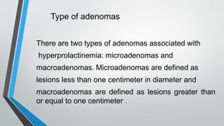
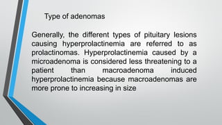
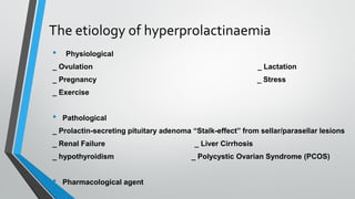
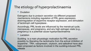
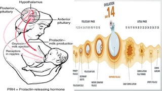
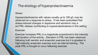
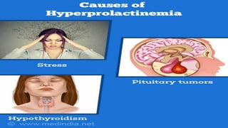
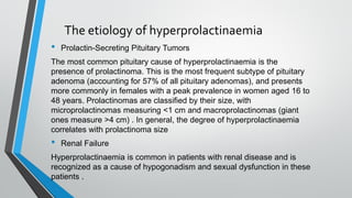
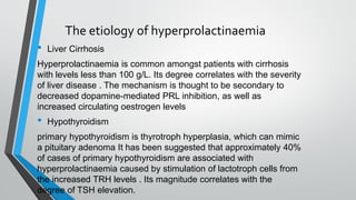
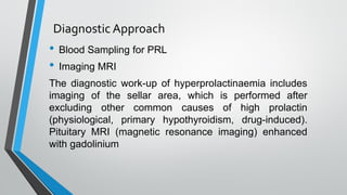
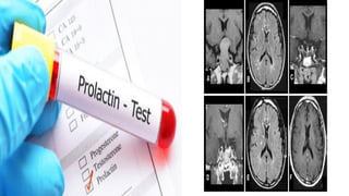
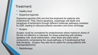
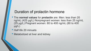
Causes and complications leading to
Hyperprolactinemia
Wurood Hasan Hadi
University of Kufa, Faculty of Science, Department of
Biology-M.sc. Zoology
Headings
I.Introduction
II.Disturbance in prolactin releasing
III.Type of adenomas
IV.Etiology
V.Aim of treatment
VI.Treatment
Introduction
Prolactin is a hormone produced in the pituitary
gland, named because of its role in lactation. It
also has other wide ranging functions in the body,
from acting on the reproductive system to
influencing behavior and regulating the immune
system.
Introduction
Prolactin was first discovered between the late 1920s
and early 1930s after different authors independently
demonstrated lobular-alveolar development and lactation
in rabbits after injection of an anterior pituitary extracts .
In 1933, It was first separated from growth hormone (GH)
in human pituitary glands by Friesen et al.
Introduction
Secretion of Prolactin by the pituitary gland has a
circadian rhythm with higher levels during sleep and
lower during wakefulness . Increased concentrations are
also seen during ovulation . Wheares, inhibiting by many
factors include dopamine, gamma-aminobutyric acid
(GABA), and somatostatin. Dopamine is the main
inhibitory factor involved in Prolactin regulation
Disturbance in prolactin releasing
Hyperprolactinemia is a common problems in clinical
endocrinology specially in prolactin hormone level in the
blood cause of menstrual disturbances affecting young
women.
There is a diversity of causes, It relates with various etiologies
(physiological, pathological, pharmacological), such as
pregnancy as a physiological factor, hypothyroidism or other
tumors in the hypothalamus-pituitary region, as well as
macroprolactinemia can also be considered as a pathological
factor.
Pharmalogical such as treatment with dopaminergic agonists
Medications and sellar/parasellar masses
(adenomas) _The sellar region includes the
sella turcica and the pituitary gland, together with
the ventral adenohypophysis and dorsal
neurohypophysis. The parasellar region
encompasses the cavernous sinuses,
suprasellar cistern, hypothalamus, and ventral
inferior third ventricle_ are the most common
causes of pathological hyperprolactinaemia.
Type of adenomas
There are two types of adenomas associated with
hyperprolactinemia: microadenomas and
macroadenomas. Microadenomas are defined as
lesions less than one centimeter in diameter and
macroadenomas are defined as lesions greater than
or equal to one centimeter .
Generally, the different types of pituitary lesions
causing hyperprolactinemia are referred to as
prolactinomas. Hyperprolactinemia caused by a
microadenoma is considered less threatening to a
patient than macroadenoma induced
hyperprolactinemia because macroadenomas are
more prone to increasing in size
Type of adenomas
The etiology of hyperprolactinaemia
• Physiological
_ Ovulation _ Lactation
_ Pregnancy _ Stress
_ Exercise
• Pathological
_ Prolactin-secreting pituitary adenoma “Stalk-effect” from sellar/parasellar lesions
_ Renal Failure _ Liver Cirrhosis
_ hypothyroidism _ Polycystic Ovarian Syndrome (PCOS)
• Pharmacological agent
The etiology of hyperprolactinaemia
• Ovulation
Estrogens due to prolacin secretion via different proposed
mechanisms including regulation of PRL gene expression,
downregulation of dopamine receptor expression, and stimulation
of lactotroph cell hyperplasia .
Accordingly, PRL levels are influenced by the menstrual cycle,
menopause, and pregnancy, and any high estrogen state (e.g.,
pregnancy) is a potential cause hyperprolactinaemia
• Lactation
Suckling is a main physiologic motivation for PRL secretion
possibly by liberating the lactotrophs from the activat inhibition of
dopamine . TRH, vasopressin, oxytocin, and salsolinol have also
been proposed as factors involved in the suckling-induced PRL
release
The etiology of hyperprolactinaemia
Stress
Hyperprolactinaemia with values usually up to 100 g/L may be
observed as a response to stress , It has been postulated that
stress-induced changes in dopamine and serotonin may a
ect PRL release contributing to hyperprolactinaemia in this setting
Exercise
Exercise increases PRL in a magnitude proportional to ​the intensity
and duration of the activity . Elevation in PRL has been observed
following both aerobic and anaerobic exercise but it is greatest in
high-intensity anaerobic exercise such as interval training . The
peak PRL is thought to occur following exercise.
The etiology of hyperprolactinaemia
• Prolactin-Secreting Pituitary Tumors
The most common pituitary cause of hyperprolactinaemia is the
presence of prolactinoma. This is the most frequent subtype of pituitary
adenoma (accounting for 57% of all pituitary adenomas), and presents
more commonly in females with a peak prevalence in women aged 16 to
48 years. Prolactinomas are classified by their size, with
microprolactinomas measuring <1 cm and macroprolactinomas (giant
ones measure >4 cm) . In general, the degree of hyperprolactinaemia
correlates with prolactinoma size
• Renal Failure
Hyperprolactinaemia is common in patients with renal disease and is
recognized as a cause of hypogonadism and sexual dysfunction in these
patients .
The etiology of hyperprolactinaemia
• Liver Cirrhosis
Hyperprolactinaemia is common amongst patients with cirrhosis
with levels less than 100 g/L. Its degree correlates with the severity
of liver disease . The mechanism is thought to be secondary to
decreased dopamine-mediated PRL inhibition, as well as
increased circulating oestrogen levels
• Hypothyroidism
primary hypothyroidism is thyrotroph hyperplasia, which can mimic
a pituitary adenoma It has been suggested that approximately 40%
of cases of primary hypothyroidism are associated with
hyperprolactinaemia caused by stimulation of lactotroph cells from
the increased TRH levels . Its magnitude correlates with the
degree of TSH elevation.
The etiology of hyperprolactinaemia
• Polycystic Ovarian Syndrome
Hyperprolactinaemia has been reported in 7 to 52% of women with
polycystic ovarian syndrome
(PCOS) . Its pathogenesis has not been elucidated; oestrogen-
mediated stimulation of lactotrophs and relative dopamine
deficiency have been proposed . Given the presence of PRL
receptors in the adrenal gland [8], it has also been postulated that
hyperprolactinaemia stimulates adrenal androgen production
contributing to the hyperandrogenism of PCOS.
Diagnostic Approach
• Blood Sampling for PRL
• Imaging MRI
The diagnostic work-up of hyperprolactinaemia includes
imaging of the sellar area, which is performed after
excluding other common causes of high prolactin
(physiological, primary hypothyroidism, drug-induced).
Pituitary MRI (magnetic resonance imaging) enhanced
with gadolinium
Aim of treatment
Hypogonadism and galactorrhea are well-recognized
manifestations of hyper prolactin , such as it is also implicating on
bone health, metabolism and immune system . The therapeutic
targets in prolactinomas are to reduce tumor size, resolve clinical
manifestations of hyperprolactinemia and of mass effects
(particularly visual disturbances) and prevent progression or
recurrence of the tumor
Treatment mainly aims at restoration and maintenance of normal
gonadal function/fertility, and prevention of osteoporosis.
Treatment
• Healthy food
• Dopamine Agonists
Dopamine agonists (DA) are first line treatment for patients with
prolactinoma They induce apoptosis, autophagic cell death and
paraptosis of lactotrophs through different molecular pathways mediated
by D2R, leading to reduced prolactin secretion and tumor shrinkage
• Surgery
Surgery could be considered for prolactinomas where maximum doses of
DA are not effective or tolerated, for those presenting with pituitary
apoplexy with visual deterioration, when there are cystic tumor
components compressing the visual pathway, or if cerebrospinal fluid
leak develops . Surgery may also be an option for young patients with
macroprolactinoma
• Radiotherapy
Duration of prolactin hormone
• The normal values for prolactin are: Men: less than 20
ng/mL (425 µg/L) Nonpregnant women: less than 25 ng/mL
(25 µg/L) Pregnant women: 80 to 400 ng/mL (80 to 400
µg/L)
• Half life 20 minuets
• Metabolized at liver and kidney
Reference
1. Tucker, H.A. Hormones, mammary growth, and lactation: A 41-year
perspective. J. Dairy Sci. 2000, 83, 874–884.
2. Trott, J.F.; Vonderhaar, B.K.; Hovey, R.C. Historical perspectives of
prolactin and growth hormone as mammogens, lactogens and
galactagogues—Agog for the future! J. Mammary Gland Biol. Neoplasia
2008, 13,
3. Friesen, H.; Guyda, H.; Hardy, J. Biosynthesis of Human Growth Hormone
and Prolactin. J. Clin. Endocrinol. Metab. 1970, 31, 611–624.
4. Capozzi, A.; Scambia, G.; Pontecorvi, A.; Lello, S. Hyperprolactinemia:
Pathophysiology and therapeutic approach. Gynecol. Endocrinol. 2015, 31,
506–510.
5. Teilum, K.; Hoch, J.C.; Gon, V.; Kinet, S.; Martial, J.A.; Kragelund, B.B.
Solution structure of human prolactin. J. Mol. Biol. 2005, 351, 810–823.
Reference
6. Cabrera-Reyes, E.A.; Limon-Morales, O.; Rivero-Segura, N.A.;
Camacho-Arroyo, I.; Cerbin, M. Prolactin function and putative
expression in the brain. Endocrine 2017, 57, 199–213.
7. Levine, S.; Muneyyirci-Delale, O. Stress-Induced Hyperprolactinemia:
Pathophysiology and Clinical Approach. Obs. Gynecol. Int. 2018,
2018, 9253083.
8. Irene Samperi ,Kirstie Lithgow and Niki Karavitaki,
Hyperprolactinaemia. Journal Clinical Medicine, 2019.
Hyperprolactinoma

Hyperprolactinoma

  • 1.
    Causes and complicationsleading to Hyperprolactinemia Wurood Hasan Hadi University of Kufa, Faculty of Science, Department of Biology-M.sc. Zoology
  • 2.
    Headings I.Introduction II.Disturbance in prolactinreleasing III.Type of adenomas IV.Etiology V.Aim of treatment VI.Treatment
  • 3.
    Introduction Prolactin is ahormone produced in the pituitary gland, named because of its role in lactation. It also has other wide ranging functions in the body, from acting on the reproductive system to influencing behavior and regulating the immune system.
  • 4.
    Introduction Prolactin was firstdiscovered between the late 1920s and early 1930s after different authors independently demonstrated lobular-alveolar development and lactation in rabbits after injection of an anterior pituitary extracts . In 1933, It was first separated from growth hormone (GH) in human pituitary glands by Friesen et al.
  • 5.
    Introduction Secretion of Prolactinby the pituitary gland has a circadian rhythm with higher levels during sleep and lower during wakefulness . Increased concentrations are also seen during ovulation . Wheares, inhibiting by many factors include dopamine, gamma-aminobutyric acid (GABA), and somatostatin. Dopamine is the main inhibitory factor involved in Prolactin regulation
  • 6.
    Disturbance in prolactinreleasing Hyperprolactinemia is a common problems in clinical endocrinology specially in prolactin hormone level in the blood cause of menstrual disturbances affecting young women. There is a diversity of causes, It relates with various etiologies (physiological, pathological, pharmacological), such as pregnancy as a physiological factor, hypothyroidism or other tumors in the hypothalamus-pituitary region, as well as macroprolactinemia can also be considered as a pathological factor. Pharmalogical such as treatment with dopaminergic agonists
  • 7.
    Medications and sellar/parasellarmasses (adenomas) _The sellar region includes the sella turcica and the pituitary gland, together with the ventral adenohypophysis and dorsal neurohypophysis. The parasellar region encompasses the cavernous sinuses, suprasellar cistern, hypothalamus, and ventral inferior third ventricle_ are the most common causes of pathological hyperprolactinaemia.
  • 8.
    Type of adenomas Thereare two types of adenomas associated with hyperprolactinemia: microadenomas and macroadenomas. Microadenomas are defined as lesions less than one centimeter in diameter and macroadenomas are defined as lesions greater than or equal to one centimeter .
  • 9.
    Generally, the differenttypes of pituitary lesions causing hyperprolactinemia are referred to as prolactinomas. Hyperprolactinemia caused by a microadenoma is considered less threatening to a patient than macroadenoma induced hyperprolactinemia because macroadenomas are more prone to increasing in size Type of adenomas
  • 10.
    The etiology ofhyperprolactinaemia • Physiological _ Ovulation _ Lactation _ Pregnancy _ Stress _ Exercise • Pathological _ Prolactin-secreting pituitary adenoma “Stalk-effect” from sellar/parasellar lesions _ Renal Failure _ Liver Cirrhosis _ hypothyroidism _ Polycystic Ovarian Syndrome (PCOS) • Pharmacological agent
  • 11.
    The etiology ofhyperprolactinaemia • Ovulation Estrogens due to prolacin secretion via different proposed mechanisms including regulation of PRL gene expression, downregulation of dopamine receptor expression, and stimulation of lactotroph cell hyperplasia . Accordingly, PRL levels are influenced by the menstrual cycle, menopause, and pregnancy, and any high estrogen state (e.g., pregnancy) is a potential cause hyperprolactinaemia • Lactation Suckling is a main physiologic motivation for PRL secretion possibly by liberating the lactotrophs from the activat inhibition of dopamine . TRH, vasopressin, oxytocin, and salsolinol have also been proposed as factors involved in the suckling-induced PRL release
  • 13.
    The etiology ofhyperprolactinaemia Stress Hyperprolactinaemia with values usually up to 100 g/L may be observed as a response to stress , It has been postulated that stress-induced changes in dopamine and serotonin may a ect PRL release contributing to hyperprolactinaemia in this setting Exercise Exercise increases PRL in a magnitude proportional to ​the intensity and duration of the activity . Elevation in PRL has been observed following both aerobic and anaerobic exercise but it is greatest in high-intensity anaerobic exercise such as interval training . The peak PRL is thought to occur following exercise.
  • 15.
    The etiology ofhyperprolactinaemia • Prolactin-Secreting Pituitary Tumors The most common pituitary cause of hyperprolactinaemia is the presence of prolactinoma. This is the most frequent subtype of pituitary adenoma (accounting for 57% of all pituitary adenomas), and presents more commonly in females with a peak prevalence in women aged 16 to 48 years. Prolactinomas are classified by their size, with microprolactinomas measuring <1 cm and macroprolactinomas (giant ones measure >4 cm) . In general, the degree of hyperprolactinaemia correlates with prolactinoma size • Renal Failure Hyperprolactinaemia is common in patients with renal disease and is recognized as a cause of hypogonadism and sexual dysfunction in these patients .
  • 16.
    The etiology ofhyperprolactinaemia • Liver Cirrhosis Hyperprolactinaemia is common amongst patients with cirrhosis with levels less than 100 g/L. Its degree correlates with the severity of liver disease . The mechanism is thought to be secondary to decreased dopamine-mediated PRL inhibition, as well as increased circulating oestrogen levels • Hypothyroidism primary hypothyroidism is thyrotroph hyperplasia, which can mimic a pituitary adenoma It has been suggested that approximately 40% of cases of primary hypothyroidism are associated with hyperprolactinaemia caused by stimulation of lactotroph cells from the increased TRH levels . Its magnitude correlates with the degree of TSH elevation.
  • 17.
    The etiology ofhyperprolactinaemia • Polycystic Ovarian Syndrome Hyperprolactinaemia has been reported in 7 to 52% of women with polycystic ovarian syndrome (PCOS) . Its pathogenesis has not been elucidated; oestrogen- mediated stimulation of lactotrophs and relative dopamine deficiency have been proposed . Given the presence of PRL receptors in the adrenal gland [8], it has also been postulated that hyperprolactinaemia stimulates adrenal androgen production contributing to the hyperandrogenism of PCOS.
  • 18.
    Diagnostic Approach • BloodSampling for PRL • Imaging MRI The diagnostic work-up of hyperprolactinaemia includes imaging of the sellar area, which is performed after excluding other common causes of high prolactin (physiological, primary hypothyroidism, drug-induced). Pituitary MRI (magnetic resonance imaging) enhanced with gadolinium
  • 20.
    Aim of treatment Hypogonadismand galactorrhea are well-recognized manifestations of hyper prolactin , such as it is also implicating on bone health, metabolism and immune system . The therapeutic targets in prolactinomas are to reduce tumor size, resolve clinical manifestations of hyperprolactinemia and of mass effects (particularly visual disturbances) and prevent progression or recurrence of the tumor Treatment mainly aims at restoration and maintenance of normal gonadal function/fertility, and prevention of osteoporosis.
  • 21.
    Treatment • Healthy food •Dopamine Agonists Dopamine agonists (DA) are first line treatment for patients with prolactinoma They induce apoptosis, autophagic cell death and paraptosis of lactotrophs through different molecular pathways mediated by D2R, leading to reduced prolactin secretion and tumor shrinkage • Surgery Surgery could be considered for prolactinomas where maximum doses of DA are not effective or tolerated, for those presenting with pituitary apoplexy with visual deterioration, when there are cystic tumor components compressing the visual pathway, or if cerebrospinal fluid leak develops . Surgery may also be an option for young patients with macroprolactinoma • Radiotherapy
  • 25.
    Duration of prolactinhormone • The normal values for prolactin are: Men: less than 20 ng/mL (425 µg/L) Nonpregnant women: less than 25 ng/mL (25 µg/L) Pregnant women: 80 to 400 ng/mL (80 to 400 µg/L) • Half life 20 minuets • Metabolized at liver and kidney
  • 26.
    Reference 1. Tucker, H.A.Hormones, mammary growth, and lactation: A 41-year perspective. J. Dairy Sci. 2000, 83, 874–884. 2. Trott, J.F.; Vonderhaar, B.K.; Hovey, R.C. Historical perspectives of prolactin and growth hormone as mammogens, lactogens and galactagogues—Agog for the future! J. Mammary Gland Biol. Neoplasia 2008, 13, 3. Friesen, H.; Guyda, H.; Hardy, J. Biosynthesis of Human Growth Hormone and Prolactin. J. Clin. Endocrinol. Metab. 1970, 31, 611–624. 4. Capozzi, A.; Scambia, G.; Pontecorvi, A.; Lello, S. Hyperprolactinemia: Pathophysiology and therapeutic approach. Gynecol. Endocrinol. 2015, 31, 506–510. 5. Teilum, K.; Hoch, J.C.; Gon, V.; Kinet, S.; Martial, J.A.; Kragelund, B.B. Solution structure of human prolactin. J. Mol. Biol. 2005, 351, 810–823.
  • 27.
    Reference 6. Cabrera-Reyes, E.A.;Limon-Morales, O.; Rivero-Segura, N.A.; Camacho-Arroyo, I.; Cerbin, M. Prolactin function and putative expression in the brain. Endocrine 2017, 57, 199–213. 7. Levine, S.; Muneyyirci-Delale, O. Stress-Induced Hyperprolactinemia: Pathophysiology and Clinical Approach. Obs. Gynecol. Int. 2018, 2018, 9253083. 8. Irene Samperi ,Kirstie Lithgow and Niki Karavitaki, Hyperprolactinaemia. Journal Clinical Medicine, 2019.