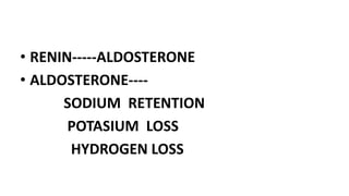
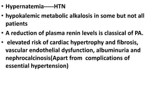
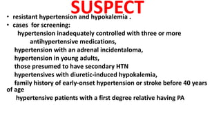
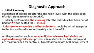
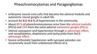
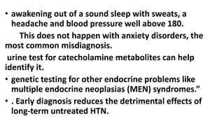
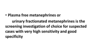
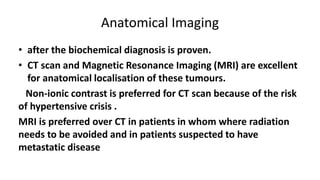
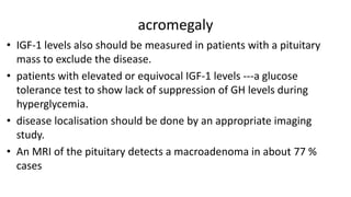
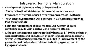
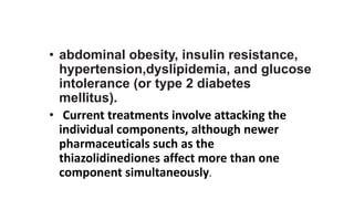
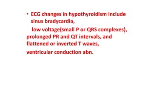
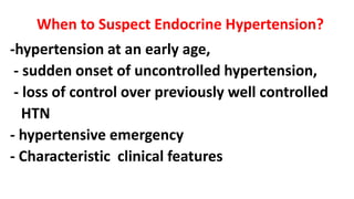
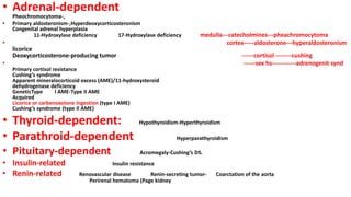
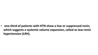
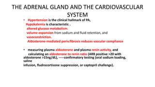
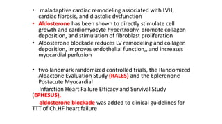
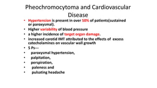
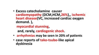
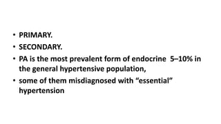
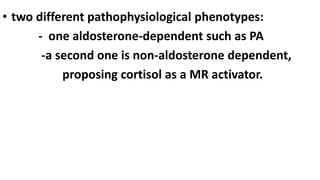
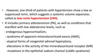
6. Endocrine hypertension
• the second most common cause of secondary hypertension after renal ds.
• a major aetiological factor for resistant hypertension.
• A diagnosis of endocrine hypertension may be obvious :
a typical clinical features of the underlying condition like acromegaly,
Cushing syndrome, hyperthyroidism, hypothyroidism and features of
virilisation in congenital adrenal hyperplasia .
a typical history e.g. paroxysmal palpitations and orthostatic
hypotension in phaeochromocytoma .
Occasionally routine laboratory tests in a patient with hypertension may
raise the suspicion of an endocrine cause e.g. hypokalemia in
mineralcorticoid excess
7. When to Suspect Endocrine Hypertension?
-hypertension at an early age,
- sudden onset of uncontrolled hypertension,
- loss of control over previously well controlled
HTN
- hypertensive emergency
- Characteristic clinical features
9. • one-third of patients with HTN show a low or suppressed renin,
which suggests a systemic volume expansion, called as low-renin
hypertension (LRH).
10. THE ADRENAL GLAND AND THE CARDIOVASCULAR
SYSTEM
• Hypertension is the clinical hallmark of PA,
Hypokalemia is characteristic .
altered glucose metabolism.
volume expansion from sodium and fluid retention, and
vasoconstriction.
Aldosterone-mediated periv.fibrosis reduces vascular compliance
• measuring plasma aldosterone and plasma renin activity, and
calculating an aldosterone to renin ratio (ARR positive >20 with
aldosterone >15ng/dL), -----confirmatory testing (oral sodium loading,
saline
infusion, fludrocortisone suppression, or captopril challenge).
11. • maladaptive cardiac remodeling associated with LVH,
cardiac fibrosis, and diastolic dysfunction
• Aldosterone has been shown to directly stimulate cell
growth and cardiomyocyte hypertrophy, promote collagen
deposition, and stimulation of fibroblast proliferation
• Aldosterone blockade reduces LV remodeling and collagen
deposition, improves endothelial function,, and increases
myocardial perfusion
• two landmark randomized controlled trials, the Randomized
Aldactone Evaluation Study (RALES) and the Eplerenone
Postacute Myocardial
Infarction Heart Failure Efficacy and Survival Study
(EPHESUS),
aldosterone blockade was added to clinical guidelines for
TTT of Ch.HF heart failure
12. Pheochromocytoma and Cardiovascular
Disease
• Hypertension is present in over 50% of patients(sustained
or paroxysmal).
• Higher variability of blood pressure
• a higher incidence of target organ damage.
• increased carotid IMT attributed to the effects of excess
catecholamines on vascular wall growth
• 5 Ps---
• paroxysmal hypertension,
• palpitation,
• perspiration,
• paleness and
• pulsating headache
13. • Excess catecholamine causer
cardiomyopathy (DCM,HCM,26%),, ischemic
heart disease(VC, increased cardiac oxygen
demand. ),
myocardial stunning,
and, rarely, cardiogenic shock.
• arrhythmias may be seen in 20% of patients
• case reports of tako-tsubo-like apical
dyskinesia
15. • PRIMARY.
• SECONDARY.
• PA is the most prevalent form of endocrine 5–10% in
the general hypertensive population,
• some of them misdiagnosed with “essential”
hypertension
16. • two different pathophysiological phenotypes:
- one aldosterone-dependent such as PA
-a second one is non-aldosterone dependent,
proposing cortisol as a MR activator.
17. • . However, one-third of patients with hypertension show a low or
suppressed renin, which suggests a systemic volume expansion,
called as low-renin hypertension (LRH).
• It includes primary aldosteronism (PA), as well as conditions that
manifest with low aldosterone levels, such as
- endogenous hypercortisolism,
- syndrome of apparent mineralocorticoid excess (AME),
- atypical forms of congenital adrenal hyperplasia,
-alterations in the activity of the mineralocorticoid receptor (MR)
-mutations in the epithelial sodium channel (Liddle syndrome)
19. Primary Mineralocorticoid Excess
• Primary Aldosteronism (PA)
5–13 % of cases of hypertension between 30-60 years
About 10 % of cases in hypertension clinics.
4 % in the community .
20 % with resistant hypertension .
.
Nearly 60–65 % of cases of PA are from idiopathic hyperaldosteronism
(IHA)
30–35 % aldosterone producing adrenal adenoma (APA)
5 % of cases of PA are familial [FH-I, II and III]) with clear
genetic background .
21. • Hypernatemia-----HTN
• hypokalemic metabolic alkalosis in some but not all
patients
• A reduction of plasma renin levels is classical of PA.
• elevated risk of cardiac hypertrophy and fibrosis,
vascular endothelial dysfunction, albuminuria and
nephrocalcinosis(Apart from complications of
essential hypertension)
22. SUSPECT
• resistant hypertension and hypokalemia .
• cases for screening:
hypertension inadequately controlled with three or more
antihypertensive medications,
hypertension with an adrenal incidentaloma,
hypertension in young adults,
those presumed to have secondary HTN
hypertensives with diuretic-induced hypokalemia,
family history of early-onset hypertension or stroke before 40 years
of age
hypertensive patients with a first degree relative having PA
23. Diagnostic Approach
• Initial Screening
estimation of plasma aldosterone and renin levels with the calculation
of aldosterone to renin ratio (ARR).
ideally performed in the morning after the individual has been out of
bed for 2 h and seated for 5–15 min
Aldosterone antagonists and beta-blockers should be withdrawn prior
to the test as they disproportionately affect the ARR.
Antihypertensives such as verapamil(slow release), hydralazine and
alpha-adrenergic blockers possess minimal effects on RAA system and
are recommended for control of hypertension before ARR measurement
24. • Biochemical Confirmation
four confirmatory tests :
1-oral sodium loading with measurement of urinary aldosterone excretion,
2-intravenous saline infusion with measurement of plasma aldosterone levels
3- fludorcortisone suppression test
4-captopril challenge test.
3 And 4 tests are not routinely used now,
1 and 2 tests may become necessary in cases with mild PA, where the ARR is only
marginally elevated in the absence of severe hypertension or heart failure.
25. • Adrenal Imaging
• Computed tomography (CT) scan of the adrenal glands is
recommended in all cases of PA for localisation of the disease and
to exclude the small possibility of an adrenocortical carcinoma
• Adrenal CT is preferred over magnetic resonance imaging (MRI) as
an imaging modality for localisation purpose because it is more
economical and has better spatial resolution.
• However, adrenal imaging had a low sensitivity of 58.6 % to detect
unilateral disease
26. Adrenal Venous Sampling (AVS)
• recommended in most cases for lateralisation of the abnormal
adrenal gland if a surgical cure is contemplated.
BUT:
lack of availability in many centres and
higher complication rates (about 5 %).
avoiding AVS in the following situations:
Age is < 40 years with marked PA in presence of a typical
unilateral adrenal adenoma and normal appearance of the opposite
adrenal on CT,
suspicion of adrenocortical carcinoma from adrenal imaging,
high risk cases for adrenalectomy,
proven cases of FH-I and FH-III .
27. MANEGMENT
• Laparoscopic adrenalectomy
• Medical Therapy
bilateral disease
cases not appropriate for adrenalectomy.
MR antagonist spironolactone is the first line of treatment. With a starting
dose of 12.5–100 mg daily) .
• Eplerenone, a selective MR antagonist, is started at a dose of 25 mg once or
twice daily.
• Caution must be taken in chronic kidney disease stage 3
• Amiloride/triamterene are useful alternative.
• PA resulting from FH-I (GRA) should be managed with glucocorticoids in adults
and MR antagonists in paediatric cases (because of the effect of steroids on
growth retardation) until they reach adulthood
28. Pheochromocytomas and Paragangliomas
• embryonic neural crest cells that become the adrenal medulla and
autonomic neural ganglia in adult life.
• account for 0.2–0.6 % of hypertension in the community
• About 85 % of pheochromocytomas arise from the adrenal medulla
(PCCs) and 15% from the extra-adrenal autonomic ganglia (PGLs).
• intense vasospasm and hypertension through α-adrenergic effect,
and vasodilatation, diaphoresis and tachycardia from the β-
adrenergic effect .
• Severe orthostatic hypotension with syncopal episodes can
occasionally result from unbalanced effects of α.
29. • awakening out of a sound sleep with sweats, a
headache and blood pressure well above 180.
This does not happen with anxiety disorders, the
most common misdiagnosis.
urine test for catecholamine metabolites can help
identify it.
• genetic testing for other endocrine problems like
multiple endocrine neoplasias (MEN) syndromes.”
• . Early diagnosis reduces the detrimental effects of
long-term untreated HTN.
30. • Plasma free metanephrines or
urinary fractionated metanephrines is the
screening investigation of choice for suspected
cases with very high sensitivity and good
specificity
31. Anatomical Imaging
• after the biochemical diagnosis is proven.
• CT scan and Magnetic Resonance Imaging (MRI) are excellent
for anatomical localisation of these tumours.
Non-ionic contrast is preferred for CT scan because of the risk
of hypertensive crisis .
MRI is preferred over CT in patients in whom where radiation
needs to be avoided and in patients suspected to have
metastatic disease
32. Functional Imaging
• After anatomical diagnosis is established.
• to exclude the possibility of metastasis and multisite disease in cases of
PGLs.
• 123I-metaiodobenzylguanidine (MIBG) scitigraphy is the usual functional
imaging modality utilized in most centers.
• The sensitivity and specificity of 123IMIBG scintigraphy is around 85 % in
PCCs.
• A variety of different radio-pharmaceutical agents can be used for
functional imaging in cases with a negative 123I-MIBG scintigraphy
• Genetic Testing
33. Peri-Operative Management
• manipulation of the tumour during surgery results in hypertensive crisis because of the
massive release of catecholamines to circulation
• Prompt control of hypertension and appropriate preoperative preparation.
non-selective α-adrenergic blockers such as phenoxybenzamine (10 mg BD to a
maximum of 1 mg/kg/day) or
α-1 selective agent doxazosin (2–32 mg/day) 10–14 days prior to the surgery along with
liberal intake of fluids and salt to replenish volume depletion is recommended
• Addition of a β-adrenergic blocker such as propranolol or atenolol may be necessary after
few days of starting α-blockers.
• Other antihypertensive medications such as calcium channel antagonists and metyrosine
may be necessary for optimal control of BP in some cases
• The target BP control should be < 130/80 mm Hg while seated and > 90 mm systolic while
standing and a heart rate 70–80 per minute
36. Cushing’s Syndrome and Cardiovascular
Disease
Hypercortisolism is associated with
- HTN,
-central obesity,-----BUFFALO BUTTOCK LEMON
-insulin resistance, diabetes
-dyslipidemia
-acne,
-menstrual disorders,hirsutism
-alterations in clotting and platelet function
37. • Cushing’s syndrome has been associated with
LVH, concentric remodeling, diastolic
dysfunction, and subclinical LV systolic
dysfunction.
• patients may continue to exhibit exercise
intolerance due to steroid-induced myopathy .
39. • initially:
24-hour urinary free cortisol (at least 2 samples),
late night salivary cortisol,
1-mg overnight dexamethasone suppression test (DST) or low-dose DST (0.5 mg QDS for 48 h).
Random plasma cortisol measurement has no value for screening cases of CS because of the marked
variability of levels depending of many factors.
Confirmation :
. Measurement of corticotrophin (ACTH) levels is the next step in diagnosis.
Suppressed level of ACTH (adrenal/iatrogenic )-------a contrast CT scan of the adrenal glands should
be done.
If ACTH levels are high or high normal, ------- MRI of pituitary should be done.
A high-dose DST (using 2 mg QDS for 48 h) and/or CRH stimulation tests if the pituitary mass is smaller or
absent, in presence of raised ACTH levels.
A suppressible cortisol with high-dose DST or 20 % rise in cortisol following CRH administration indicates
pituitary-driven corticotrophin excess is well recognised.
, imaging of thorax and/or abdomen and pelvis should be performed to identify an ectopic source of ACTH.
Finally, an octreotide scan may be necessary if all other confirmatory tests are negative
41. -pts with prolactinoma may be treated for
decades thus increasing risks of valvulopathy,
including T.R., M.R,, and A.R..
-SO,clinicians are advised to use the lowest
possible doses of dopamine agonists(including
cabergoline, bromocriptime, and quinagolide) ,
with Echocardiographic monitoring.
-Peripartum cardiomyopathy is a rare clinical
entity. It has been suggested that a 16 kDa
prolactin fragment may play a role in its
pathophysiology.
-Case reports have described the use of
bromocriptine in addition to standard heart failure
therapy in peripartum cardiomyopathy.
42. Growth Hormone
• It acts directly on tissues via interaction with the GH receptor,
and indirectly via stimulation of insulin-like growth factor type
1 (IGF-1) synthesis.
• In virtually all cell types, IGF-1 promotes glucose uptake and
cellular protein synthesis. GH and IGF-1 regulate somatic
growth, including cardiac development and function
43. • GH Defiency is associated with
increased body fat and central adiposity,
dyslipidemia ,low HDLc,high total cholesterol, and
high LDLc, endothelial dysfunction, and insulin
resistance and Increased carotid arterial intima-media
thickness (IMT).
GH replacement therapy can result in increased
lean body mass and decreased visceral adipose
tissue,and may decrease total and LDLc levels,
Several studies have shown that GH replacement
therapy improves cardiac performance and increases
LV mass, LVEDV, and stroke volume
44. Acromegaly :-
Hypertension occurs in 20%-50%
increased prevalence of diabetes mellitus.
Cardiac histological abnormalities in acromegaly include myocyte
hypertrophy, interstitial fibrosis, inflammatory cell infiltration, reduced capillary
density, myofibril derangement, and extracellular collagen deposition.
early stage of acromegaly, there is enhanced myocardial contractility
overall increased cardiac performance.
intermediate stage, after about 5 years of active disease, there is
biventricular hypertrophy, diastolic dysfunction, and impaired exertional cardiac
performance.
Late-stage acromegalic cardiomyopathy is characterized by systolic and
diastolic dysfunction, increased myocardial mass, increased systemic vascular
resistance.
• Acromegalic cardiomyopathy is frequently present at diagnosis in two thirds
of patients including about half of all normotensive acromegalics.
• Patients with severe cardiomyopathy may progress to heart failure, with
heart failure seen in 3%-10% of patients.
45. • Cardiac valve disease (aortic and mitral regurgitation)
is frequent in acromegaly(myxomatous valvulopathy. )
and often persist despite treatment.
• cardiac rhythm abnormalities in acromegaly.
severity of vent. arrhythmias correlates with LV mass.
• Resting ECG changes include left axis deviation,
increased QT intervals, septal Q-waves, and ST-T wave
depression.
• Somatostatin analogs have been shown to reduce QT
intervals, and to improve the arrhythmic profile in
acromegalic patients.
46. acromegaly
• IGF-1 levels also should be measured in patients with a pituitary
mass to exclude the disease.
• patients with elevated or equivocal IGF-1 levels ---a glucose
tolerance test to show lack of suppression of GH levels during
hyperglycemia.
• disease localisation should be done by an appropriate imaging
study.
• An MRI of the pituitary detects a macroadenoma in about 77 %
cases
47. Iatrogenic Hormone Manipulation
• development of/or worsening of hypertension.
• Glucocorticoid administration is the most common cause .
• Prevalence of Steroid-induced hypertension increases with age,
• new-onset hypertension was observed in 22 % of cases receiving
long term steroids .
• hormone replacement in post-menopausal women showed
conflicting results with positive , and negative effects .
• Although testosterone can theoretically increase BP by the effects of
vasoconstriction and stimulation of renin-angiotensinaldosterone
system , testosterone replacement resulted in improvement of the
parameters of metabolic syndrome including hypertension in
hypogonadal men
48. • abdominal obesity, insulin resistance,
hypertension,dyslipidemia, and glucose
intolerance (or type 2 diabetes
mellitus).
• Current treatments involve attacking the
individual components, although newer
pharmaceuticals such as the
thiazolidinediones affect more than one
component simultaneously.
49. Agenda
-Introduction
-Endocrine Signals and the Cardiovascular System
-Nuclear Receptors
-Second Messenger Signaling Systems
-Clinical Endocrinology:-
A. Hypertension
B. Metabolic syndrome
C. Obesity
D. Dyslipidemia
E. Thyroid disease
F. Cushing’s syndrome
G. Diabetes and cardiovascular disease
VI. Hormone Replacement Therapy
51. • Both and hyperthyroidism have deleterious effects on
CVS.
• In hypothyroidism:-
elevations of LDL, hypertension,cholesreol
pericardial effusion , cardiomyopathy and heart failure.
Hyperthyroidism can lead to hypertension, atrial
arrhythmias, including A.F., precipitate or exacerbate angina
pectoris and myocardial infarction, and heart failure.
In addition, up to 15% of women over age of 60 have
subclinical hypothyroidism(elevations of LDL?) .
Furthermore, subclinical hyperthyroidism, may be
associated with an increased incidence of atrial fibrillation.
management with or without H F……????
53. cardiovascular hemodynamic changes in overt hyperthyroidism
• decreased systemic vascular resistance (SVR),
• increased H.R.,
• increased cardiac preload, and increased cardiac
output.
(The decrease in SVR activates RAAS, leading to
increased plasma volume and increased cardiac
preload.
Thyroid hormone also promotes an increase in blood
volume via upregulation of erythropoietin secretion,
further enhancing cardiac preload.)
• the increase in cardiac output, which may be 50%-
300% higher than normal .
• Treatment of hyperthyroidism reverses these
hemodynamic changes.
54. • Hyperthyroid patients often present with
palpitations, sinus tachycardia, atrial fibrillation,
systoli hypertension, widened pulse pressure,
exercise intolerance, and exertional dyspnea.
• Systolic hypertension may be seen in up to 30%
of hyperthyroid patients.
• Sinus tachycardia occurs in approximately 40% of
cases of overt hyperthyroidism.
• Atrial fibrillation is the second most common
arrhythmia in overt hyperthyroidism, and occurs
in 10%-15% of patients, its prevalence increasing
with age
55. • Hypothyroid patients may present with
mild increased SVR, diastolic hypertension
(20%), narrowed pulse pressure, and
bradycardia.
• Cardiac output may be reduced by up to
30%-40% as a result of decreased stroke
volume and heart rate.
56. • In hypothyroidism there is resting LV
diastolic dysfunction, and both systolic and
diastolic dysfunction with exertion.
• Pericardial effusions occur in up to 25% of
patients with overt hypothyroidism
• hese pericardial effusions accumulate slowly
---SO,rare cases of cardiac tamponade
• resolve after 2-3 months of thyroid
hormone replacement
therapy.
57. • Overt hypothyroidism is associated with
accelerated atherosclerosis and CAD .
hypercholesterolemia,
increased LDLc (90% ).
increased Apo B and lipoprotein(a)
VLDL(TG) normal to increased,
HDL are variable
Overt hypothyroidism has also been
associated with hyperhomocysteinemia,
increased C-reactive protein levels, and altered
58. • ECG changes in hypothyroidism include
sinus bradycardia,
low voltage(small P or QRS complexes),
prolonged PR and QT intervals, and
flattened or inverted T waves,
ventricular conduction abn.
59. • Amiodarone, causes thyroid dysfunction in
15%–20% of treated patients, either causing
hypothyroidism or thyrotoxicosis
60. • Amiodarone-induced thyrotoxicosis (AIT) is present in
two forms:
type 1 AIT, or iodine-induced hyperthyroidism,
type 2 AIT, or destructive thyroiditis.
Type 1 AIT ---synthesis and release of excess thyroid
hormone,
Type 2 AIT ---- release of preformed thyroid hormone from
the inflamed thyroid gland.
Differentiating between the two forms can be difficult,
Type 1 AIT is managed with antithyroid drugs and possibly
potassium perchlorate.
Type 2 AIT is managed with glucocorticoids, beta-blockade,
and rarely thyroidectomy.
61. • Baseline thyroid function tests and
thyroid peroxidase antibodies should be
performed prior to initiating
amiodarone, and monitored every 6
months for the duration of
amiodarone therapy.
62. Congestive Heart Failure and Thyroid Hormone
• A low serum T3 is the most common thyroid function
abnormality in patients with heart failure(10%–30% ).
• It remains unclear whether this reduction in T3 is an
adaptive or maladaptive process.
• role of thyroid hormone therapy remains unclear in
patients with heart failure and low serum T3 level
• Current areas of research include thyroid hormone
replacement with T3 and/or T4, use of thyroid hormone
analogs (e.g. diiodothyropropionic acid), and gene therapy
to modify thyroid hormone receptor or deiodinase
expression and activity.
•
64. • LVH
Calcifications of the aortic valve, mitral valve, and
myocardium
Hypercalcemia, particularly serum calcium >12 mg/dL,
ECG findings include shortened QT and QTc intervals,
increased QRS amplitude, early peaking and gradual
down slope of the descending limb of the T wave,
biphasic T waves, and shortened ST segment intervals.
65. Hypoparathyroidism and cardiovascular disease
decreased myocardial performance,
dilated cardiomyopathy,
congestive heart failure
QT prolongation is ECG hallmark of
hypocalcemia,
ST segment elevation, possibly due to
coronary artery spasm.
68. Agenda
-Introduction
-Endocrine Signals and the Cardiovascular System
-Nuclear Receptors
-Second Messenger Signaling Systems
-Clinical Endocrinology:-
A. Hypertension
B. Metabolic syndrome
C. Obesity
D. Dyslipidemia
E. Thyroid disease
F. Cushing’s syndrome
G. Diabetes and cardiovascular disease
VI. Hormone Replacement Therapy
69. Agenda
-Introduction
-Endocrine Signals and the Cardiovascular System
-Nuclear Receptors
-Second Messenger Signaling Systems
-Clinical Endocrinology:-
A. Hypertension
B. Metabolic syndrome
C. Obesity
D. Dyslipidemia
E. Thyroid disease
F. Cushing’s syndrome
G. Diabetes and cardiovascular disease
VI. Hormone Replacement Therapy
70. Agenda
-Introduction
-Endocrine Signals and the Cardiovascular System
-Nuclear Receptors
-Second Messenger Signaling Systems
-Clinical Endocrinology:-
A. Hypertension
B. Metabolic syndrome
C. Obesity
D. Dyslipidemia
E. Thyroid disease
F. Cushing’s syndrome
G. Diabetes and cardiovascular disease
VI. Hormone Replacement Therapy
71. Agenda
-Introduction
-Endocrine Signals and the Cardiovascular System
-Nuclear Receptors
-Second Messenger Signaling Systems
-Clinical Endocrinology:-
A. Hypertension
B. Metabolic syndrome
C. Obesity
D. Dyslipidemia
E. Thyroid disease
F. Cushing’s syndrome
G. Diabetes and cardiovascular disease
VI. Hormone Replacement Therapy
73. • Relation with C.V. diseases: 2-4 FOLDS
M.I.
CVA
HF
CHD
PVD
MORTALIRT AND MORBIDITY
74. Agenda
-Introduction
-Endocrine Signals and the Cardiovascular System
-Nuclear Receptors
-Second Messenger Signaling Systems
-Clinical Endocrinology:-
A. Hypertension
B. Metabolic syndrome
C. Obesity
D. Dyslipidemia
E. Thyroid disease
F. Cushing’s syndrome
G. Diabetes and cardiovascular disease
VI. Hormone Replacement Therapy
76. • both estrogens and androgens have
major effects on the cardiovascular
system.
• there is controversy whether
deficiencies of both of these classes of
hormones, as occurs after menopause
and andropause, result in an increased
risk for developing cardiovascular
complications.
•
77. • Most clinicians agree that replacement of
androgens is indicated in men with testosterone
deficiency.
But estrogen replacement therapy is controversial.
• recent studies using estrogen and progestin
replacement therapy failed to demonstrate
improvement in cardiovascular risk in
postmenopausal women.
• Because estrogens have complex actions on other
tissues, including bone, uterus, breast, and the
central nervous system, the decision to initiate
estrogen replacement therapy has become more
complex.
78. Agenda
-Introduction
-Endocrine Signals and the Cardiovascular System
-Nuclear Receptors
-Second Messenger Signaling Systems
-Clinical Endocrinology:-
A. Hypertension
B. Metabolic syndrome
C. Obesity
D. Dyslipidemia
E. Thyroid disease
F. Cushing’s syndrome
G. Diabetes and cardiovascular disease
VI. Hormone Replacement Therapy
VII.PITUIRY GLAND
79. CONCLUSIONS
• Endocrine dysfunction may have a significant
impact on the cardiovascular system.
• Restoration of normal endocrine function
often results in reversal of cardiovascular
changes.
• Hormone-mediated cardiac changes should
be considered when evaluating endocrine
and cardiac patients